

Copyrights: Asghar Sepahvand, Hossein Eliasy, Mehdi Mohammadi, Ali Safarzadeh, Kimia Azarbaijani, Somayeh Shahsavari, Mohsen Alizadeh, Fatemeh Beyranvand, 2018. License: This work is licensed under a Creative Commons Attribution 4.0 International License.
Abstract
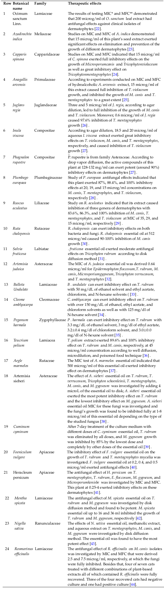
Fungi can evade the immune system via different processes, including recombination, mitosis, and expression of genes involved in oxidative stress responses. These processes can lead to chronic fungal diseases. Despite the growth of health care facilities, the incidence rate of fungal infections is still considerably high. Dermatophytes represent the main cause of cutaneous diseases. Dermatophytes attack keratinized tissues, such as nail, hair, and stratum corneum, due of their gravitation towards keratin, which leads to dermatophytosis. Medicinal plants have long been used to treat different diseases, and in the recent years, use of plant-based products to fight fungal, bacterial, and parasitic infections have attracted extensive attention. This is because the use of medicinal plants has many advantages, such as decreased costs and fewer side effects. This review article was conducted to report medicinal plants with anti-dermatophytosis properties. Seventy-six articles were retrieved from databases Google Scholar, PubMed, ScienceDirect, and Scopus. After exclusion of duplicate and irrelevant articles, 54 articles were selected. Of the remaining articles, 23 articles were screened and included in this study. According to the findings, Azadirachta indica, Capparis spinosa, Anagallisarvensis, Juglans regia, Inula viscosa, Phagnalon rupestre, Plumbago europaea, Ruscus aculeatus, Ruta chalepensis, Salvia fruticosa, Artemisia judaica, Ballota undulate, Cleome amblyocarpa, Peganum harmala, Teucrium polium, Aegle marmelos, Artemisia sieberi, Cuminum cyminum, Foeniculum vulgare, Heracleum persicum, Mentha spicata, Nigella sativa, and Rosmarinus officinalis are the most effective plants against dermatophytes which have been identified to date.
Background
Fungal infections are divided into two types: primary and opportunistic. Opportunistic infections occur mainly in immunocompromised hosts, but primary infections may also occur in hosts with a healthy immune system. Besides that, fungal infections can be systemic or local [1]. Fungi can evade immune system responses via different processes, including recombination, mitosis, and expression of genes involved in oxidative stress responses, and, therefore, can cause chronic fungal diseases. Despite the expansion of health care facilities, the incidence rate of fungal infections is still very high. For example, fungal infections are the fourth leading skin diseases worldwide. In 1984, 984 million people suffered from fungal skin infections. Secondary infection, deafness, tinea, and skin lesions are some of the complications due to fungal infections [2].
Dermatophytes represent the main cause of cutaneous diseases. Dermatophytes attack keratinized tissues, such as nail, hair, and stratum corneum, causing dermatophytosis [1]. Dermatophytosis is very common and can be life-threatening for the elderly and immunocompromised people. The highest prevalence of this disease is seen in people between the ages of 20-31 years. Dermatophytes cause skin scaling, create gray loops in the skin, and cause hair loss and loosening and nail deformities [3]. Dermatophytes have three genera: Microsporum, Tricophyton, and Epidermophyton, whose reservoirs are soil, animals, and humans [4].
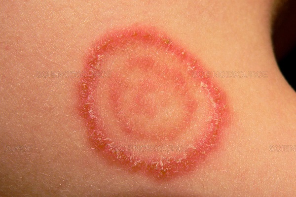
The cloning process of dermatophytes is associated with the release of proteolytic enzymes and spontaneous stimulation of the host inflammatory responses, and causes dermatophytosis or tinea (ringworm). Inflammatory symptoms at the infection site include redness, swelling, and alopecia. Swelling causes transmission of infection to other parts of the body that causes circular lesions. The severity of infection due to dermatophytes depends on age, surrounding temperature, humidity level, and health and social conditions [5]. Different genera of dermatophytes have many phenotypic and genotypic similarities that make their detection challenging. Colony testing, microscopic examination of morphology, genotypic tests, and in some cases, detection of nutritional requirements, temperature tolerance, and urease production are used to detect dermatophytes [6]. Some parts of the body, such as nail subdistal region and area between the fingers, are more prone to dermatophytosis because they are exposed to the fungus in the long-term and provide certain factors, such as sugar and pH, that are required for the fungus [5].
Dermatophytes can induce increased immediate, delayed, or mediated cell susceptibility. In infected people with a normal immune system, response to increased susceptibility is induced within 30 days and is spontaneously recovered after 50 days. Dermatophyte distribution is different across the world [5]. In the past half-century, Tricophyton rubrum has been the most common dermatophyte, and in poor developing countries, Mycosis is endemic and affects a large number of children. Epidermophyton floccosum was common during two decades, i.e. 1980-1990, and among the isolated dermatophytes, is the leading dermatophyte in Iran with 31.4% prevalence [7][8]. In eastern and southern Europe, low quality of life has caused increased infection with animal-friendly dermatophytes. As well, urbanism, contacts, and travels have led to increased prevalence of T. rubrum [9].
Medicinal plants have long been used to treat different diseases in developed and developing countries [10]-[13]. In recent years, medicinal plants have also attracted much attention because their uses have had many benefits, such as decreased expenses and fewer side effects [14]-[17]. Use of plant-based products to fight fungal, bacterial, and parasitic infections has also been considered as an effective approach [7][8]. Moreover, certain measures can be taken to produce drugs by identifying the active compounds of the plants [18][19]. The antifungal effects of some plants, such as ginger, Narcissus tazetta, Myrtus communis, dill, cilantro, garlic, onions, henna, oak, black beans and thyme, on fungal infections have already been demonstrated. Flavonoids, alkaloids, tannins, citronellol , geraniol, thymoquinone, and phenolic compounds are some of the antifungal or other microbial active compounds found in these plants [20][21]. This review article was conducted to report medicinal plants with anti-dermatophytosis properties.
Materials and Methods
We searched the keywords “Medicinal plants”, “Traditional medicine” with the keyword “Dermatophytosis” in the Google Scholar, PubMed, Science Direct, and Scopus databases.
After the first search, seventy-six articles were found to be relevant.
After exclusion of duplicates and irrelevant articles by checking article topics, 54 articles were selected. After reviewing abstracts of the remaining articles, 23 articles were included in this study.
Results
Fungal diseases are known as mycoses, according to the National Institute of Allergies and Infectious Diseases. Mycoses can affect various parts of the body, including body hair, lungs, nervous system, nails, and skin. The most common types of fungal infections are tinea infections and fungal infections of hair, skin or nails; these include athlete’s foot, jock itch, candida, and vaginal yeast infections. Fungal skin infections usually involve itching, skin discoloration, and changes in skin texture in the affected area.
Certain herbs are known as fungicides, or agents that destroy fungi and their spores. Since fungal infections are often tenacious and difficult to eliminate, several different medicines may be necessary to provide therapeutic relief. On the other hand, the routine fungicides like azoles have several side effects.
According to findings of the study herein, Azadirachta indica, Capparis spinosa, Anagallisavensis, Juglans regia, Inula viscosa, Phagnalon rupestre, Plumbago europaea, Ruscus aculeatus, Ruta chalepensis, Salvia fruticosa, Artemisia judaica, Ballota undulate, Cleome amblyocarpa, Peganum harmala, Teucrium polium, Aegle marmelos, Artemisia sieberi, Cuminum cyminum, Foeniculum vulgare, Heracleum persicum, Mentha spicata, Nigella sativa, and Rosmarinus officinalis are the most effective plants on dermatophytes that have been identified to date. Various form of extracts and essences of these plants were found to have growth inhibition or killing effect on dermatophytosis agents and their pathogenicity. Most of the plants belong to family Lamiaceae. All the plants have very good effects against dermatophytosis agents in either minimum inhibitory concentration test (MIC) or in minimum fungicidal concentration test (MFC), and other tests. Even some clinical dermatophytosis isolates show drug resistance from these plants. Table 1 shows further information about the botanical names, studied doses, and effects of these plants.
Discussion
According to the findings of the present study, A. indica, C. spinosa, A . avensis, J. regia, I. viscosa, P. rupestre, P. europaea, R. aculeatus, R. chalepensis, S. fruticosa, A. judaica, B. undulate, C. amblyocarpa, P. harmala, T. polium, A. marmelos, A. sieberi, C. cyminum, F. vulgare, H. persicum, M. spicata, N. sativa, and R. officinalis are the most effective plants on dermatophytes that have been identified to date. Overall, many of the sulfuric compounds, phenolic compounds, flavonoids, tannins, and anthocyanins in the plants cause antifungal effects. Here, in this review article, we touched on the active compounds of the reported plants according to phytochemical investigations. Eugenol is the main antifungal compound of O. sanctum [45]. Tetranortriterpenoid has been demonstrated to be main antifungal compound present in A. indica, while some other compounds, such as 6-deacetylnimbin, azadiradione, nimbin, salannin and epoxyazadiradione, are considered main active compounds of A. indica with antifungal activity [46]. Moreover, rutin, tocopherols, carotenoids, and vitamin C have been confirmed to be the antimicrobial and antifungal compounds of C. spinosa. Glycosidic saponin is the antifungal active compound of A. arvensis [47]. A study on C. amblyocarpa demonstrated that this plant contained a number of valuable fatty acids, such as stearic acid, oleic acid, and linoleic acid. In addition, C. amblyocarpa contained some other compounds, including vitamin C, gallic acid, gallotannins and iridoid, that have many properties, namely antifungal [48]. In A. judaica, certain antifungal active compounds were identified, including camphor, piperitone, and ethyl cinnamate [49], while 3-hydroxy-(5- beta)-androst-2-en-17-one, 4-(1,1-dimethyl)-1,2-benzenediol, and desoxy-dihydro-isostevilo were reported to be the active compounds of B. undulate [16]. Limonene, 2,4-di-tetr-butyl, and p-cymene were the most important antifungal compounds of T. polium [14]. Moreover, 2-isopropenyl- 4-methyl-1-oxa-cyclopenta[b]anthracene-5,10-dione, imperatorin, plumbagin, and b-sitosterol glucoside are some of the main antifungal compounds of A. marmelos [15].
Furthermore, alkaloids, flavonoids, glycosides, and tannins are the main antifungal compounds of P. harmala [50]. Investigations have demonstrated that the main active antifungal compounds present in J. regia are naphthoquinones and flavonoids [51]. Carvacrol and thymol are the most important active compounds of P. rupestre [52]. Azoles and flavonoids, especially sesquiterpenes, are the most important antifungal compounds of I. viscosa [53]. Carvacrol and thymol are the active compounds of S. fruticosa [8], and 2-nonanone and 2-undecanone are the active compounds of R. chalepensis [54]. The active compounds of P. europaea are plumbagin, 1- octane-ly3-acetate, limonene, and nonanal [55]. Certain active compounds, such as vitexin, rutin, isoquercitrine, nicotiflorin, and schaftoside, are present in R. aculeatus [56].
Thymoquinone with antifungal activity has been shown to be one of the important active compounds of N. sativa [57], while spearmint oil and (S)-(-)-carvone are the active compounds of M. spicata [58]. Additional studies have demonstrated that cuminaldehyde and pinenes were among the active compounds of C. cyminum [59]. In F. vulgare, certain compounds, like (E)- anethole and fenchone, had antifungal properties [60], in A. sieberi, α-thujone and β-thujone were found to be antifungal active compounds [61], and in H. persicum, hexyl butyrate and p-cymene were the important antifungal compounds [62]. Meanwhile, the active compounds of R. officinalis
were 1.8-cineole and α-pinene [63]. As can be seen, most of the effective components of these plants are from phenolic compounds, which besides having antifungal activities have mostly antioxidant properties [64]-[69]. Antioxidants are beneficial compounds which are effective against a wide variety of diseases [70]-[75]. Hence, each individual plant, other than for treating fungal infections, might be used for other diseases, such as gastrointestinal, cardiovascular and/or immunological disorders [76]-[81]. These group of plants also may have antidotal activity [82].
Due to a high prevalence of dermatophytosis and high speed of acquiring this disease around the world, especially in low-income regions and developing countries, it is necessary to investigate these drugs as well as pharmaceutical and antifungal agents with anti-dermatophytes effects. Indeed, there are life-threatening side effects due to these chemical drugs. The findings of this study may have many implications, such as in the development of new drugs or in paving the way to conduct additional studies, particularly related to mechanism.
Conclusion
Common medications used to treat fungal infections, especially those with systemic use, have many proven side effects. There is also drug resistance in many fungal species to commonly used drugs, and this resistance has grown mostly in recent years. Medicinal plants are valuable sources of effective antifungal and therapeutic agents which can be useful in the treatment of various diseases, including fungal infections. These plants have a broad use in traditional medicine and complementary medicine, in the unprocessed form, and in modern medicine, either in processed or purified active substance form. Further research studies are required for developing new drugs from these valuable sources.
Open Access
This article is distributed under the terms of the Creative Commons Attribution License (CC-BY 4.0) which permits any use, distribution, and reproduction in any medium, provided the original author(s) and the source are credited.
List of abbreviations
MIC: Minimum inhibitory concentration; microg/mL: Microgram per milliliter
Ethics approval and consent to participate
Not to be applied
Competing interests
The authors declare that they have no conflicts of interest.
Funding
Not to be applied
Authors’ contributions
All of the authors have participated in manuscript preparation, Manuscript review, Design, Literature search, Manuscript editing. All authors read and approved the final version of manuscript.
References
-
LD
Morrow.
Management of feline dermatophytosis in the rescue shelter environment. Companion Animal.
2016;
21
:
634-639
.
View Article Google Scholar -
Faway
Émilie,
L
Cambier,
B
Mignon,
Y
Poumay,
CL
de Rouvroit.
Modeling 8 dermatophytosis in reconstructed human epidermis: A new tool to study infection mechanisms and to test antifungal agents. Medical mycology.
2016;
55
:
485-494
.
View Article Google Scholar -
AO
Akinboro,
OA
Olayinka,
O
Onayemi,
A
Oguntola,
AI
Ajibola.
Prediction of Dermatophyte Culture by Clinical Features: Saving Time and Cost in Resource-Poor Settings. Ibnosina Journal of Medicine and Biomedical Sciences.
2013;
5
:
189-195
.
View Article Google Scholar -
P
Nenoff,
M
Erhard,
JC
Simon,
GK
Muylowa,
J
Herrmann,
W
Rataj,
Y
Gräser.
MALDI-TOF mass spectrometry-a rapid method for the identification of dermatophyte species. Medical mycology.
2013;
51
:
17-24
.
View Article PubMed Google Scholar -
NM
Martinez-Rossi,
NT
Peres,
A
Rossi.
Pathogenesis of dermatophytosis: sensing the host tissue. Mycopathologia.
2017;
182
:
215-227
.
View Article PubMed Google Scholar -
V
Sharma,
TK
Kumawat,
A
Sharma,
R
Seth,
S
Chandra.
Dermatophytes: Diagnosis of dermatophytosis and its treatment. African Journal of Microbiology Research.
2015;
9
:
1286-1293
.
View Article Google Scholar -
M
Rafieian-Kopaei,
M
Bahmani,
A
Sepahvand,
H
Hassanzadazar,
A
Abaszadeh,
R
Rafieian,
S
Soroush.
Candidiasis phytotherapy: An overview of the most important medicinal plants affecting the Candida albicans. Journal of Chemical and Pharmaceutical Sciences.
2016;
9
:
1284-1293
.
-
A
Sepahvand,
Z
Eftekhari,
M
Rafieian-Kopaei,
S
Soroush.
Phytotherapy in Aspergillus: An overview of the most important medicinal plants affecting Aspergillus. International Journal of PharmTech Research.
2016;
9
:
274-281
.
-
C
Seebacher,
JP
Bouchara,
B
Mignon.
Updates on the epidemiology of dermatophyte infections. Mycopathologia.
2008;
166
:
335-352
.
View Article PubMed Google Scholar -
F
Jamshidi-Kia,
Z
Lorigooini,
H
Amini-Khoei.
Medicinal plants: past history and future perspective. Journal of herbmed pharmacology.
2018;
1
:
1-7
.
-
S
Kazemi,
H
Shirzad,
M
Rafieian-Kopaei.
Recent findings in molecular basis of inflammation and anti-inflammatory plants. Current pharmaceutical design.
2018
.
-
M
Bahmani,
A
Sarrafchi,
H
Shirzad,
M
Rafieian-Kopaei.
Autism: Pathophysiology and promising herbal remedies. Current pharmaceutical design.
2016;
22
:
277-285
.
View Article PubMed Google Scholar -
M
Rahimi-Madiseh,
P
Karimian,
M
Kafeshani,
M
Rafieian-Kopaei.
The effects of ethanol extract of Berberis vulgaris fruit on histopathological changes and biochemical markers of the liver damage in diabetic rats. Iranian journal of basic medical sciences.
2017a;
20
:
552
.
PubMed PMC Google Scholar -
M
Rahimi-Madiseh,
Z
Lorigoini,
H
Zamani-gharaghoshi,
M
Rafieian-kopaei.
Berberis vulgaris: specifications and traditional uses. Iranian journal of basic medical sciences.
2017b;
20
:
569
.
PubMed PMC Google Scholar -
A
Sarrafchi,
M
Bahmani,
H
Shirzad,
M
Rafieian-Kopaei.
Oxidative stress and Parkinson's disease: New hopes in treatment with herbal antioxidants. Current pharmaceutical design.
2016;
22
:
238-246
.
View Article PubMed Google Scholar -
S
Karami,
M
Roayaei,
E
Zahedi,
M
Bahmani,
L
Mahmoodnia,
H
Hamzavi,
M
Rafieian-Kopaei.
Antifungal effects of Lactobacillus species isolated from local dairy products. International journal of pharmaceutical investigation.
2017;
7
:
77
.
View Article PubMed PMC Google Scholar -
M
Rahimi-Madiseh,
E
Heidarian,
S
Kheiri,
M
Rafieian-Kopaei.
Effect of hydroalcoholic Allium ampeloprasum extract on oxidative stress, diabetes mellitus and dyslipidemia in alloxan-induced diabetic rats. Biomedicine & Pharmacotherapy.
2017;
86
:
363-367
.
View Article PubMed Google Scholar -
N
Tavakolli,
M
Ghanadian,
G
Asghari,
H
Sadraei,
A
Borjlou,
M
Tabakhian.
Development of a validated HPLC method for determination of an active component in Pycnocycla spinosa and tablets prepared from its extract. Journal of Herbmed Pharmacology.
2017;
6
.
-
EA
Ayeni,
GIA
Abubakar,
ZMV
Atinga.
Phytochemical, nutraceutical and antioxidant studies of the aerial parts of Daucus carota L.(Apiaceae). Journal of Herbmed Pharmacology.
2018;
7
:
68-73
.
View Article Google Scholar -
F
Alizadeh,
A
Khodavandi,
S
Esfandyari,
S
Nouripour-Sisakht.
Analysis of ergosterol and gene expression profiles of sterol ▵ 5, 6-desaturase (ERG3) and lanosterol 14$a$- demethylase (ERG11) in Candida albicans treated with carvacrol. Journal of Herbmed Pharmacology.
2018;
7
.
-
A
Nikpay,
M
Soltani.
In vitro anti-parasitic activities of Pulicaria dysenterica and Lycopus europaeus methanolic extracts against Trichomonas gallinae. Journal of Herbmed Pharmacology.
2018;
7
:
112-118
.
View Article Google Scholar -
S
Balakumar,
S
Rajan,
T
Thirunalasundari,
S
Jeeva.
Antifungal activity of Ocimum 9 sanctum Linn. (Lamiaceae) on clinically isolated dermatophytic fungi. Asian Pacific journal of tropical medicine.
2011;
4
:
654-657
.
View Article Google Scholar -
V
Natarajan,
PV
Venugopal,
T
Menon.
Effect of Azadirachta indica (neem) on the growth pattern of dermatophytes. Indian journal of medical microbiology.
2003;
21
:
98
.
PubMed Google Scholar -
MS
Ali-Shtayeh,
SIA
Ghdeib.
Antifungal activity of plant extracts against dermatophytes. mycoses.
1999;
42
:
665-672
.
-
B
Taye,
M
Giday,
A
Animut,
J
Seid.
Antibacterial activities of selected medicinal plants in traditional treatment of human wounds in Ethiopia. Asian Pacific Journal of Tropical Biomedicine.
2011;
1
:
370-375
.
View Article Google Scholar -
MS
Ali-Shtayeh,
SIA
Ghdeib.
Antifungal activity of plant extracts against dermatophytes. mycoses.
1999;
42
:
665-672
.
-
MI
Gandhi,
S
Ramesh.
Antifungal and haemolytic activities of organic extracts of Tecoma stans (Bignoniaceae). Journal of Ecobiotechnology.
2010;
2
.
-
EA
Mazzio,
KF
Soliman.
Topical treatment for dyshidrosis (pompholyx) and dry skin disorders. 2008
.
-
J
Reuter,
U
Wölfle,
HC
Korting,
C
Schempp.
Which plant for which skin disease? Part 2: Dermatophytes, chronic venous insufficiency, photoprotection, actinic keratoses, vitiligo, hair loss, cosmetic indications. JDDG: Journal der Deutschen Dermatologischen Gesellschaft.
2010;
8
:
866-873
.
View Article PubMed Google Scholar -
M
Khoury,
D
Stien,
N
Ouaini,
V
Eparvier,
NA
Apostolides,
ME
Beyrouthy.
Chemical composition and antimicrobial activity of the essential oil of Ruta chalepensis L. growing wild in Lebanon. Chemistry & biodiversity.
2014;
11
:
1990-1997
.
-
K
Adam,
A
Sivropoulou,
S
Kokkini,
T
Lanaras,
M
Arsenakis.
Antifungal activities of Origanum vulgare subsp. hirtum, Mentha spicata, Lavandula angustifolia, and Salvia fruticosa essential oils against human pathogenic fungi. Journal of Agricultural and Food Chemistry.
1998;
46
:
1739-1745
.
-
S
Kordali,
A
Cakir,
A
Mavi,
H
Kilic,
A
Yildirim.
Screening of chemical composition and antifungal and antioxidant activities of the essential oils from three Turkish Artemisia species. Journal of agricultural and food chemistry.
2005;
53
:
1408-1416
.
View Article PubMed Google Scholar -
M
Hashem.
Antifungal properties of crude extracts of five Egyptian medicinal plants against dermatophytes and emerging fungi. Mycopathologia.
2011a;
172
:
37-46
.
-
M
Hashem.
Antifungal properties of crude extracts of five Egyptian medicinal plants against dermatophytes and emerging fungi. Mycopathologia.
2011b;
172
:
37-46
.
-
J
Asgarpanah,
F
Ramezanloo.
Chemistry, pharmacology and medicinal properties of Peganum harmala L. African Journal of Pharmacy and Pharmacology.
2012;
6
:
1573-1580
.
View Article Google Scholar -
M
Vahdani,
P
Faridi,
MM
Zarshenas,
S
Javadpour,
Z
Abolhassanzadeh,
N
Moradi,
Z
Bakzadeh,
A
Karmostaji,
A
Mohagheghzadeh,
Y
Ghasemi.
Major compounds and antimicrobial activity of essential oils from five Iranian endemic medicinal plants. Pharmacognosy Journal.
2011;
3
:
48-53
.
View Article Google Scholar -
BB
Mishra,
DD
Singh,
N
Kishore,
VK
Tiwari,
V
Tripathi.
Antifungal constituents isolated from the seeds of Aegle marmelos. Phytochemistry.
2010;
71
:
230-234
.
View Article PubMed Google Scholar -
M
Mahboubi,
N
Kazempour.
The antifungal activity of Artemisia sieberi essential oil from different localities of Iran against dermatophyte fungi. Journal de mycologie medicale.
2015;
25
:
e65-e71
.
View Article PubMed Google Scholar -
C
Romagnoli,
E
Andreotti,
S
Maietti,
R
Mahendra,
D
Mares.
Antifungal activity of essential oil from fruits of Indian Cuminum cyminum. Pharmaceutical biology.
2010;
48
:
834-838
.
View Article PubMed Google Scholar -
M
Patra,
SK
Shahi,
G
Midgely,
A
Dikshit.
Utilization of essential oil as natural antifungal against nail-infective fungi. Flavour and fragrance journal.
2002;
17
:
91-94
.
View Article Google Scholar -
RA
Khosravi,
H
Shokri,
Z
Farahnejat,
R
Chalangari,
M
Katalin.
Antimycotic efficacy of Iranian medicinal plants towards dermatophytes obtained from patients with dermatophytosis. Chinese journal of natural medicines.
2013;
11
:
43-48
.
-
KK
Aggarwal,
SPS
Khanuja,
A
Ahmad,
TRS
Kumar,
VK
Gupta,
S
Kumar.
Antimicrobial activity profiles of the two enantiomers of limonene and carvone isolated from the oils of Mentha spicata and Anethum sowa. Flavour and Fragrance Journal.
2002;
17
:
59-63
.
View Article Google Scholar -
H
Mahmoudvand,
A
Sepahvand,
S
Jahanbakhsh,
B
Ezatpour,
SA
Mousavi.
Evaluation of antifungal activities of the essential oil and various extracts of Nigella sativa and its main component, thymoquinone against pathogenic dermatophyte strains. Journal de Mycologie Médicale/Journal of Medical Mycology.
2014;
24
:
e155-e161
.
-
L
Mugnaini,
S
Nardoni,
L
Pinto,
L
Pistelli,
M
Leonardi,
F
Pisseri,
F
Mancianti.
In vitro and in vivo antifungal activity of some essential oils against feline isolates of Microsporum canis. Journal de Mycologie Médicale/Journal of Medical Mycology.
2012;
22
:
179-184
.
-
A
Kumar,
R
Shukla,
P
Singh,
NK
Dubey.
Chemical composition, antifungal and antiaflatoxigenic activities of Ocimum sanctum L. essential oil and its safety assessment as plant based antimicrobial. Food and Chemical Toxicology.
2010;
48
:
539-543
.
-
TR
Govindachari,
G
Suresh,
G
Gopalakrishnan,
B
Banumathy,
S
Masilamani.
Identification of antifungal compounds from the seed oil of Azadirachta indica. Phytoparasitica.
1998;
26
:
109-116
.
View Article Google Scholar -
JR
Qasem.
Fungitoxic properties of scarlet pimpernel (Anagallis arvensis) against Helminthosporium sativum and Fusarium oxysporum. Allelopathy Journal.
2011;
28
:
251-258
.
-
R
Upadhyay.
Cleome viscosa Linn: A natural source of pharmaceuticals and pesticides. International Journal of Green Pharmacy.
2015;
9
:
71
.
View Article Google Scholar -
MS
Abu-Darwish,
C
Cabral,
MJ
Goncalves,
C
Cavaleiro,
MT
Cruz,
A
Zulfiqar,
IA
Khan,
T
Efferth,
L
Salgueiro.
Chemical composition and biological activities of Artemisia judaica essential oil from southern desert of Jordan. Journal of ethnopharmacology.
2016;
191
:
161-168
.
View Article PubMed Google Scholar -
AM
Saadabi.
Antifungal activity of some Saudi plants used in traditional medicine. Asian J Plant Sci.
2006;
5
:
907-909
.
View Article Google Scholar -
H
Mansour-Djaalab,
F
Kahlouche-Riachi,
Z
Djerrou,
M
Serakta-Delmi,
S
Hamimed,
W
Trifa,
I
Djaalab,
YH
Pacha.
In vitro evaluation of antifungal effects of Lawsonia inermis, Pistacia lentiscus and Juglans regia. International Journal of Medicinal and Aromatic Plants.
2012;
2
:
263-268
.
-
DD
Orhan,
N
Orhan.
Novel antidermatophytic drug candidates from nature. In Antimicrobials: Synthetic and Natural Compounds. Taylor & Francis.
2015
.
-
ND
Laurentis,
V
Losacco,
MA
Milillo,
O
Lai.
Chemical investigations of volatile constituents of Inula viscosa (L.) Aiton (Asteraceae) from different areas of Apulia, Southern Italy. Delpinoa.
2002;
44
:
115-119
.
-
EB
Bnina,
S
Hammami,
M
Daamii-remadi,
HB
Jannet,
Z
Mighri.
Chemical composition and antimicrobial effects of Tunisian Ruta chalepensis L. essential oils. Journal de la Société Chimique de Tunisie.
2010;
12
:
1-9
.
-
MN
Navaei,
M
Mirza,
M
Dini.
Chemical composition of the essential oil of Plumbago europaea L. roots from Iran. Flavour and fragrance journal.
2005;
20
:
213-214
.
-
N
Hadzifejzovic,
J
Kukic´-Markovic´,
S
Petrovic´,
M
Sokovic´,
GlamoJ
D
Stojkovic´,
A
Nahrstedt.
Bioactivity of the extracts and compounds of Ruscus aculeatus L. and Ruscus hypoglossum L. Industrial crops and products.
2013;
49
:
407-411
.
-
MAU
Khan,
MK
Ashfaq,
HS
Zuberi,
MS
Mahmood,
AH
Gilani.
The in vivo antifungal activity of the aqueous extract from Nigella sativa seeds. Phytotherapy Research.
2003;
17
:
183-186
.
View Article PubMed Google Scholar -
J
Singh,
AK
Dubeyd,
NN
Tripathi.
Antifungal activity of Mentha spicata. International journal of pharmacognosy.
1994;
32
:
314-319
.
-
MB
Pai,
GM
Prashant,
KS
Murlikrishna,
KM
Shivakumar,
al
Chandu GN et.
Antifungal efficacy of Punica granatum, Acacia nilotica, Cuminum cyminum and Foeniculum vulgare on Candida albicans: an in vitro study. Indian Journal of Dental Research.
2010;
21
:
334
.
View Article PubMed Google Scholar -
N
Mimica-Dukic´,
S
Kujundc´,
M
Sokovic´,
M
Couladis.
Essential oil composition and antifungal activity of Foeniculum vulgare Mill. obtained by different distillation conditions. Phytotherapy Research.
2003;
17
:
368-371
.
View Article PubMed Google Scholar -
M
Farzaneh,
M
Ahmadzadeh,
J
Hadian,
AS
Tehrani.
Chemical composition and antifungal activity of the essential oils of three species of Artemisia on some soil-borne phytopathogens. Communications in agricultural and applied biological sciences.
2006;
71
:
1327-1333
.
PubMed Google Scholar -
H
Atefeh,
M
Azamia,
S
Abd0lham1'dAngaj.
Medicinal effects of Heracleum persicum (Golpar). Middle-East Journal of Scientific Research.
2010;
5
:
174-176
.
-
A
Angioni,
A
Barra,
E
Cereti,
D
Barile,
JD
Coisson,
M
Arlorio,
S
Dessi,
V
Coroneo,
P
Cabras.
Chemical composition, plant genetic differences, antimicrobial and antifungal activity investigation of the essential oil of Rosmarinus officinalis L. Journal of agricultural and food chemistry.
2004;
52
:
3530-3535
.
View Article PubMed Google Scholar -
S
Asgharzade,
M
Rafieian-kopaei,
A
Mirzaeian,
S
Reiisi,
L
Salimzadeh.
Aloe vera toxic effects: expression of inducible nitric oxide synthase (iNOS) in testis of Wistar rat. Iranian journal of basic medical sciences.
2015;
18
:
967
.
PubMed PMC Google Scholar -
Z
Rabiei,
S
Naderi,
M
Rafieian-Kopaei.
Study of antidepressant effects of grape seed oil in male mice using tail suspension and forced swim tests. Bangladesh Journal of Pharmacology.
2017;
12
:
397-402
.
View Article Google Scholar -
M
Bahmani,
A
Sarrafchi,
H
Shirzad,
S
Asgari,
M
Rafieian-Kopaei.
Cardiovascular Toxicity of Cyclooxygenase Inhibitors and Promising Natur al Substitutes. Current pharmaceutical design.
2017;
23
:
952-960
.
View Article PubMed Google Scholar -
H
Rouhi-Boroujeni,
E
Heidarian,
H
Rouhi-Boroujeni,
F
Deris,
M
Rafieian-Kopaei.
Medicinal plants with multiple effects on cardiovascular diseases: A systematic review. Current pharmaceutical design.
2017;
23
:
999-1015
.
View Article PubMed Google Scholar -
E
Shayganni,
M
Bahmani,
S
Asgary,
M
Rafieian-Kopaei.
Inflammaging and cardiovascular disease: Management by medicinal plants. Phytomedicine.
2016;
23
:
1119-1126
.
View Article PubMed Google Scholar -
A
Karimi,
M
Mohammadi-Kamalabadi,
M
Rafieian-Kopaei,
al
Amjad L et.
Determination of antioxidant activity, phenolic contents and antiviral potential of methanol extract of Euphorbia spinidens Bornm (Euphorbiaceae). Tropical Journal of Pharmaceutical Research.
2016;
15
:
759-764
.
View Article Google Scholar -
Z
Hosseini,
Z
Lorigooini,
M
Rafieian-Kopaei,
HA
Shirmardi,
K
Solati.
A review of botany and pharmacological effect and chemical composition of Echinophora species growing in Iran. Pharmacognosy research.
2017;
9
:
305
.
View Article PubMed PMC Google Scholar -
M
Asadi-Samani,
N
Bagheri,
M
Rafieian-Kopaei,
H
Shirzad.
Inhibition of Th1 and Th17 cells by medicinal plants and their derivatives: A systematic review. Phytotherapy Research.
2017
.
-
S
Karami,
M
Roayaei,
H
Hamzavi,
M
Bahmani,
H
Hassanzad-Azar,
M
Leila,
M
Rafieian-Kopaei.
Isolation and identification of probiotic Lactobacillus from local dairy and evaluating their antagonistic effect on pathogens. International journal of pharmaceutical investigation.
2017;
7
:
137
.
View Article PubMed PMC Google Scholar -
Z
Rabiei,
M
Gholami,
M
Rafieian-Kopaei.
Antidepressant effects of Mentha pulegium in mice. Bangladesh Journal of Pharmacology.
2016;
11
:
711-715
.
View Article Google Scholar -
L
Jalaly,
G
Sharifi,
M
Faramarzi,
A
Nematollahi,
M
Rafieian-Kopaei,
M
Amiri,
F
Moattar.
Comparison of the effects of Crataegus oxyacantha extract, aerobic exercise and their combination on the serum levels of ICAM-1 and E-Selectin in patients with stable angina pectoris. DARU Journal of Pharmaceutical Sciences.
2015;
23
:
54
.
View Article PubMed PMC Google Scholar -
Z
Rabiei,
M
Rafieian-Kopaei,
S
Mokhtari,
M
Shahrani.
Effect of dietary ethanolic extract of Lavandula officinalis on serum lipids profile in rats. Iranian journal of pharmaceutical research: IJPR.
2014;
13
:
1295
.
PubMed Google Scholar -
M
Bahmani,
A
Zargaran,
M
Rafieian-Kopaei.
Identification of medicinal plants of Urmia for treatment of gastrointestinal disorders. Revista Brasileira de Farmacognosia.
2014;
24
:
468-480
.
View Article Google Scholar -
S
Asgary,
A
Sahebkar,
MR
Afshani,
M
Keshvari,
S
Haghjooyjavanmard,
M
Rafieian-Kopaei.
Clinical evaluation of blood pressure lowering, endothelial function improving, hypolipidemic and anti-inflammatory effects of pomegranate juice in hypertensive subjects. Phytotherapy Research.
2014;
28
:
193-199
.
View Article PubMed Google Scholar -
A
Baradaran,
H
Nasri,
M
Rafieian-Kopaei.
Erythropoietin and renal protection. DARU Journal of Pharmaceutical Sciences.
2013;
21
:
78
.
View Article PubMed PMC Google Scholar -
S
Asgari,
M
Setorki,
M
Rafieian-kopaei,
N
Shahinfard,
R
Ansari,
al
Forouz Z et.
Postprandial hypolipidemic and hypoglycemic effects of Allium hertifolium and Sesamum indicum on hypercholesterolemic rabbits. African Journal of Pharmacy and Pharmacology.
2012;
6
:
1131-1135
.
-
M
Setorki,
B
Nazari,
S
Asgary,
L
Azadbakht,
M
Rafieian-Kopaei.
Anti atherosclerotic effects of verjuice on hypocholesterolemic rabbits. African Journal of Pharmacy and Pharmacology.
2011;
5
:
1038-1045
.
-
H
Shirzad,
M
Shahrani,
M
Rafieian-Kopaei.
Comparison of morphine and tramadol effects on phagocytic activity of mice peritoneal phagocytes in vivo. International immunopharmacology.
2009;
9
:
968-970
.
View Article PubMed Google Scholar -
E
Heidarian,
M
Rafieian-Kopaei.
Protective effect of artichoke (Cynara scolymus) leaf extract against lead toxicity in rat. Pharmaceutical biology.
2013;
51
:
1104-1109
.
View Article PubMed Google Scholar
Comments
Downloads
Article Details
Volume & Issue : Vol 5 No 6 (2018)
Page No.: 2378-2388
Published on: 2018-06-28
Citations
Copyrights & License
This work is licensed under a Creative Commons Attribution 4.0 International License.
Search Panel
- HTML viewed - 26726 times
- Download PDF downloaded - 3197 times
- View Article downloaded - 0 times
