Changes blood protein composition under experimental chemical burns of esophageal development in rats
Abstract
Exogenous poisoning with alkalis takes the leading position among causes of acute poisoning. Esophageal burns as a result of accidental swallowing of caustic material are seen frequently in children ages 1 to 8 years. A burn wound is perhaps the most intense stress that a human body can suffer. In the place of chemical trauma localization the processes of synthesis and degradation of proteins increase. As a result the structure and functions of vital organs and immune system suffer. The inflammation process after burn injury is determines the changes in protein expression. In our research we have shown that chemical burn of the esophagus is characterized by decreased level of total proteolytic activity, total protein and development of endogenous intoxication of the body as indicated by elevated MMM level. Obtained results suggest that the development of chemical burns of the esophagus grade 2 is characterized by more substantial changes in the protein composition of blood compared with grade 1. An in-depth study of this disease has a crucial scientific and practical importance for development of pathogenically oriented methods of wound process healing after the chemical esophageal burns depending on the stage and extent of the burn process
Introduction
According to the World Health Organization noted a steady increase in number of chemical burns of the esophagus, which is directly connected with a large number of relatively available technical and household aggressive liquids synthesized in a result of scientific and technological progress of mankind Weigert and Black, 2005. According to the American Association of Poison Control Centers, only in 2010 there were more than 1.6 million of occurrences when children used potentially hazardous chemicals, among which 18-46% of cases was using of household substances of alkaline nature Reinberg, 2013. There are many pathologies and complications following an acid esophageal burns: swelling of the larynx, toxic shock syndrome Dakshesh H. Parikh,2009Deng et al., 2008Moerman et al., 2000, necrosis of esophageal and stomach tissue, dysmotility Tiryaki et al., 2005, scar stricture, esophageal deformations, corrosive esophagitis, gastroesophageal reflux Mutaf et al., 1996Ramasamy and Gumaste, 2003, candidiasis Zwischenberger et al., 2002, malignization in a remote period Marzaro et al., 2006 and others.
One of the major symptoms of chemical burns is intoxication that comes from the first minutes after the injury and lasts during all phases of the burn disease Koziniec G.P.,2005. Burn injury is always accompanied by the development of intoxication, which is caused by many non-specific toxic metabolites and biological agents Li et al., 2011. Higher concentrations of these compounds provoke a number of negative effects: violation of the structure and function of plasma- and intracellular membranes, inhibiting the activity of many seroenzymes, protein tissues modification Navarro-Yepes et al.,2014Yavorskaya V.A., 2000.
Literature data indicate multidirectional pathogenic changes after the formation of the damage area in a result of the burn Zahs et al., 2012. Burn disease causes dysfunction of all tissues and organs Jeschke, 2013. Despite the large amount of researches of this problem, the issues of fluctuations of protein fractions at various development stages of chemical burns of the esophagus and proteolysis processes in the body are studied insufficiently.
Material and methods
In our experiments we used nonlinear immature white rats (1 month) weighing 90-110g, which were kept on a standard vivarium diet. The animals were experimentally simulated with the alkali esophageal burn with 10% (grade 1) and 20% (grade 2) solvent of NaOH Raetska Ya.B., 2013. Serum and plasma of blood for research were collected at 1st, 7th, 15th, and 21st day. Level of low and medium mass molecules were measured by method Gabrielyan with modifications Gabrielyan N.I., 1985. To the centrifuge tubes added 1 ml serum and 0.5 ml of trichloroacetic acid (100 g/L), stirred and centrifuged for 30 min. at 3000 rev./min. The supernatant was collected and transferred to a test tube with 4.5 ml of distilled water. The content of the tube was stirred and measured at λ 254 nm. Number of oligopeptides was estimated by the level of protein in the supernatant using the method of Bradford.
Separation of protein fractions serum rats were determined by method Lemmli with modifications with sodium dodecyl sulfate (SDS) Laemmli, 1970. Polyacrylamide gels (PAGE) were 10%. After the electrophoresis gels were kept in solution of Coumassie R.
For estimate the results of electrophoresis used the program Totallab 2.01. Total protein was measured by the method of Bradford Bradford, 1976. The total proteolytic activity analyzed by method of caseinolytic activity Hummel, 1959.
ІgG fraction was isolated by affinity chromatography on Sepharose A from blood serum experimental rats.
The statistical analysis of the obtained results was performed using the methods of variation statistics and correlation analysis using the computer program Excel. To determine the reliability of the differences between the two samples we used the Student test (t). Whereby differences P < 0.05 were deemed reliable.
Results
As of today, the majority of authors consider middlemass molecules (MMM) and oligopeptides as a universal marker of endogenous intoxication Chmielewski et al.,2014Karyakin, 2004Krotenko N.M., 2012Nikolskaya V. A., 2013Sukhomlyn T.A.,2011. The toxic effect of MMM is caused by their ability to alter the permeability of membranes and membrane transport, disrupt processes of DNA synthesis, tissue respiration and phosphorylation. MMM have cardio-, vazo- and immunosuppressive properties Laemmli, 1970. We have determined the oligopeptides and MMM levels in serum of rats after experimental alkali burns of the esophagus (ABE) grade 1 ( Table 1 ). The study on the first day of experimental burn grade 1 revealed elevated level of markers of endogenous intoxication (oligopeptides and MMM) in serum of rats by 20% compared with control values. On the 15th and 21st day was observed reduction in MMM and oligopeptides levels, which coincides with the data of individual researchers Fistal E.Y., 2005. The study on the first day of experimental burn grade 2 revealed elevated level of markers of endogenous intoxication (MMM) in serum of rats by 33% compared with control values. On the 1st and 21st day oligopeptides level increased by 34% and 104% respectively ( Table 2 ).
We also determined the total protein level in serum of rats after experimental ABE grade 1. Thus, on the first day, total protein level decreased by 28% compared with control values. Shown a significant reduction in total protein level by 32% on the 21st day of the experiment. The total protein level in serum of rats after experimental ABE grade 2 a significant reduction in all days of the experiment
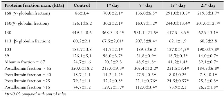
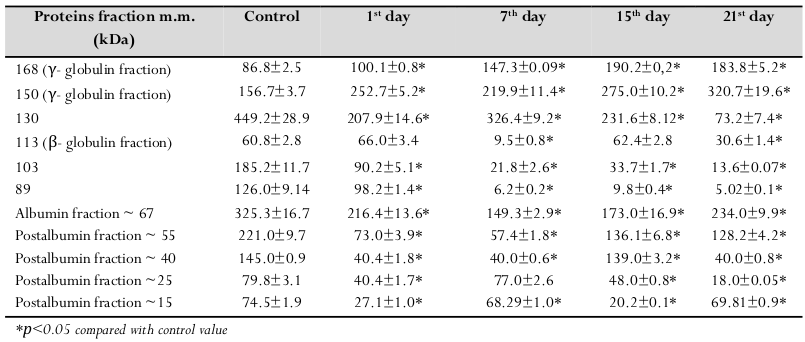
Electrophoretic analysis of serum protein composition of experimental animals showed in all studied samples, both in control and in conditions of chemical burns of the esophagus grade 1 and 2, presence of protein fractions with molecular masses from 15 to 168 kDa ( Table 3 and Table 4 ).
Conducted researches did not show qualitative changes of protein levels in serum, but it is possible to determine their quantitative changes. Thus, we have shown a growth of protein levels in fractions with m.m. 167 kDa and 150 kDa on the 7th, 15th and 21st day of experimental alkali burns grade I and 2 compared with control values. These protein stripes correspond to IgG fraction, and in view of obtained data on fluctuations of their levels, of some interest are analysis of their levels and a more detailed study. Obtained data coincide with our results of determination of the antibodies level in serum of experimental rats. We have isolated IgG fractions by method of affinity chromatography from serum of rats, by which was experimentally simulated ABE ( Table 5 ). After simulation of alkali burns grade 1, IgG levels increased on the 1st, 7th and 15th day. Obtained data coincide with our results of the analysis of electrophoretograms, the level the antibodies increase in all days of the experiment after simulation of chemical burns grade 1.
Analysis of electrophoretograms showed elevated levels of protein fraction m.m. 130 kDa on the 7th day by 107% after simulation of chemical burns grade 1. We have shown increased levels of the fraction m.m. 113 kDa on the 7th day 1 by 244%. This fraction can correspond C-reactive protein, which is a central component of markers of the acute phase of inflammation. Elevated levels of the shown fraction on the 7th day indicate addition of bacterial infection or inflammation caused by non-infectious parts of necrotic tissues.
Shown reduction in level of albumin fraction (67 kDa) after simulation of chemical burns grade 1 and 2 in all days of the experiment. We have determined reduction of fraction 55 kDa after simulation of alkali burns grade 1 and 2, which corresponds prealbumin.
The results of investigations showed that in experimental animals after simulation of ABE grade 1, the level of postalbumin fraction ~ 40 kDa is reduced on the 1st day of the experiment by 31%. On the 7th, 15th and 21st day level of this fraction increased by 49%, 133% and 139% respectively compared with control values. Level of postalbumin fraction ~ 25 kDa was significantly decreased on the 1st, 7th, 15th and 21st day by 144%, 259%, 224% and 211% respectively after simulation of alkali burns grade 1. We have determined elevated level of postalbumin fraction ~ 15 kDa on the 1st and 7th day by 115% and 51% respectively and reduction of this fraction by 179% on the 21st day after simulation of ABE grade 1.
Analysis of electrophoretograms showed significantly decreased postalbumin fraction ~ 40 kDa throughout the experiment by 258% after simulation ABE of grade 2. Level of postalbumin ~ 25 kDa fraction was significantly decreased on the 1st, 15th and 21st day by 97%, 65%, and 343% respectively. We have determined elevated level of postalbumin fraction ~ 15 kDa on the 1st and 15th day by 174% and 263% respectively day after simulation of alkali burns grade 2.
Research of the total proteolytic activity in plasma of rats with chemical burns of the esophagus showed its reduction during the whole period of the experiment compared with control values ( Figure 1 ). On the first day after simulation of alkaline chemical burns of the esophagus grade 1, overall proteolytic activity decreased by 3.9 times compared with control values. Experimental burn of the esophagus caused decrease of proteolytic activity in plasma on the 7th, 15th and 21st day by 2.7; 2.65 and 3.09 times respectively. We have shown reduction in level of proteolytic activity decrease at all day of experiment after simulation of ABE grade 2.
Discussion
Middle-mass molecules are a convenient clinical indicant characterizing the pathologic processes development dynamics. It is known that the end-stage of dyscrasia irrespective of ethiology and pathogenesis of the primary disease is accompanied by endogenous intoxication progression. The core of the abovementioned state is determined by congestion of excessive amount of biologically active components in blood. When the metabolism shifts to retrograde reactions a great amount of metabolic-waste products and metabolites appears in blood in unusually high concentrations of different biologically active substances, organs and tissues destruction products, bacterial toxins, protein and lipid hydroperoxides, etc. Generally this pool of substances in blood is distributed between the plasma and erythrocytes, and characterizes the notion of intoxication from the point of biochemistry Tupikova and Osipovich, 1990Val'dman et al., 1991. For the past years sufficient data was collected proving that accumulation of these substances in the wound leads to lysis of tissues and increase of toxic products in blood (urea, uric acid, creatinine). MMM pool is considered to be the basic biochemical marker reflecting the level of pathological protein metabolism.
MMM is a type of combinations with the molecular mass up to 5,000D. MMM is divided into two big groups – average molecular mass substances and oligopeptides. Retrograde MMM pool – non-protein derivatives of different nature: urea, creatinine, uric acid, glucose, lactic and other organic acids, amino acids, aliphatic acids, cholesterol, phospholipids, lipid peroxidation products, intermediary metabolites of nucleoproteid exchange etc., accumulating in the body and exceeding the normal concentrations Chmielewski et al., 2014Nikolskaya V. A., 2013. The other part of MMM – oligopeptides – is represented by substances of peptide nature fulfilling different functions, in particular – regulatory ones. At that, the maximum peak of intoxication occurs due to the molecules with molecular mass of 1,000-1,200 Daltons. Their accumulation is associated to insufficient activity of exopeptidase performing degradation of these peptides normally Val'dman et al., 1991.
A number of authors note that progression of substantial disturbances of tissue respiration revealed at the early stages of burn disease show the existence of general mechanisms of energy metabolism disorder and do not rule out the fact that average molecular components of peptide nature play an important role in determining of the course of many biochemical processes in the bodies of burnt patients and in forming of toxemic disorders specific of burn disease Karyakin, 2004Krotenko N.M., 2012.
Notwithstanding that in the cases of esophageal burns there takes place an increase of MMM level as products of blood protein catabolism, the MMM synthesis for homeostasis stabilisation should not be excluded Krotenko N.M., 2012Sukhomlyn T.A., 2011. Consequently probable use detoxification therapy in acuity burn disease will be reduced risk of developing complications after burn disease.
Electrophoretic separation of protein fractions had shown a significant reduction of albumin fraction and a gradual increase of globulin fraction after simulation of chemical burns grade 1 and 2. The burning injury regeneration increases the protein, carbohydrates and nucleic acids metabolism level locally as well as in the whole body. Serum albumin is the most plentiful plasma protein. In humans, albumin is the most abundant plasma protein, accounting for 55–60% of the measured serum protein. Unlike other plasma proteins which tend to have single, specific functions, albumin has been assigned numerous physiological roles. It is the principal agent responsible for the osmotic pressure of blood, for transport of fatty acids, and for the sequestration and transportation of bilirubin Norbury et al., 2008. Other less well-defined functions included transport of tryptophan, cysteine, and various hormones and it is a source of amino acids for peripheral tissues Lu et al., 2007. It is synthesized almost exclusively by the liver. The normal halflife of circulating albumin is 19 days. This corresponds to daily degradation of about 10 per cent of the total protein synthesized by the liver Nicholson et al., 2000. Rothschild et al. had shown the loss of albumin In severely burned patients, due to increased capillary permeability and overall reduction in protein synthesis in the liver Akbal et al., 2012. Quantitative changes in postalbumin factions may be associated with increase of degradation processes of tissue proteins due to increase of activity of proteolytic enzymes of different specificity or traumatic effect of chemical factors. So, after simulation of chemical burns of the esophagus one may talk of hypoproteinemia, which occurs mainly due to reduction in the amount of albumins.
In terms of tissue destruction and due to effect of alkaline substances, own proteins may undergo certain structural modifications, which in turn can be an impulse for the formation of a certain number of autoantibodies to these "defective" protein molecules
Obtained data of level decreased total protein, albumin and prealbumin may indicate suppression of protein- synthetic function of the liver due to burn injuries.
In the place of chemical trauma localization the processes of synthesis and degradation of proteins increase. As a result the structure and functions of vital organs such as skeletal muscles, skin, and immune system suffer. The inflammation process after burn injury is determined by the changes in protein expression. It is known that the reparation process of the injured tissue proceeds in several stages. The first stage is the inflammation reaction the aim of which consists in activation of endoproteases and freeing of the injured area from the necrotized tissues, infection agents. If for some reason that wouldn’t happen, the damage won’t be able to heal spontaneously and the process will acquire a chronic form. The inflammation reaction is accompanied by deep changes in cellular as well as humoral link of immune system, which is a favourable factor in development of different complications of mostly infectious origin Jeschke et al., 2008Messingham et al., 2000. The superfamily of immunoglobulins plays an important role in embryogenesis, in healing of wounds and immune response. The functions of immunoglobulin family consist in tying of fluid-phase ligands and surface ligands of cells. The immunoglobulin molecules also play an important role in the processes of activation and differentiation of cells, greatly contributing to fulfilment of the cellular interaction Bariar et al., 1996Rapaport and Bachvaroff, 1976. The increase of immunoglobulin level after esophageal burns accords with a number of researchers who claim that the amount of Blymphocytes in the blood of burn patients increases significantly Bariar et al., 1996Lehnhardt et al., 2005. These changes were revealed with 79% of the examined patients at that the growth of B-cells population was accompanied by growth of concentration of the corresponding antibodies. The burn patients have an increase of immunoglobulin levels during the first weeks after the burn Fayazov et al., 2009. Therefore, the burn trauma is characterized by disorders in immune system activity as a result of damaging of skin and mucous coat barrier functions, powerful stress influence, increased antigen load determined by the denatured tissue proteins. It should be mentioned that the electrophoresis is available and rapid method for assessing gross appearance of protein serum after chemical burns of the esophagus.
The progress of the wound process after chemical burns is characterized by cooperation of different metabolic systems of the body as well as contacting tissue, and is controlled by different types of biochemical homeostasis support. A very important role in the wound process belongs to the proteolytic enzymes catalysing protein molecules breakdown. Thus the enzymes through a non-specific proteolysis take part in elimination of necrotized tissues of damaged proteins from the wound. The essential role in physiological balancing of synthesis and proteolysis belongs to the protease inhibitors. These specific proteins prevent abnormal destruction of protein compounds allowing to timely slow down or stop any biological process Neely et al., 1997. A disorder in proteaseantiprotease system may be a reason as well as a consequence of a pathologic condition. But an abnormal activation of proteolysis leads to damaging of native (“undamaged”) tissue proteins, favours to the inflammation processes, the progress if which is connected to an intensive destruction of extracellular matrix and migration processes in cells. On the other hand, the insufficient activity of proteinases accompanied by a prolonged and abnormal build-up of matrix components leads to slowing down of healing of the wound and nascence of a coarse tissue of the scar due to abnormal build-up of collagen Doucet and Overall, 2008Ulrich et al., 2010. We hypothesized that reduction of total proteolytic activity during the whole period of the experiment can be associated with the imbalance between activity of proteolytic enzymes and level of their inhibitors. So the further decrease of proteolytic enzymes activity may result in pathological healing of the wound process. Research the balance of proteolytic enzymes and antiproteases is the important for the study of the pathogenic mechanisms of post-burn process and also for possibility of using the proteolytic enzymes and their inhibitors in the complex therapy after esophageal burns injury.
The abovementioned data shows that the progress of the after-burn wounds healing process is characterized by interrelated processes, the most significant of which are as follows: proteolysis, endogenous intoxication, immunological reactivity of the organism. Therefore it’s important to study the cooperation of these basic systems of burn process at its early and late stages on the basis of studying of biochemical, pathological and immunological values. The solution of these problems has a crucial scientific and practical importance for development of pathogenically oriented methods of wound process healing after the chemical esophageal burns depending on the stage and extent of the burn process.
Conclusion
In summary, chemical burn of the esophagus is characterized by decreased level of total proteolytic activity and development of endogenous intoxication of the body as indicated by elevated MMM level. Obtained results suggest that the development of chemical burns of the esophagus grade 2 is characterized by more substantial changes in the protein composition of blood compared with ABE grade 1. Further researches of features of blood protein changes will contribute to a better understanding of the body’s response mechanism against the damaging effects of alkaline substances in chemical burns of the esophagus and can be used as markers of the burn injury.
Abbreviations
MMM- middle-mass molecules; ABE- alkali esophageal burn.
References
-
E.
Akbal,
S.
Koklu,
G.
Karaca,
H.M.
Astarci,
E.
Kocak,
A.
Tas,
Y.
Beyazit,
G.
Topcu,
I.C.
Haznedaroglu.
Beneficial effects of Ankaferd Blood Stopper on caustic esophageal injuries: an experimental model. Dis Esophagus.
2012;
25
:
188-194
.
-
L.M.
Bariar,
A.
Bal,
A.
Hasan,
V.
Sharma.
Serum levels of immunoglobulins in thermal burns. Journal of the Indian Medical Association.
1996;
94
:
133-134
.
-
M.M.
Bradford.
A rapid and sensitive method for the quantitation of microgram quantities of protein utilizing the principle of protein-dye binding. Anal Biochem.
1976;
72
:
248-254
.
-
M.
Chmielewski,
G.
Cohen,
A.
Wiecek,
J.
Jesus Carrero.
The peptidic middle molecules: is molecular weight doing the trick?. Semin Nephrol.
2014;
34
:
118-134
.
-
H. Parikh
Dakshesh,
D.C.G.C.,
W. Auldist
Alexander,
S. Rothenberg
Steven.
Pediatric Thoracic Surgery. Springer.
2009
.
-
B.
Deng,
R.W.
Wang,
Y.G.
Jiang,
T.Q.
Gong,
J.H.
Zhou,
Y.D.
Lin,
Y.P.
Zhao,
Y.
He,
Q.Y.
Tan.
Prevention and management of complications after colon interposition for corrosive esophageal burns. Dis Esophagus.
2008;
21
:
57-62
.
-
A.
Doucet,
C.M.
Overall.
Protease proteomics: revealing protease in vivo functions using systems biology approaches. Molecular aspects of medicine.
2008;
29
:
339-358
.
-
A.D.
Fayazov,
S.I.
Shukurov,
B.I.
Shukurov,
B.C.
Sultanov,
A.N.
Namazov,
D.A.
Ruzimuratov.
Disorders of the immune system in severely burned patients. Ann Burns Fire Disasters.
2009;
22
:
121-130
.
-
E.Y.
Fistal,
G.G.
Koziniec,
G.E.
Samojlenko.
Combustiology. Donetsk.
2005
.
-
N.I. L.E.R.
Gabrielyan,
A.A.
Dmitriev.
Screening method of middle molecules in biological fluids. In Medicine methodical recommendations Medicine, Moscow.
1985
.
-
Hummel.
Canadian Journal of Biochemistry and Physiology. 1959;
:
1393-1995
.
-
M.
Jeschke.
Pathophysiology of Burn Injury. In Burn Care and Treatment, M.G. Jeschke, L.-P. Kamolz, and S. Shahrokhi, eds.. Springer Vienna.
2013;
:
13-29
.
-
M.G.
Jeschke,
D.L.
Chinkes,
C.C.
Finnerty,
G.
Kulp,
O.E.
Suman,
W.B.
Norbury,
L.K.
Branski,
G.G.
Gauglitz,
R.P.
Mlcak,
D.N.
Herndon.
THE PATHOPHYSIOLOGIC RESPONSE TO SEVERE BURN INJURY. Annals of surgery.
2008;
248
:
387-401
.
-
B.S.V.
Karyakin.
The middle mass molecule as an integral indicator of metabolic disorders. Clinical Laboratory Services.
2004;
:
3-8
.
-
G.P. S.S.V.
Koziniec,
A.P.
Razdihovsky.
Burn intoxication: pathogenesis, clinical, principles of treatment. MEDpress-Moscow.
2005
.
-
N.M. B.A.S.
Krotenko,
Ye.M.
Yepanchintseva,
S.A
Ivanova.
Parameters oxidative stress and endogenous intoxication of peripheral blood in patients with exogenous organic disorders in dynamic of the pharmacotherapy. Bulletin of Siberian Medicine.
2012;
:
179-185
.
-
U.K.
Laemmli.
Cleavage of structural proteins during the assembly of the head of bacteriophage T4. Nature.
1970;
227
:
680-685
.
-
M.
Lehnhardt,
H.J.
Jafari,
D.
Druecke,
L.
Steinstraesser,
H.U.
Steinau,
W.
Klatte,
R.
Schwake,
H.H.
Homann.
A qualitative and quantitative analysis of protein loss in human burn wounds. Burns.
2005;
31
:
159-167
.
-
X.
Li,
S.
Akhtar,
E.J.
Kovacs,
R.L.
Gamelli,
M.A.
Choudhry.
Inflammatory response in multiple organs in a mouse model of acute alcohol intoxication and burn injury. Journal of burn care & research : official publication of the American Burn Association.
2011;
32
:
489-497
.
-
Z.
Lu,
Y.
Zhang,
H.
Liu,
J.
Yuan,
Z.
Zheng,
G.
Zou.
Transport of a cancer chemopreventive polyphenol, resveratrol: interaction with serum albumin and hemoglobin. Journal of fluorescence.
2007;
17
:
580-587
.
-
M.
Marzaro,
S.
Vigolo,
B.
Oselladore,
M.T.
Conconi,
D.
Ribatti,
S.
Giuliani,
B.
Nico,
G.
Perrino,
G.G.
Nussdorfer,
P.P.
Parnigotto.
In vitro and in vivo proposal of an artificial esophagus. J Biomed Mater Res A.
2006;
77
:
795-801
.
-
K.A.
Messingham,
C.V.
Fontanilla,
A.
Colantoni,
L.A.
Duffner,
E.J.
Kovacs.
Cellular immunity after ethanol exposure and burn injury: dose and time dependence. Alcohol.
2000;
(Fayetteville
:
NY) 22, 35-44
.
-
M.B.J.
Moerman,
K.G.W.
Bouche,
X.
Branquaer,
H.F.E.
Vermeersch.
Colon interposition in a patient with total postcricoid stenosis after caustic ingestion and preservation of full laryngeal function. European Archives of Oto-Rhino-Laryngology.
2000;
257
:
27-29
.
-
O.
Mutaf,
A.
Genc,
O.
Herek,
M.
Demircan,
C.
Ozcan,
A.
Arikan.
Gastroesophageal reflux: a determinant in the outcome of caustic esophageal burns. Journal of pediatric surgery.
1996;
31
:
1494-1495
.
-
J.
Navarro-Yepes,
M.
Burns,
A.
Anandhan,
O.
Khalimonchuk,
L.M.
del Razo,
B.
Quintanilla-Vega,
A.
Pappa,
M.I.
Panayiotidis,
R.
Franco.
Oxidative stress, redox signaling, and autophagy: cell death versus survival. Antioxidants & redox signaling.
2014;
21
:
66-85
.
-
A.N.
Neely,
R.L.
Brown,
C.E.
Clendening,
M.M.
Orloff,
J.
Gardner,
D.G.
Greenhalgh.
Proteolytic activity in human burn wounds. Wound repair and regeneration : official publication of the Wound Healing Society [and] the European Tissue Repair Society.
1997;
5
:
302-309
.
-
J.P.
Nicholson,
M.R.
Wolmarans,
G.R.
Park.
The role of albumin in critical illness. British journal of anaesthesia.
2000;
85
:
599-610
.
-
V. A.
Nikolskaya,
Z.N.
Memetova.
Level of middle mass molecules in serum and mouth liquid in pregnant women in a state of hyperinsulinism and gestational diabetes mellitus. Scientific Notes of-Taurida National V.I.. Vernadsky University eries “Biology and Chemistry”.
2013;
26
:
132-137
.
-
W.B.
Norbury,
D.N.
Herndon,
L.K.
Branski,
D.L.
Chinkes,
M.G.
Jeschke.
Urinary cortisol and catecholamine excretion after burn injury in children. The Journal of clinical endocrinology and metabolism.
2008;
93
:
1270-1275
.
-
Ya.B. I.T.V.
Raetska,
O.I.
Dzhus,
O.M.
Savchuk,
L.I.
Ostapchenko.
Experimental modeling of 1st and 2nd degrees alkali esophageal burn in immature rats. Biological system.
2013;
:
116-120
.
-
K.
Ramasamy,
V.V.
Gumaste.
Corrosive ingestion in adults. J Clin Gastroenterol.
2003;
37
:
119-124
.
-
F.T.
Rapaport,
R.J.
Bachvaroff.
Kinetics of humoral responsiveness in severe thermal injury. Ann Surg.
1976;
184
:
51-59
.
-
O.
Reinberg.
Esophageal Replacements in Children. In Pediatric Thoracic Surgery, M. Lima , ed.. Springer Milan.
2013;
:
145-157
.
-
T.A. N.u.L.G.
Sukhomlyn.
Pathogenetic mechanisms of lung’s damage by the burn disease. Journal World of Medicine and Biology.
2011;
:
184-189
.
-
T.
Tiryaki,
Z.
Livanelioglu,
H.
Atayurt.
Early bougienage for relief of stricture formation following caustic esophageal burns. Pediatr Surg Int.
2005;
21
:
78-80
.
-
Z.A.
Tupikova,
V.K.
Osipovich.
[The effect of middle molecules isolated from the serum of burn patients on the state of lipid peroxidation in animal tissues]. Voprosy meditsinskoi khimii.
1990;
36
:
24-26
.
-
D.
Ulrich,
F.
Ulrich,
F.
Unglaub,
A.
Piatkowski,
N.
Pallua.
Matrix metalloproteinases and tissue inhibitors of metalloproteinases in patients with different types of scars and keloids. Journal of.
2010;
plastic
:
reconstructive & aesthetic surgery : JPRAS 63, 1015-1021
.
-
B.M.
Val'dman,
I.A.
Volchegorskii,
A.S.
Puzhevskii,
B.G.
Iarovinskii,
R.I.
Lifshits.
[Middle-molecular peptides in blood as endogenous regulators of lipid peroxidation in the normal state and during thermal burns]. Voprosy meditsinskoi khimii.
1991;
37
:
23-26
.
-
A.
Weigert,
A.
Black.
Caustic ingestion in children. Continuing Education in.
2005;
Anaesthesia
:
Critical Care & Pain 5, 5-8
.
-
V.A. B.A.M.
Yavorskaya,
A.N.
Mohammed.
Research of middle mass molecules levels and lipid peroxidation in the blood of patients with different forms of stroke. Journal of Neurology and Psychiatry.
2000;
1
:
48-51
.
-
A.
Zahs,
M.D.
Bird,
L.
Ramirez,
J.R.
Turner,
M.A.
Choudhry,
E.J.
Kovacs.
Inhibition of long myosin light-chain kinase activation alleviates intestinal damage after binge ethanol exposure and burn injury. American journal of physiology Gastrointestinal and liver physiology.
2012;
303
:
G705-712
.
-
J.B.
Zwischenberger,
C.
Savage,
A.
Bidani.
Surgical aspects of esophageal disease: perforation and caustic injury. American journal of respiratory and critical care medicine.
2002;
165
:
1037-1040
.
Comments

Downloads
Article Details
Volume & Issue : Vol 2 No 04 (2015)
Page No.: 241-249
Published on: 2015-04-12
Citations
Copyrights & License

This work is licensed under a Creative Commons Attribution 4.0 International License.
Search Panel
Pubmed
Google Scholar
Pubmed
Google Scholar
Pubmed
Google Scholar
Pubmed
Search for this article in:
Google Scholar
Researchgate
- HTML viewed - 5221 times
- Download PDF downloaded - 1673 times
- View Article downloaded - 5 times
 Biomedpress


