
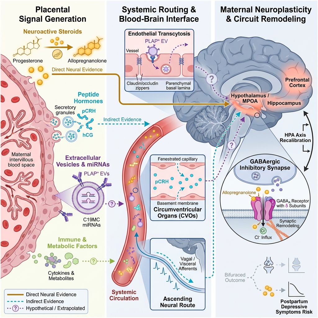


Placental Signalling as a Potential Component of Maternal Brain Adaptation During Pregnancy: A Critical Narrative Review of Candidate Mechanisms and Levels of Evidence
- Department of Human Anatomy, College of Medicine and Health Sciences, Afe Babalola University, Ado-Ekiti, Nigeria
- Stress & Neuroimmunology Group, Department of Human Anatomy, College of Medicine and Health Sciences, Afe Babalola University, Ado-Ekiti, Nigeria
- Department of Radiology, ABUAD Multisystem Hospital, Ado-Ekiti, Nigeria
- Department of Human Physiology, College of Medicine and Health Sciences, Afe Babalola University, Ado-Ekiti, Nigeria
Abstract
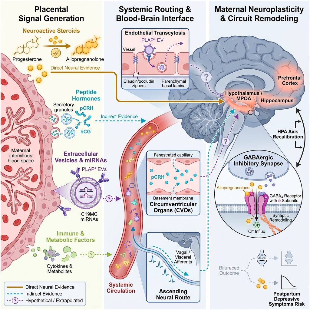
Pregnancy induces profound physiological and neurobiological adaptations that support maternal behaviour and the transition to parenthood. Human neuroimaging studies have documented consistent pregnancy-associated changes in maternal brain structure and function within neural networks subserving social cognition, emotional processing, and behavioural adaptation. Although these adaptations have largely been attributed to maternal ovarian hormones and established neuroendocrine pathways, gestation also involves sustained signalling from the placenta, a transient endocrine organ that regulates maternal metabolic, vascular, immune, and hormonal homeostasis. This critical narrative review evaluates whether placenta-derived endocrine, extracellular vesicle, immune, and metabolic-vascular signals contribute to the physiological milieu in which pregnancy-associated maternal neuroplasticity occurs. Literature published between January 1990 and June 2026 was identified through structured searches of PubMed/MEDLINE, Scopus, and Web of Science, supplemented by Google Scholar and citation tracking. The evidence supporting each candidate pathway was synthesised as a mechanistic hypothesis and graded as direct, indirect, or extrapolated. Across the four hypotheses examined, evidence confirming placental origin and maternal systemic action is substantial, whereas evidence demonstrating direct action on maternal neural tissue remains predominantly indirect. The strongest mechanistic candidates are placental steroidogenic output—specifically progesterone and its neuroactive metabolite allopregnanolone—alongside placental corticotropin-releasing hormone, human chorionic gonadotropin acting at central luteinizing hormone/chorionic gonadotropin receptors, and syncytiotrophoblast-derived extracellular vesicles. Active instructive placental signalling is distinguished from allostatic remodelling of the maternal brain in response to the physiological workload of the feto-placental unit, and four anatomically plausible routes of central nervous system access are specified. Finally, placenta-specific molecular profiling strategies capable of isolating placental signals from the maternal systemic background are outlined as essential methodological preconditions for testing this hypothesis in human pregnancy.
Graphical abstract
Maternal brain plasticity during pregnancy: an unresolved biological question
Pregnancy is associated with substantial neurobiological adaptations that prepare the maternal organism for the physiological and behavioural demands of motherhood. Human neuroimaging studies have demonstrated pronounced pregnancy-associated changes in maternal brain structure and function, particularly within regions governing social cognition, emotional processing, motivation, and maternal caregiving behaviour. A landmark prospective study comparing women before conception and after their first pregnancy reported reductions in grey matter volume across regions subserving social cognition; these reductions persisted for at least two years postpartum and accurately distinguished parous from nulliparous women1. Subsequent population-based neuroimaging work extended these observations by demonstrating that parity markers are associated with distinct structural brain signatures decades after childbirth, indicating that gestational neural adaptations leave enduring rather than merely transient traces2. More recently, dense-sampling precision neuroimaging tracking an individual from preconception through two years postpartum mapped the gestational trajectory of these changes at high temporal resolution, revealing progressive reductions in cortical grey matter volume and thickness alongside concomitant increases in white matter microstructural integrity3. The morphometric profile of these gestational adaptations closely parallels that of adolescence—a second developmental window of intensive steroid-driven cortical reorganisation6—and extends to subcortical reward circuitry, where volumetric reductions in the ventral striatum predict postpartum maternal neural responsiveness to infant cues7. Longitudinal postpartum investigations further indicate that several of these structural alterations evolve non-linearly, with gestational grey matter reductions in certain regions followed by partial volumetric recovery or expansion across cortical and subcortical networks during the early postpartum weeks8,9, though the extent to which specific adaptations persist or reverse over subsequent years remains an active question10. Collectively, these findings establish that pregnancy entails dynamic, functionally relevant neural reorganisation linked to the transition to caregiving rather than passive exposure to endocrine fluctuations11,12.
Current models of maternal brain plasticity have centred largely on maternal ovarian hormones and classical neuroendocrine pathways; while these models account for major features of maternal neuroadaptation, they remain incomplete. Gestational surges in oestradiol, progesterone, prolactin, oxytocin, and glucocorticoids modulate neural circuits governing stress reactivity, reward processing, and maternal responsiveness, providing an essential foundation for understanding pregnancy-associated neuroadaptation13,14 and shaping female brain health across the lifespan15. However, gestation also involves sustained biochemical communication from the maternal-foetal interface. The placenta functions as a high-output transient endocrine organ that releases steroid and peptide hormones, growth factors, immune mediators, and extracellular vesicles into the maternal circulation. These placenta-derived signals orchestrate maternal metabolic, cardiovascular, immunological, and endocrine adaptations throughout gestation4,5, while parallel cross-talk across the same interface shapes foetal neurodevelopment and metabolic programming16. Despite its established role in governing maternal systemic physiology, the potential contribution of placenta-derived signalling to maternal brain plasticity remains insufficiently integrated into contemporary models of pregnancy neurobiology—an omission recently highlighted as a priority for longitudinal, multi-system investigation17.
This article provides a critical narrative review of the evidence addressing a central biological question: whether placenta-derived signals contribute to the physiological environment supporting maternal brain adaptation during pregnancy. It is structured as a critical review rather than a purely conceptual perspective because direct in vivo evidence in human pregnancy remains too limited to sustain an uncritical hypothesis-driven model. Four candidate pathways are examined sequentially: placental endocrine signalling, extracellular vesicle-mediated communication, immune modulation, and metabolic-vascular regulation. Each pathway is formulated as a mechanistic hypothesis and appraised across a standardised evidentiary sequence: biological rationale, confirmation of placental origin, evidence of maternal systemic action, direct evidence within the maternal brain, indirect or extrapolated evidence from foetal, animal, or pathological contexts, and remaining methodological uncertainties. Table 1 summarises this evidence appraisal across the four candidate pathways, and Figure 2 illustrates the anatomical routes by which placenta-derived signals could access the maternal central nervous system (CNS). The integrative placenta–maternal brain framework presented in Figure 1 and discussed in the closing sections thus emerges as a testable synthesis that complements, rather than displaces, established maternal neuroendocrine mechanisms.
Comparative appraisal of candidate placenta-derived signalling pathways, physiological settings, and levels of evidence for maternal brain adaptation during pregnancy.
| Candidate pathway & primary ligands | Confirmed placental origin | Direct evidence (maternal brain in vivo or neural tissue) | Indirect evidence (maternal systemic physiology) | Extrapolated evidence (foetal compartment, non-human models, or pathology) | Principal methodological uncertainties |
|---|---|---|---|---|---|
| 1. Endocrine signalling: Progesterone / allopregnanolone, oestrogens, pCRH, hCG, hPL, hPGH | Syncytiotrophoblast steroidogenesis and peptide hormone secretion into maternal blood | Gestational allopregnanolone remodels maternal brain GABAA receptor subunits in rats | Drives maternal insulin resistance, lipid mobilisation, cardiovascular expansion | Central LH/hCG receptor expression and behavioural effects of hCG in cycling female rats | Shared maternal ovarian/pituitary and placental hormone pools confound source attribution in human neuroimaging studies; rodent models lack pCRH and rely on luteal progesterone |
| 2. Extracellular vesicle (EV) signalling: Syncytiotrophoblast exosomes and microvesicles (PLAP+, C19MC miRNAs, proteins, lipids) | Trophoblast-specific C19MC miRNA cargo and surface PLAP expression confirm syncytiotrophoblast origin | None reported in healthy maternal brain tissue or in vivo during physiological pregnancy. | Circulating PLAP+ exosomes increase progressively across the first trimester | EVs from pre-eclamptic plasma and hypoxic placentae disrupt BBB integrity in experimental models (attenuated by magnesium sulfate) | Pathological EV shedding in pre-eclampsia differs quantitatively and qualitatively from healthy gestation |
| 3. Immune signalling: Trophoblast-derived cytokines, chemokines, and immunomodulatory factors | Active trophoblast secretion and immune regulation at the maternal-foetal interface | Region-specific alterations in microglial density, ramification, and central cytokine expression in pregnant and postpartum female rats (placental causality unproven) | Shifts maternal systemic inflammatory balance and peripheral leukocyte priming across gestation | Maternal immune activation models demonstrate cytokine-mediated disruption of foetal neurodevelopment | No experimental study has selectively perturbed placental immune output while measuring maternal central neuroimmune or behavioural outcomes. |
| 4. Metabolic & vascular signalling: Placental nutrient sensors, metabolic hormones, angiogenic factors (PlGF, sFlt-1) | Trophoblast nutrient-sensing pathways and angiogenic factor secretion | None demonstrating direct action of placenta-specific metabolic or angiogenic ligands on maternal neural circuits. | Orchestrates maternal uterine and systemic vascular remodelling, plasma volume expansion, and substrate allocation | Pathological anti-angiogenic imbalance in pre-eclampsia compromises maternal cerebrovascular endothelial function and BBB integrity | Difficult to separate direct instructive placental signalling from secondary allostatic cerebrovascular accommodation to systemic haemodynamic workload. |
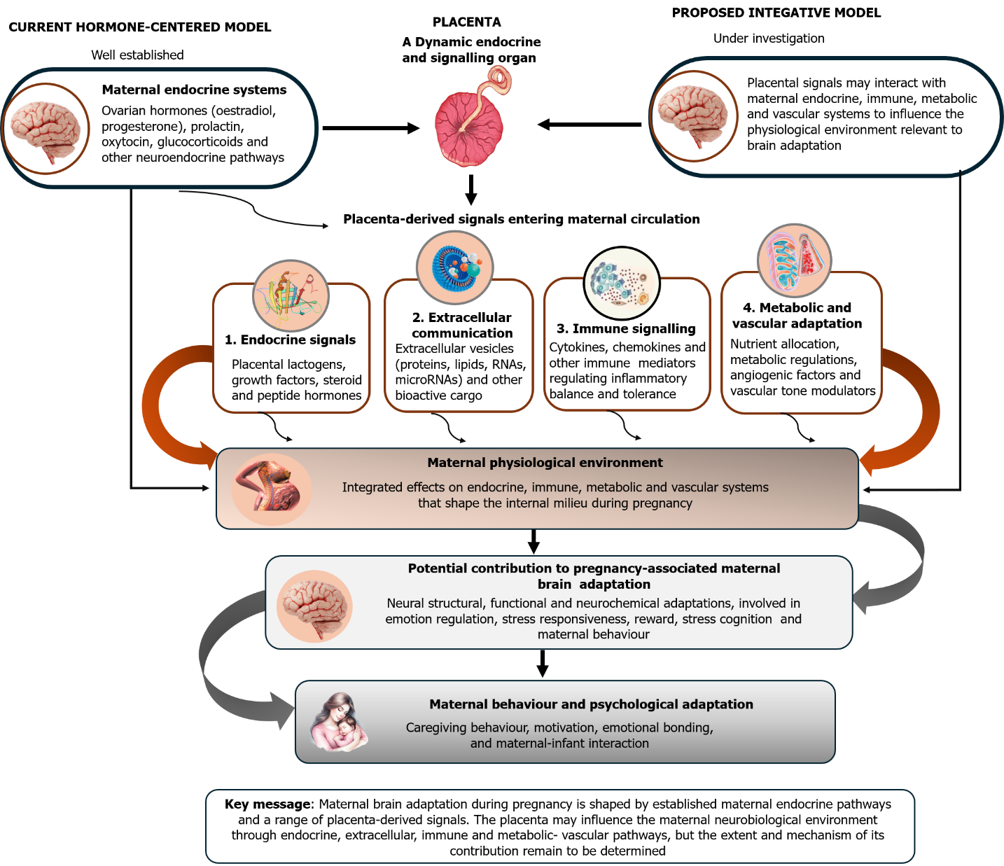
Conceptual framework integrating placenta-derived signalling pathways with established maternal neuroendocrine mechanisms during pregnancy. The schematic compares the classical hormone-centred model of maternal brain plasticity (top left)—which attributes gestational neuroadaptation primarily to maternal ovarian steroids (oestradiol and progesterone), pituitary prolactin, oxytocin, and glucocorticoids—with a proposed integrative placenta-inclusive model (top right). In the integrative framework, the placenta acts as a dynamic endocrine and signalling organ that releases four major classes of bioactive mediators into the maternal circulation (middle panels): (1) endocrine signals (placental lactogens, growth factors, and steroid and peptide hormones); (2) extracellular communication vesicles transporting proteins, lipids, messenger RNAs (mRNAs), and microRNAs; (3) immune mediators (cytokines and chemokines regulating systemic inflammatory balance and feto-maternal tolerance); and (4) metabolic and vascular regulators (nutrient-sensing signals, angiogenic factors, and vascular tone modulators). Solid black arrows denote established physiological pathways; curved orange and grey arrows denote coordinated multi-system interactions shaping the maternal internal milieu, which in turn supports structural, functional, and neurochemical adaptations in maternal brain circuits governing emotion regulation, stress responsiveness, reward processing, social cognition, and caregiving behaviour (bottom panels). Candidate anatomical routes across the maternal blood-brain interface are detailed in
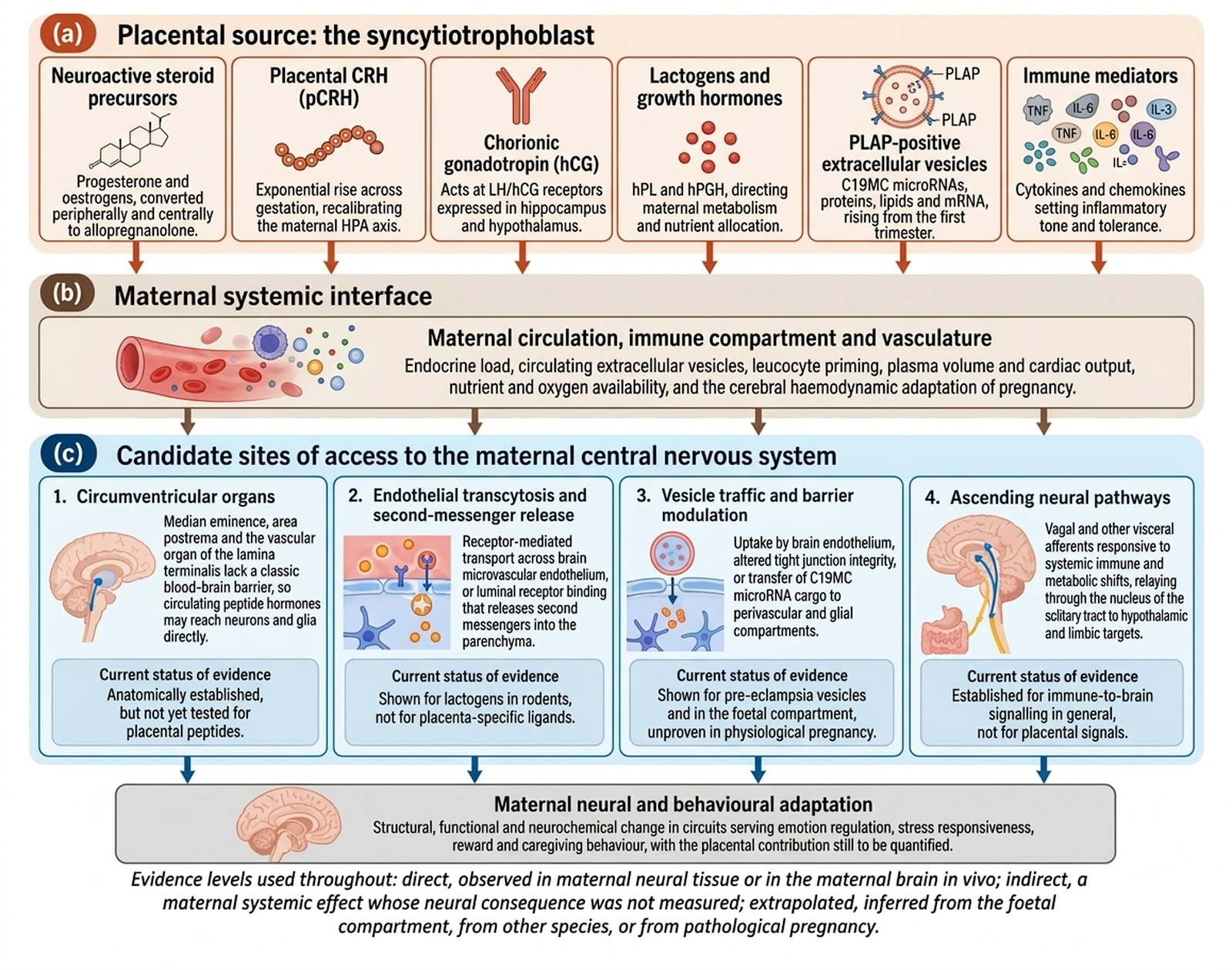
Candidate anatomical routes and cellular mechanisms mediating placental signal access to the maternal central nervous system. The diagram illustrates the sequential pathway from syncytiotrophoblast secretion to maternal neural adaptation across three biological compartments, alongside the current level of empirical evidence supporting each route. (a) Placental source (the syncytiotrophoblast): Bioactive signals released into maternal blood include neuroactive steroid precursors (progesterone and oestrogens metabolised peripherally and centrally to allopregnanolone), placental corticotropin-releasing hormone (pCRH), human chorionic gonadotropin (hCG), human placental lactogen (hPL) and human placental growth hormone (hPGH), placental alkaline phosphatase (PLAP)-positive extracellular vesicles (EVs) carrying chromosome 19 microRNA cluster (C19MC) microRNAs, proteins, lipids, and mRNA, and immunomodulatory cytokines and chemokines (e.g., TNF, IL-6, IL-1, IL-8). (b) Maternal systemic interface: Circulating placental factors modulate maternal endocrine load, leukocyte priming, plasma volume, cardiac output, nutrient and oxygen availability, and gestational cerebral haemodynamics. (c) Candidate sites of access to the maternal central nervous system: Four anatomically plausible access routes are depicted: (1) direct humoral access via fenestrated capillaries at circumventricular organs (median eminence, area postrema, and organum vasculosum of the lamina terminalis), which lack a classical blood-brain barrier (anatomically established, but untested for placental peptides); (2) saturable receptor-mediated endothelial transcytosis or luminal receptor binding with abluminal second-messenger release across the cerebral microvasculature (demonstrated for lactogens in rodents, but untested for placenta-specific ligands in humans); (3) endothelial uptake of PLAP-positive EVs, tight junction modulation, or perivascular/glial transfer of C19MC microRNA cargo (demonstrated for pre-eclamptic vesicles and in the foetal compartment, but unproven in healthy human pregnancy); and (4) ascending neural signalling via vagal and visceral sensory afferents relaying through the nucleus of the solitary tract to hypothalamic and limbic structures (established for systemic immune-to-brain communication, but untested for placental signals). Evidence tiers follow Section 2 definitions (direct, indirect, and extrapolated). Abbreviations: C19MC, chromosome 19 microRNA cluster; CRH, corticotropin-releasing hormone; EV, extracellular vesicle; hCG, human chorionic gonadotropin; HPA, hypothalamic-pituitary-adrenal; hPGH, human placental growth hormone; hPL, human placental lactogen; IL, interleukin; LH, luteinizing hormone; mRNA, messenger ribonucleic acid; pCRH, placental corticotropin-releasing hormone; PLAP, placental alkaline phosphatase; TNF, tumour necrosis factor.
Review methodology and literature selection
Literature for this review was identified through structured searches of PubMed/MEDLINE, Scopus, and Web of Science, supplemented by Google Scholar and backward and forward citation tracking of seminal articles, covering the period from January 1990 to June 2026 without language restrictions at initial screening. Search queries combined three concept blocks using Boolean operators. The first block targeted the maternal brain using the terms "maternal brain", "matrescence", and "pregnancy" paired with "neuroplasticity", "grey matter", "gray matter", "neuroimaging", or "maternal behaviour". The second block targeted the placenta and its secretory products using the terms "placenta", "syncytiotrophoblast", "placental hormone", "placental lactogen", "chorionic gonadotropin", "placental corticotropin-releasing hormone", "allopregnanolone", "extracellular vesicle", "exosome", and "C19MC". The third block targeted candidate CNS access and communication pathways using the terms "blood-brain barrier", "circumventricular organ", "transcytosis", "neuroimmune", "cytokine", "nutrient sensing", and "cerebral haemodynamics". Reference lists of retrieved review articles were manually screened to capture additional primary studies not indexed by the database queries.
Articles were selected based on their evidentiary contribution to one or more of the four mechanistic hypotheses rather than for exhaustive coverage of each subdiscipline, with priority given to primary human studies, mechanistic animal investigations where human data were unavailable, and authoritative reviews synthesising mature physiological literature. Records were retained if they established a placental origin for a signalling molecule, demonstrated a maternal systemic effect of that molecule, or characterised an action of that molecule (or its molecular class) on neural tissue; purely clinical obstetric outcome studies lacking a signalling or neurobiological component were excluded. Consistent with a critical narrative review design, no formal risk-of-bias scoring instrument or quantitative meta-analysis was applied; literature screening and study selection were conducted by the first author, independently verified by the co-authors, and reconciled through consensus discussion. To maintain strict evidentiary transparency, every mechanistic claim in the subsequent sections is explicitly graded according to three predefined levels of evidence:
-
Direct evidence: Empirical observation within maternal neural tissue or in the maternal brain in vivo.
-
Indirect evidence: Demonstration of a maternal systemic physiological effect whose downstream neural consequence was not directly measured.
-
Extrapolated evidence: Inference derived from the foetal compartment, non-human experimental models with divergent placentation, or pathological pregnancy states (such as pre-eclampsia).
These same three evidentiary tiers structure the comparative appraisal presented in Table 1.
Beyond maternal hormones: expanding the endocrine framework of maternal brain adaptation
Maternal brain plasticity during pregnancy has traditionally been interpreted through the actions of hormones synthesised by maternal endocrine glands—a framework that remains well supported but physiologically incomplete. Circulating concentrations of oestradiol, progesterone, prolactin, oxytocin, and glucocorticoids undergo marked gestational fluctuations and modulate neural circuits governing stress responsiveness, social cognition, reward motivation, and maternal caregiving13,14. Extensive preclinical and clinical evidence confirms that these endocrine adaptations are central to gestational neurobiology, with circulating hormones binding to cognate receptors across hypothalamic, limbic, and cortical networks to remodel neuronal excitability, synaptic architecture, and behavioural output18,19.
Nevertheless, the endocrine milieu of pregnancy extends beyond maternal glandular secretion to encompass a transient, anatomically distinct feto-placental source. Throughout gestation, the placental syncytiotrophoblast secretes substantial quantities of steroid hormones, peptide hormones, growth factors, and bioactive signalling molecules directly into the maternal circulation, coordinating the metabolic, cardiovascular, immunological, and reproductive adaptations required to sustain pregnancy4,5,20. Crucially, the placenta functions not merely as a constitutive secretory conduit, but as an active metabolic sensor that dynamically adjusts its growth, nutrient transport capacity, and endocrine output in response to maternal nutrient and oxygen availability21,22. Because placental endocrine secretion varies continuously with maternal metabolic and nutritional status, any downstream placental influence on maternal neuroplasticity would likewise be expected to operate as a context-dependent, dynamic signal rather than a fixed gestational programme.
The most compelling endocrine candidate linking placental function to maternal neural adaptation is placental steroidogenesis. During the second and third trimesters of human pregnancy, the placenta becomes the dominant source of circulating progesterone and oestrogens, converting maternal and foetal steroid precursors in large quantities. Progesterone is metabolised both peripherally and within the CNS into the neuroactive steroid allopregnanolone, a potent positive allosteric modulator of gamma-aminobutyric acid type A (GABAA) receptors23. Preclinical rodent studies provide direct neural evidence that the gestational rise and peripartum withdrawal of brain allopregnanolone drive structural and functional reorganisation of GABAA receptor subunit composition in the maternal brain24. In human pregnancy, lower mid-gestational circulating allopregnanolone concentrations have been prospectively associated with postpartum depressive symptoms, and the abrupt loss of placental progesterone synthesis at parturition provides a coherent mechanistic explanation for the acute postpartum drop in central neurosteroid tone25. Because the human placenta supplies the overwhelming majority of the progesterone substrate from which allopregnanolone is synthesised during the second half of gestation, placental steroidogenic output currently represents the strongest candidate for a placenta-dependent modulator of maternal neural function.
Two additional placenta-derived peptide hormones warrant classification as primary candidates for central maternal signalling because both possess verified receptor targets within the mammalian brain: placental corticotropin-releasing hormone (pCRH) and human chorionic gonadotropin (hCG). Anthropoid primates, including humans, are unique among eutherian mammals in that the syncytiotrophoblast secretes pCRH into the maternal circulation at exponentially rising concentrations across gestation, progressively recalibrating the maternal hypothalamic-pituitary-adrenal (HPA) axis and stress responsiveness26; moreover, accelerated mid-gestational pCRH trajectories have been linked prospectively to postpartum depressive symptomatology27. Similarly, hCG binds to functional luteinizing hormone/chorionic gonadotropin (LH/hCG) receptors expressed in multiple neural regions, including the hippocampus and hypothalamus28. In cycling female rats, peripheral and intracerebroventricular administration of hCG crosses the blood-brain barrier in small but intact fractions and alters hippocampus-dependent exploratory and neophobic behaviours29, while circulating maternal hCG concentrations in human gestation vary systematically with foetal sex and maternal physiological characteristics30. Both pCRH and hCG therefore satisfy the criteria of confirmed placental origin, established maternal systemic action, and plausible central receptor targets, setting them apart from placental factors whose neurobiological relevance relies solely on systemic inference.
Several additional placenta-derived hormones exhibit biological properties pertinent to maternal neurobiology, although the supporting evidence remains more indirect. Human placental lactogen (hPL) and placental growth hormone (hPGH) regulate maternal insulin sensitivity, lipolysis, and nutrient partitioning—processes increasingly recognised as modulators of central energy homeostasis and behaviour5,31. In rodents, lactogenic signalling exerts direct central actions: activation of central prolactin receptors stimulates forebrain neurogenesis during gestation32, and circulating lactogens enter the rodent brain via a saturable transport mechanism that operates independently of the prolactin receptor itself33. However, whether placenta-derived hPL, as distinct from maternal pituitary prolactin, crosses the human blood-brain barrier to engage maternal neural circuits remains unverified in vivo, rendering this extrapolated evidence. Similarly, placental modulation of maternal immune and vascular pathways may shape neural adaptation indirectly by altering the systemic physiological environment in which gestational neuroplasticity unfolds34,35. On the foetal side of the interface, conditional genetic ablation of placental allopregnanolone synthesis in mice disrupts cerebellar white matter myelination and induces autism-like social deficits in male offspring36, providing definitive proof-of-principle that placental endocrine output can sculpt mammalian brain development while leaving open whether the adult maternal brain exhibits comparable sensitivity. Furthermore, because placental hormone secretion varies across gestational age and differs according to foetal sex20, the maternal systemic milieu supporting neuroadaptation is unlikely to be uniform across pregnancies.
Before appraising the four individual pathways in detail, it is essential to distinguish between two conceptual accounts of how placental activity could relate to maternal brain adaptation. These accounts are not mutually exclusive, but they entail markedly different evidentiary thresholds:
-
Active instructive signalling: In this model, placenta-specific molecules engage defined receptors or cellular targets within maternal neural circuits to direct neurochemical, structural, or behavioural adaptations (e.g., priming caregiving motivation or affective regulation). Placental neurosteroid precursors (via allopregnanolone), pCRH, and hCG represent the primary candidates for instructive signalling because each possesses identified molecular targets in neural tissue.
-
Allostatic or homeostatic remodelling: In this model, the maternal brain undergoes structural and metabolic reorganisation as a secondary adaptation to the substantial haemodynamic, immunological, and metabolic workload imposed by the feto-placental unit. Here, neural plasticity reflects central homeostatic accommodation to systemic physiological demand rather than purposive molecular instruction from the placenta.
Much of the empirical literature appraised below is fully compatible with allostatic remodelling. Maintaining a rigorous distinction between instructive signalling and allostatic adaptation prevents the misinterpretation of secondary systemic adjustments as directed feto-placental programming of the maternal brain. Establishing active instructive signalling requires demonstrating that a placenta-specific ligand accesses a defined maternal neural target and that selective perturbation of that ligand alters maternal neural or behavioural outcomes—an evidentiary standard not yet met by any placental signal in human pregnancy.
An expanded neuroendocrine framework of pregnancy should therefore integrate both maternal glandular hormones and placenta-derived signals without attributing greater explanatory weight to placental communication than current evidence warrants. Reviews of female brain ageing have similarly proposed that the profound immunoendocrine transitions of pregnancy, alongside menopause, shape long-term trajectories of neurological health and neurodegenerative risk37. Defining how placenta-derived signals interact with established maternal hormonal pathways is the critical step required to transform this conceptual framework into a falsifiable neurobiological model.
Candidate mechanistic pathways and the levels of evidence supporting them
Determining whether placenta-derived signals contribute to maternal brain adaptation requires specifying the biological pathways linking the maternal-foetal interface to maternal physiological systems and grading the empirical evidence supporting each step. Throughout gestation, the syncytiotrophoblast releases steroid and peptide hormones, extracellular vesicles, immunomodulatory cytokines, and metabolic-vascular regulators into the maternal circulation4,5. Below, four candidate pathways are formulated as mechanistic hypotheses and evaluated across a uniform evidentiary sequence: biological rationale, placental origin, maternal systemic effect, direct evidence in the maternal brain, indirect or extrapolated evidence, and remaining uncertainties. Table 1 synthesises this pathway-by-pathway appraisal.
Hypothesis 1: Placental endocrine signalling modifies maternal neural function
The biological rationale for the endocrine hypothesis is that the human placenta functions as a high-capacity endocrine organ whose secretory products circulate in maternal blood at concentrations unmatched during any other phase of adult life; consequently, any maternal neural circuit expressing cognate receptors or steroid-metabolising enzymes would be exposed to sustained gestational stimulation. Placental origin is unequivocally established for progesterone and oestrogen synthesis, pCRH, hCG, hPL, and hPGH5,20. Maternal systemic effects are equally well documented for each hormone, encompassing gestational insulin resistance, maternal lipid mobilisation, cardiovascular volume expansion5,38, and progressive recalibration of the maternal HPA axis18,26. Direct evidence within the maternal brain is currently restricted to the neurosteroid arm of this pathway: gestational allopregnanolone dynamically remodels cortical and hypothalamic GABAA receptor subunit expression and function in pregnant rats24, and lower mid-gestational plasma allopregnanolone concentrations prospectively predict postpartum depressive symptoms in women25. For the peptide hormones (hCG and lactogens), central receptor expression, blood-brain barrier transport, and behavioural modulation have been demonstrated in rodent models28,29,32,33, but direct neural action has not been confirmed in human pregnancy. Indirect evidence is substantial, given that systemic metabolic substrate availability and HPA axis tone profoundly modulate central synaptic plasticity39, while extrapolated evidence from mice demonstrates that placenta-specific allopregnanolone synthesis is required for normal foetal cerebellar white matter development36. The principal methodological uncertainty is source attribution: because placental and maternal ovarian/pituitary sources of steroids and lactogens overlap in the maternal circulation, no human neuroimaging study has yet isolated the neural effect of a specifically placental hormone from the broader maternal endocrine background17.
Hypothesis 2: Syncytiotrophoblast-derived extracellular vesicles carry signals to maternal tissues
The biological rationale for the extracellular vesicle (EV) hypothesis is that membrane-bound vesicles (including exosomes and microvesicles) transport functional proteins, lipids, mRNAs, and microRNAs between distant organs and represent a conserved mechanism of intercellular communication, including across the neurovascular unit40. Crucially, placental origin can be established with high molecular specificity: syncytiotrophoblast-derived EVs express surface placental alkaline phosphatase (PLAP) and carry trophoblast-restricted microRNAs transcribed from the primate-specific chromosome 19 microRNA cluster (C19MC), enabling direct analytical separation of placental vesicles from maternal systemic vesicles41,42. Maternal systemic effects are well documented, with circulating PLAP-positive exosome concentrations rising progressively across the first trimester of healthy human pregnancy43 and syncytiotrophoblast microparticles contributing to the controlled systemic inflammatory priming characteristic of normal gestation44,45. However, direct evidence of placental EV action within the healthy maternal brain is currently absent. Available neurovascular evidence is entirely extrapolated from pathological pregnancy models and the foetal compartment: plasma EVs isolated from women with pre-eclampsia and EVs shed by hypoxic ex vivo human placentae disrupt blood-brain barrier tight junction integrity in rodent and endothelial models—an effect attenuated by magnesium sulfate46—while placenta-derived EVs in the foetal circulation cross developing biological barriers and modulate neurodevelopmental pathways47. The primary uncertainty is both qualitative and quantitative: pathological EV shedding in pre-eclampsia differs markedly from physiological release in particle concentration, surface antigen profile, and pro-inflammatory/anti-angiogenic cargo48. Consequently, evidence that pre-eclamptic vesicles compromise endothelial barrier integrity cannot be extrapolated as evidence that physiological placental EVs deliver instructive molecular cargo across an intact maternal blood-brain barrier during uncomplicated pregnancy.
Hypothesis 3: Placental regulation of maternal immune physiology shapes the maternal brain
The biological rationale for the neuroimmune hypothesis is that successful gestation requires precise, stage-specific immunological tolerance and priming at the maternal-foetal interface, and that peripheral immune mediators exert well-characterised regulatory effects on central glial activity, synaptic plasticity, and affective behaviour. Placental origin and regulatory control are firmly established: the trophoblast actively secretes cytokines, chemokines, and immunomodulatory factors that govern maternal leukocyte recruitment and systemic immune balance rather than acting as a passive physical barrier34,49. Maternal systemic effects include the finely tuned shift in peripheral inflammatory tone and innate immune priming across gestation, to which circulating syncytiotrophoblast microparticles directly contribute44. Within neuroscience, peripheral cytokines and immune signalling molecules are known to modulate central neural function via microglial activation, neurotransmitter metabolism, and neuroendocrine circuits50,51, while classical immune proteins (such as major histocompatibility complex class I molecules and complement components) play essential physiological roles in developmental synaptic pruning and adult synaptic plasticity independent of infection or injury52. Direct evidence involving the maternal brain itself comes from preclinical rodent studies demonstrating that late pregnancy and the early postpartum period are accompanied by region-specific reductions in microglial density, altered microglial process ramification, and shifts in central cytokine expression within the maternal prefrontal cortex, hippocampus, and amygdala53. While this constitutes direct observation of neuroimmune remodelling in the rodent maternal brain, its specific causal attribution to placental signals—as opposed to maternal sex steroids—has not been established. Because the vast majority of the pregnancy neuroimmunology literature focuses on pathological maternal immune activation and its adverse consequences for foetal neurodevelopment35, direct extrapolation to physiological maternal brain adaptation is unwarranted without targeted experiments that selectively manipulate placental immune output while measuring maternal central neuroimmune outcomes.
Hypothesis 4: Placental regulation of maternal metabolic and vascular physiology alters the cerebral environment
The biological rationale for the metabolic and vascular hypothesis is that the mammalian brain is metabolically demanding and highly dependent on continuous cerebral blood flow and regulated substrate delivery, both of which undergo profound systemic reorganisation driven by the placenta during gestation. Placental origin is well established for the nutrient-sensing pathways, metabolic hormones, and angiogenic factors (such as placental growth factor and soluble fms-like tyrosine kinase-1) that govern maternal resource allocation and vascular tone21,22. Maternal systemic effects are among the most thoroughly characterised phenomena in reproductive physiology, encompassing extensive uterine spiral artery and systemic vascular remodelling, plasma volume expansion, increased cardiac output, and gestational adjustments in glucose and lipid metabolism5,54. Evidence involving the maternal brain is indirect but physiologically significant: preclinical and clinical studies demonstrate that the maternal cerebral circulation adapts during pregnancy through enhanced pressure autoregulation, altered myogenic tone, and remodelling of cerebral arterioles and blood-brain barrier transport properties that protect the maternal brain from hyperperfusion under elevated haemodynamic load55. However, no study has yet demonstrated that these cerebrovascular adaptations are triggered directly by placenta-specific ligands acting on cerebral endothelium rather than representing an autoregulatory response to the systemic cardiovascular and endocrine state of gestation. Consequently, this pathway aligns most closely with the allostatic remodelling framework outlined in Section 3, with neural and cerebrovascular adjustments inferred from systemic physiological workload rather than direct placental instruction.
Routes by which placental signals could reach the maternal brain
A critical mechanistic question left unaddressed by most discussions of feto-maternal communication is how large, hydrophilic, or vesicle-encapsulated placental molecules could gain physical or functional access to maternal neural tissue across the blood-brain barrier. Four anatomically and physiologically plausible routes of CNS access can be specified (summarised in Figure 2):
-
Circumventricular organs (CVOs): Sensory and secretory CVOs—including the median eminence, area postrema, and organum vasculosum of the lamina terminalis (OVLT)—possess fenestrated capillaries lacking a classical tight-junction blood-brain barrier, enabling circulating peptide hormones and cytokines to interact directly with central neurons and astrocytic processes56. While anatomically established for systemic neuroendocrine regulation, this route has not yet been experimentally tested for placenta-specific peptides such as pCRH or hPL.
-
Endothelial receptor-mediated transcytosis and luminal signalling: Circulating protein hormones can cross the cerebral microvascular endothelium via saturable, receptor-mediated vesicular transport, or bind to luminal endothelial receptors to trigger abluminal release of second messengers (such as nitric oxide or prostaglandins) into the brain parenchyma without the intact ligand crossing the barrier57. The demonstration that circulating prolactin and lactogens enter the mouse brain via a choroid plexus and endothelial transport mechanism independent of the classical prolactin receptor provides direct proof-of-principle for this route33.
-
Extracellular vesicle trafficking and barrier modulation: Syncytiotrophoblast-derived EVs may be internalised by cerebral endothelial cells via endocytosis, transfer C19MC microRNA cargo into perivascular and glial compartments, or modulate tight junction permeability. At present, empirical support for this route is restricted to extrapolated pathological models showing blood-brain barrier disruption by pre-eclamptic plasma EVs46 and trans-barrier EV trafficking within the foetal compartment47.
-
Ascending visceral afferent neural pathways: Peripheral immune cytokines and metabolic signals can activate vagal and other visceral sensory afferents innervating maternal peripheral tissues, relaying neural signals through the nucleus of the solitary tract (NTS) to hypothalamic, limbic, and autonomic centres without requiring humoral transport across the blood-brain barrier50. While well established for general peripheral immune-to-brain communication, vagal relay of specifically placenta-derived immune or metabolic signals remains untested.
Taken together, the appraisal across Sections 4.1–4.5 indicates that any placental contribution to maternal neuroplasticity is far more likely to operate through coordinated, multi-system interactions across endocrine, vesicular, immune, and cerebrovascular interfaces than through a single linear pathway. Moreover, the four candidate hypotheses differ substantially in their current evidentiary maturity: evidence of placental origin and maternal systemic action is robust across all four pathways, whereas evidence of direct maternal neural action is strongest for placental steroidogenesis (via allopregnanolone), mechanistically plausible via central receptors for pCRH and hCG, and currently limited to indirect or extrapolated observations for EV, immune, and metabolic-vascular pathways (Table 1; Figure 1).
Distinguishing placental signals from maternal background: methodological requirements
The principal technical obstacle to testing any of the four hypotheses in human pregnancy is biochemical overlap: because placenta-derived and maternal endogenous molecules circulate together in maternal blood, observing a statistical correlation between a circulating hormone or cytokine and a neuroimaging metric does not establish the tissue of origin. Three molecular profiling strategies now offer the analytical specificity required to overcome this limitation in human cohort studies:
-
Placenta-specific microRNA signatures: Targeting nucleic acids expressed almost exclusively by trophoblast cells—specifically the primate-specific C19MC microRNA cluster exported within exosomes (and the orthologous chromosome 14 microRNA cluster [C14MC] in rodents for comparative preclinical studies)—provides an unambiguous molecular tag of placental origin in maternal plasma42.
-
Immunoaffinity isolation of syncytiotrophoblast EVs: Capturing PLAP-positive extracellular vesicles from maternal peripheral blood via immunoaffinity chromatography or fluorescence nanoparticle tracking physically separates syncytiotrophoblast-derived vesicles from the much larger pool of maternal platelet-, endothelial-, and leukocyte-derived EVs prior to proteomic or transcriptomic cargo analysis41.
-
Maternal plasma cell-free RNA (cfRNA) profiling: Quantifying trophoblast-specific gene transcripts within maternal circulating cfRNA already enables accurate non-invasive prediction of gestational age, preterm birth, and pre-eclampsia58,59. Pairing longitudinal cfRNA quantification of placental transcripts with serial structural and functional neuroimaging in the same pregnant participants would allow investigators to test whether fluctuations in placenta-specific transcriptional output covary with maternal cortical and subcortical remodelling while statistically controlling for maternal systemic background.
Complementing human longitudinal studies, experimental animal and ex vivo models remain indispensable for establishing causal mechanisms that cannot be ethically manipulated in pregnant women. However, pronounced interspecies divergence in placental anatomy and endocrinology dictates that experimental model selection determines translational validity. Laboratory rodents possess haemochorial placentation but express expanded families of prolactin-like proteins lacking human orthologues, rely on the maternal corpus luteum rather than the placenta for progesterone synthesis throughout most of gestation, and do not express placental CRH60,61,62. Furthermore, rodent maternal caregiving behaviour is heavily dependent on olfactory processing and the medial preoptic area, whereas human maternal adaptation engages distributed higher-order cortical networks subserving theory of mind and social cognition12,19. Consequently, experimental designs should prioritise model systems that preserve the relevant human biology:
-
Non-human primates: Anthropoid primates share both haemochorial villous placentation and exponentially rising syncytiotrophoblast pCRH secretion across gestation26,60.
-
Trophoblast-specific genetic manipulation in mice: Conditional gene targeting using trophoblast-restricted Cre recombinase drivers (such as
Cyp19a1-Cre )63 or blastocyst trophectoderm-specific lentiviral gene transfer prior to implantation64 enables selective deletion or overexpression of a placental gene while leaving the maternal genome—and maternal endogenous endocrine/immune tissues—completely intact. -
Ex vivo dual perfusion of the human placental cotyledon: The dually perfused human term placental lobule allows the directional release of hormones, cytokines, and PLAP-positive EVs into the maternal-side perfusate to be quantified in human tissue in the complete absence of maternal systemic confounding65.
The power of trophoblast-restricted genetic manipulation is exemplified by the conditional deletion of the allopregnanolone-synthesising enzyme Akr1c14 specifically within mouse trophoblast (Cyp19a1-Cre), which definitively demonstrated that placental neurosteroid output causally regulates offspring cerebellar myelination and social behaviour36. Executing the analogous placenta-specific knockout experiment while evaluating maternal brain structure, microglial morphology, GABAA receptor plasticity, and postpartum caregiving behaviour in the genetically wild-type dam represents the single most informative preclinical experiment needed to test the placenta–maternal brain hypothesis.
Defining the placenta-maternal brain hypothesis: priorities for future research
Determining whether placenta-derived signals actively shape maternal neuroplasticity requires research designs that bridge reproductive biology and maternal systems neuroscience. As our evidentiary appraisal demonstrates, biological plausibility is strongly supported by the placenta's established governance of maternal systemic physiology, whereas direct empirical proof linking placenta-derived molecules to maternal neural circuits remains largely confined to neuroactive steroids and preclinical rodent models. Future investigations should therefore move beyond isolated single-hormone correlations and examine gestation as an integrated multi-organ communication network encompassing the placenta, maternal endocrine axes, peripheral immunity, cerebrovascular autoregulation, and the CNS17.
Longitudinal human cohort studies pairing high-frequency placental molecular profiling with precision maternal neuroimaging represent the highest-priority translational step. Recent dense-sampling neuroimaging work has demonstrated that maternal cortical volume, thickness, and white matter quantitative anisotropy can be tracked week-by-week from preconception through the postpartum period3. Integrating this precision imaging paradigm with serial maternal blood sampling for PLAP-positive EV cargo, C19MC microRNAs, trophoblast-specific cfRNA transcripts, pCRH, and neurosteroid metabolomics would test whether individual trajectories of placental signalling predict the magnitude or regional specificity of maternal brain remodelling. Crucially, incorporating the placenta-specific molecular assays detailed in Section 5 is what will allow investigators to attribute observed brain–biomarker associations to placental output rather than maternal glandular activity.
Human cohort designs should also measure peripheral immune, metabolic, cardiovascular, and psychological variables concurrently with placental biomarkers, as the biologically relevant question is how placental signals interact with maternal endogenous pathways rather than whether they act in isolation. Stratification by foetal sex is particularly essential in these cohorts: first-trimester and term human placental transcriptomes exhibit pronounced sexual dimorphism66, maternal peripheral blood mononuclear cell cytokine responses across gestation vary systematically with foetal sex67, and maternal circulating hCG trajectories differ between male- and female-bearing pregnancies30. Testing whether foetal sex modifies gestational maternal brain plasticity—or alters maternal vulnerability to peripartum mood disorders—will require large, demographically diverse longitudinal cohorts12.
Finally, investigations of extracellular vesicle and neuroimmune pathways must rigorously separate the physiological signalling of uncomplicated gestation from the pathophysiological cascades of obstetric syndromes such as pre-eclampsia. In healthy pregnancy, circulating PLAP-positive EVs rise gradually and support a controlled, adaptive priming of maternal innate immunity and vascular compliance43,44. In pre-eclampsia, by contrast, hypoxic and oxidatively stressed syncytiotrophoblast tissue sheds elevated quantities of EVs enriched in anti-angiogenic factors, pro-inflammatory cytokines, and oxidized lipids that provoke systemic endothelial dysfunction, hypertension, and blood-brain barrier breakdown [45,46,48]. Because much of the experimental literature on placental EVs and cerebrovascular barrier function has been generated in the context of pre-eclampsia, care must be taken not to conflate pathological barrier injury with physiological feto-maternal neurocommunication. Studies aiming to define normative maternal brain adaptation must establish uncomplicated gestation as their primary mechanistic reference.
An integrative, placenta-inclusive framework does not replace classical neuroendocrine models of the maternal brain; rather, it embeds them within the complete reproductive physiology of pregnancy, treating the maternal-foetal interface as a candidate co-regulator of the internal milieu supporting maternal neuroplasticity while maintaining a strict distinction between instructive placental signalling and allostatic maternal remodelling.
Conclusion: towards an integrated model of maternal brain adaptation
Maternal brain plasticity during pregnancy is a robust, reproducible neurobiological phenomenon, yet the upstream physiological signals that initiate and coordinate this remodelling remain incompletely defined. While classical models have rightly emphasised maternal ovarian steroids and pituitary neuroendocrine pathways, gestation also involves continuous, high-output biochemical signalling from the placenta—a transient endocrine organ that reshapes maternal physiology through steroidogenesis, peptide hormone secretion, nutrient sensing, immunomodulation, and extracellular vesicle release.
Our critical appraisal of the literature indicates that current evidence does not yet prove that placenta-derived signals directly instruct maternal neural circuits in human pregnancy, and shows that the four candidate pathways differ substantially in their evidentiary proximity to that standard. Placental steroidogenic output holds the strongest mechanistic claim via progesterone-derived allopregnanolone, supported by direct neural remodelling evidence in rodents and prospective clinical associations in women, followed by pCRH and hCG, which engage verified central receptors but lack direct neuroimaging correlates in human pregnancy. Extracellular vesicle, neuroimmune, and metabolic-vascular pathways currently rest on indirect systemic and extrapolated pathological or foetal evidence. Integrating placental biology into models of maternal neuroplasticity provides a rigorous, biologically complete framework that complements precision human neuroimaging3,17 and generates falsifiable predictions for multidisciplinary research across reproductive biology, endocrinology, and neuroscience.
Declarations
Abbreviations
BBB: Blood-brain barrier; C14MC: Chromosome 14 microRNA cluster; C19MC: Chromosome 19 microRNA cluster; cfRNA: Cell-free ribonucleic acid; CNS: Central nervous system; CRH: Corticotropin-releasing hormone; CVO: Circumventricular organ; EV: Extracellular vesicle; GABAA: Gamma-aminobutyric acid type A; hCG: Human chorionic gonadotropin; HPA: Hypothalamic-pituitary-adrenal; hPGH: Human placental growth hormone; hPL: Human placental lactogen; LH: Luteinizing hormone; miRNA: MicroRNA; MRI: Magnetic resonance imaging; mRNA: Messenger ribonucleic acid; NTS: Nucleus of the solitary tract; OVLT: Organum vasculosum of the lamina terminalis; pCRH: Placental corticotropin-releasing hormone; PLAP: Placental alkaline phosphatase.
Acknowledgements
The authors acknowledge the contributions of researchers whose work in maternal neuroscience, placental biology, endocrinology, and reproductive physiology has informed the synthesis presented in this review, and thank the peer reviewers whose constructive comments shaped its restructuring.
Author’s contributions
EEE conceptualised the manuscript, designed and executed the structured literature search, developed the evidence appraisal framework, and prepared the initial draft. KFC, BEI, INM, and AAO independently verified the literature selection and contributed to the evidence evaluation, scientific synthesis, and critical revision of the manuscript. All authors read and approved the final manuscript.
Funding
None. The authors received no specific grant from any funding agency in the public, commercial, or not-for-profit sectors for the preparation of this manuscript.
Availability of data and materials
None. Not applicable, as this manuscript is a critical narrative review based on previously published literature and does not involve the generation or analysis of new datasets or materials.
Ethics approval and consent to participate
None. Not applicable, as this manuscript does not involve human participants, human tissue or data collection, animal experiments, or biological sample analysis.
Consent for publication
None. Not applicable, as this manuscript does not contain individual participant data, identifiable clinical information, or images requiring consent for publication.
Declaration of generative AI and AI-assisted technologies in the writing process
None. During the preparation and editing of this work, AI-assisted language editing was used solely to improve grammar, readability, reference formatting, and adherence to biomedical publishing style; the authors reviewed and edited the content as needed and take full responsibility for the integrity and scientific content of the publication.
Competing interests
None. The authors declare that they have no competing financial or non-financial interests.