


The Role of ZBTB7A in the Metabolic Landscape of t(8;21) Acute Myeloid Leukemia (AML): A Comprehensive Review of Current Evidence

- Department of Biomedical Science, Pusat Kanser Tun Abdullah Ahmad Badawi, Universiti Sains Malaysia, 13200 Kepala Batas, Pulau Pinang, Malaysia
- Analytical Biochemistry Research Centre (ABrC), Inkubator Inovasi Universiti (I2U), SAINS@USM, Universiti Sains Malaysia, 11900 Bayan Lepas, Pulau Pinang, Malaysia
- Centre for Medical Laboratory Technology Studies, Faculty of Health Sciences, Universiti Teknologi MARA, 42300 Puncak Alam, Selangor, Malaysia
Abstract
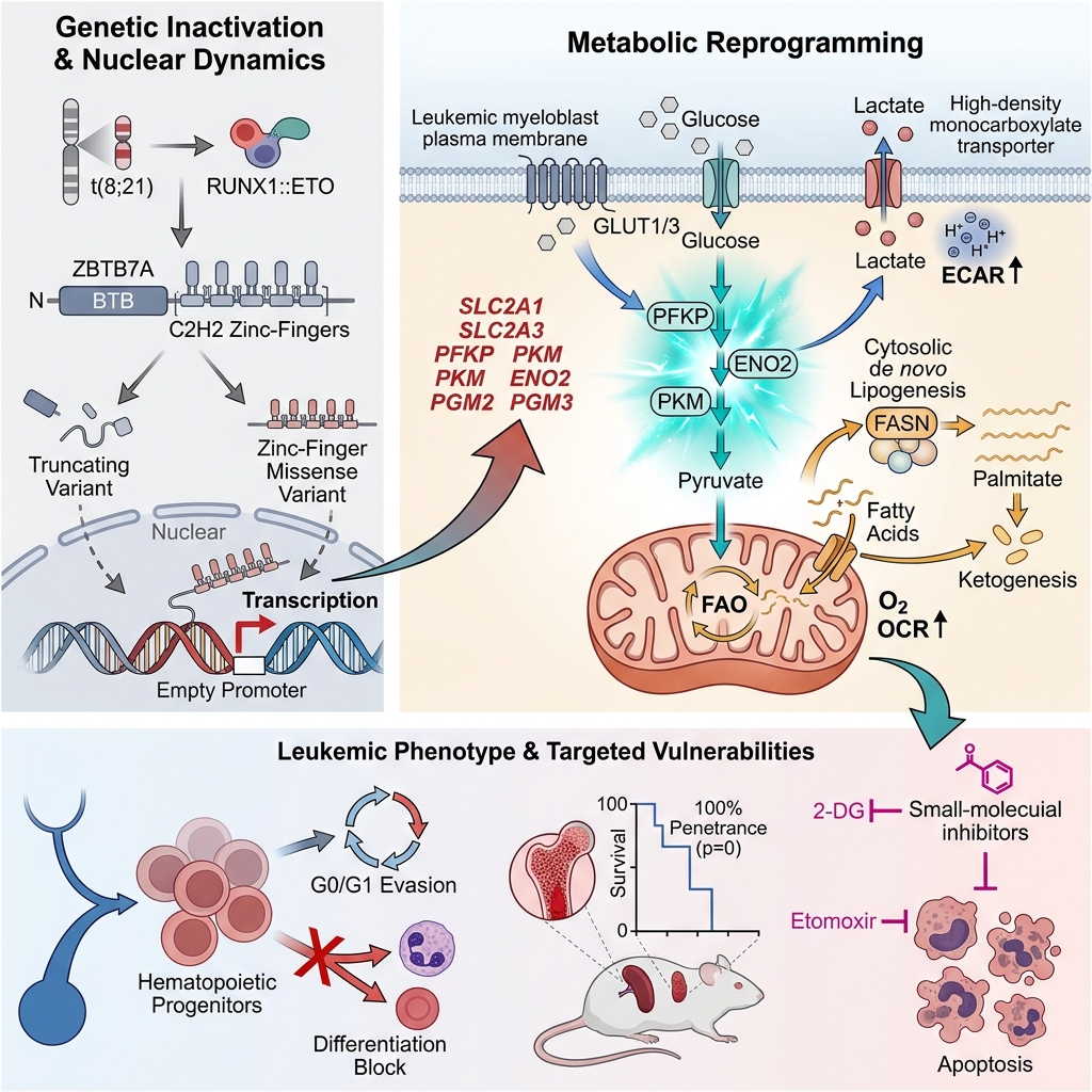
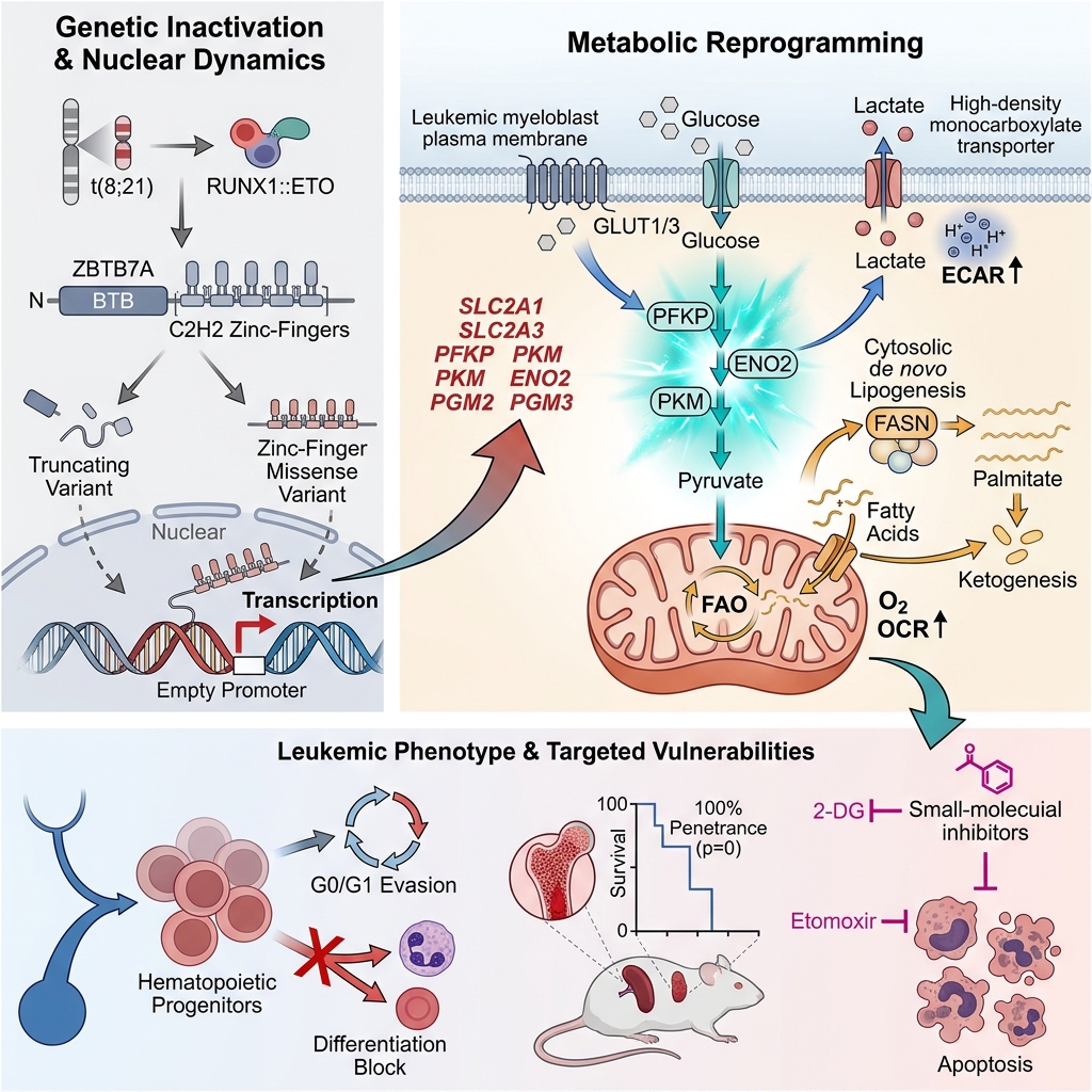
Introduction: The RUNX1::RUNX1T1 (also known as RUNX1::ETO) fusion oncogene generated by the t(8;21)(q22;q22) chromosomal translocation initiates leukemogenesis but requires additional cooperating mutations to induce overt t(8;21) acute myeloid leukemia (AML). Zinc finger and BTB domain-containing protein 7A (ZBTB7A, also known as POKEMON or LRF) is a transcriptional repressor that regulates multiple cellular processes, including hematopoiesis and energy metabolism. Somatic ZBTB7A mutations occur preferentially in t(8;21) AML compared with other AML subtypes, suggesting a distinct cooperative role in this cytogenetic subgroup. Because metabolic reprogramming is a hallmark mechanism through which leukemic cells sustain proliferation and resist therapy, defining the metabolic impact of ZBTB7A inactivation is of high clinical relevance.
Methods: A narrative literature search of PubMed and Google Scholar was conducted up to December 2025 to identify studies investigating whether ZBTB7A mutations drive metabolic reprogramming and cooperate with RUNX1::ETO in the pathogenesis of t(8;21) AML.
Results: All somatic ZBTB7A alterations characterized in t(8;21) AML act as loss-of-function or dominant-negative mutations. Evidence across in silico, in vitro, and in vivo models demonstrates that loss of ZBTB7A de-represses key glycolytic target genes—including SLC2A3 (GLUT3), PFKP, and PKM—resulting in elevated glycolytic flux that supports leukemic cell survival and clonal expansion. Furthermore, ZBTB7A deficiency alters lipid homeostasis by upregulating de novo fatty acid synthesis, fatty acid beta-oxidation, and ketogenesis, thereby providing metabolic flexibility under nutrient-restricted or hypoxic bone marrow conditions.
Conclusion: Loss-of-function mutations in ZBTB7A promote metabolic reprogramming in t(8;21) AML by increasing cellular dependence on glycolysis and lipid metabolism, which facilitates leukemic cell survival, differentiation arrest, and disease progression. These metabolic dependencies represent promising therapeutic vulnerabilities that may be exploited through targeted inhibition of glycolytic and lipid metabolic pathways.
Graphical abstract
Introduction
Acute myeloid leukemia (AML) is an aggressive hematological malignancy characterized by the arrest of myeloid differentiation and the clonal accumulation of immature myeloblasts. The balanced chromosomal translocation t(8;21)(q22;q22) defines a distinct clinical subtype of core-binding factor (CBF) AML and generates the RUNX1::RUNX1T1 (commonly termed RUNX1::ETO or AML1::ETO) fusion oncogene, which impairs erythropoiesis and blocks myeloid maturation1. However, murine models have demonstrated that expression of RUNX1::ETO alone is insufficient to induce overt leukemogenesis2. Instead, secondary genetic alterations are required to cooperate with RUNX1::ETO to drive full-blown t(8;21) AML2,3.
Leukemic cells extensively reprogram their metabolome to sustain rapid proliferation, survive within the hypoxic bone marrow niche, and mediate disease relapse4. This metabolic rewiring correlates strongly with clinical prognosis in AML, as specific metabolites and enzymatic regulators govern multiple pro-survival pathways5. In particular, metabolic adaptation is a principal mechanism through which dormant leukemic stem and progenitor cells evade chemotherapy and drive relapse following remission6. This phenomenon is especially prominent in t(8;21) AML, which exhibits a pronounced reliance on glucose metabolism mediated by key glycolytic regulators such as solute carrier family 2 member 3 (SLC2A3, encoding GLUT3) and platelet-type phosphofructokinase (PFKP)7,8,9. Notably, multiple rate-limiting glycolytic enzymes and glucose transporters are transcriptionally repressed by zinc finger and BTB domain-containing protein 7A (ZBTB7A).
ZBTB7A belongs to the ZBTB7 transcription factor subfamily, which also includes ZBTB7B and ZBTB7C, and plays an essential role in regulating gene transcription across hematopoiesis, oncogenesis, and cellular metabolism10. In normal hematopoiesis, loss of ZBTB7A reduces myeloid progenitor populations and impairs the repression of premature lymphoid differentiation11,12. Clinically, elevated ZBTB7A expression has been associated with favorable prognosis, even among patients with cytogenetically normal AML (CN-AML)13. In this review, we synthesize current evidence on how somatic ZBTB7A mutations cooperate with the RUNX1::ETO fusion oncoprotein to drive metabolic reprogramming and leukemogenesis in t(8;21) AML.
Methodology
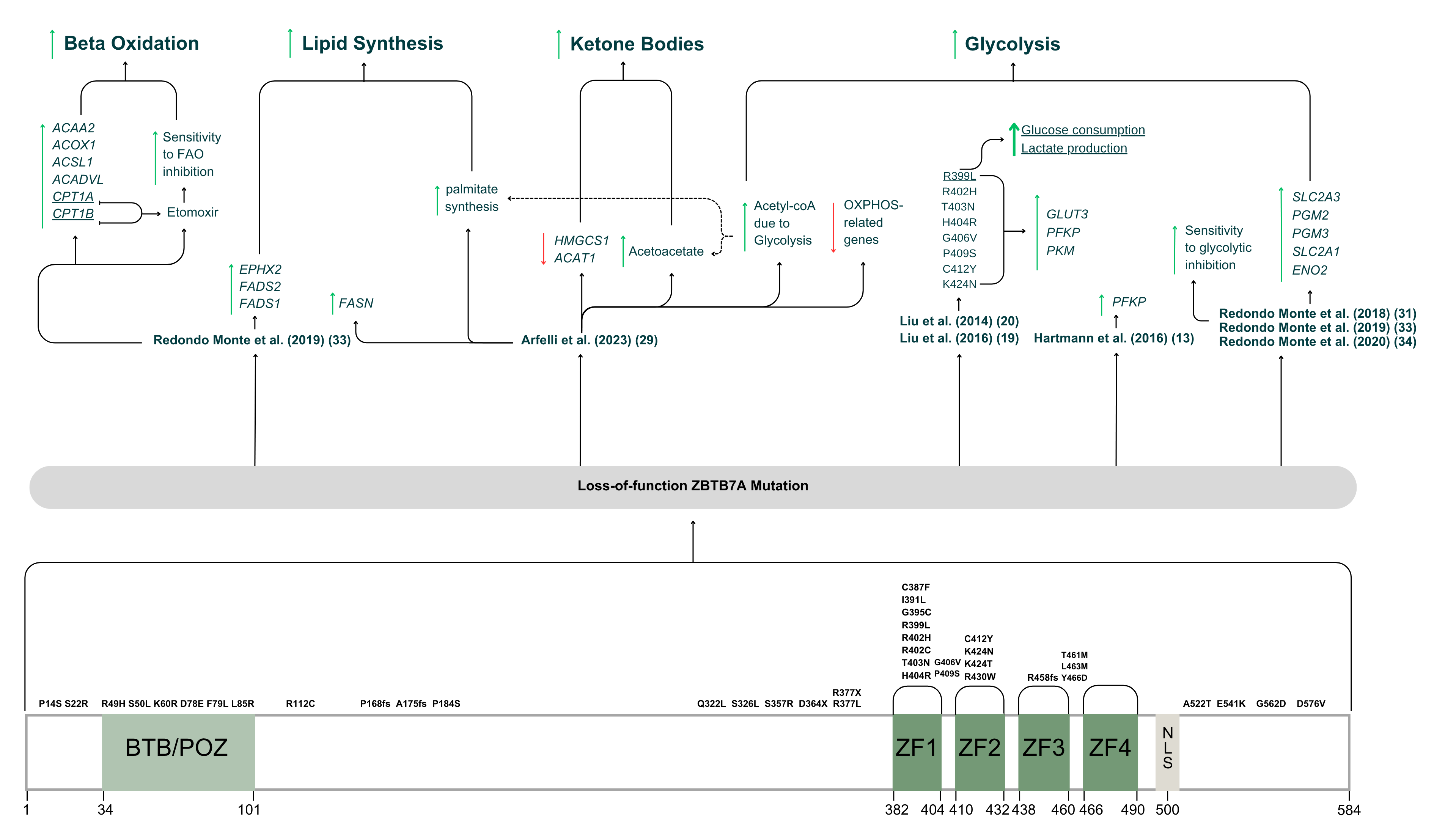
This article is a narrative review based on a comprehensive literature search conducted in PubMed and Google Scholar up to December 2025. Search terms included combinations and synonyms of "ZBTB7A" (including ZBTB7, POKEMON, LRF, and FBI-1), "metabolism", and "t(8;21) AML" (including RUNX1::RUNX1T1, RUNX1::ETO, RUNX1/RUNX1T1, RUNX1/ETO, AML1/ETO, AML1/MTG8, AML1::ETO, and AML1::MTG8). Peer-reviewed original research articles and published conference abstracts fulfilling all three search domains were selected for primary analysis. For studies matching two or fewer search domains, flexible inclusion criteria were applied to capture mechanistic findings in solid tumor or hematopoietic models with direct translational relevance to t(8;21) AML. The experimental models, specific ZBTB7A genetic alterations, metabolomic assays, and functional outcomes identified across the selected studies are summarized in Table 1 and illustrated schematically in Figure 1.
Summary of experimental models,
| Study | Reported / Studied ZBTB7A Alterations | Experimental Models | Key Metabolomic and Functional Assays | Significant Metabolic Consequences of ZBTB7A Loss | Other Phenotypic and Clinical Consequences of ZBTB7A Loss |
|---|---|---|---|---|---|
| Liu et al., 2014 | R399L; KD via sh | Gene expression profiling; glucose uptake and lactate production assays; LC-MS/MS; microarray analysis; luciferase promoter reporter assay; ChIP; cell proliferation and nude mouse subcutaneous xenograft assays with and without 2-DG | Elevated aerobic glycolysis; transcriptional de-repression and overexpression of | — | |
| Liu et al., 2016 | P14S, S22R, R49H, K60R, D78E, R112C, P184S, Q322L, S326L, S357R, R377L, C387F, I391L, R399L, R402H, T403N, H404R, G406V, P409S, C412Y, K424N, K424T, R430W, T461M, L463M, Y466D, A522T, E541K, G562D, and D576V expressed via pcDNA- | Gene expression profiling; luciferase promoter reporter assay; glucose uptake and lactate production measurements; ChIP; cell proliferation and nude mouse tumor formation assays with and without 2-DG | Loss or reduction of transcriptional repression of | Reduced steady-state protein stability in the BTB/POZ domain mutant ZBTB7A R49H | |
| Hartmann et al., 2016 | Patient-derived mutations mapped via IBS software: R49H, S50L, F79L, L85R, P168fs, A175fs, D364X, R377X, G395C, R402H, R402C, and R458fs; functional expression of A175fs, R377X, R402C, and R402H via pcDNA3.1-His- | Whole-exome and targeted amplicon sequencing; gene expression profiling; luciferase promoter reporter assay; immunofluorescence localization; cell proliferation and serial CFC assays | Elevated glycolysis; significant overexpression of | Loss of DNA-binding activity and cytoplasmic mislocalization (A175fs, R377X); loss of | |
| Redondo Monte et al., 2018 | R402C and A175fs | Lentiviral/retroviral hCD34+ transduction and clonal expansion assays; 2-DG glycolysis inhibition assay; flow cytometric granulocytic and monocytic differentiation assays | Heightened sensitivity to pharmacological glycolytic inhibition with 2-DG | Enhanced | |
| Redondo Monte et al., 2019 | CRISPR/Cas9-mediated | RNA-Seq; ChIP-Seq; extracellular flux analysis (ECAR and OCR); etomoxir FAO inhibition assay | Elevated glycolysis and mitochondrial beta-oxidation; direct de-repression of | — | |
| Redondo Monte et al., 2020 | R402C, A175fs, and CRISPR/Cas9-mediated | Myeloid differentiation assays; extracellular metabolic flux analysis; 2-DG drug sensitivity assay; hCD34+ competitive growth and cell cycle analyses | Elevated glycolytic flux via | Promotion of monocytic differentiation and blockade of granulocytic/erythroid maturation in HL60 cells; facilitation of | |
| Arfelli et al., 2023 | CRISPR/Cas9-mediated | Extracellular metabolic flux analysis; stable-isotope metabolic tracing; palmitate oxidation assay; etomoxir FAO inhibition assay; RNA-Seq | Upregulation of | Accelerated leukemogenesis in |
Structural distribution of loss-of-function
ZBTB7A Structure and Function
ZBTB7A—also referred to as POKEMON, FBI-1, LRF, and OCZF—is a transcriptional repressor encoded by the ZBTB7A gene located on human chromosome 19p13.314. The gene produces two transcript variants that encode an identical 584-amino-acid protein with an approximate molecular weight of 86 kDa15. ZBTB7A is a member of the BTB/POZ and Krüppel-type (POK) family of transcription factors, which are structurally defined by two conserved functional regions: an N-terminal broad-complex, tramtrack, and bric-à-brac/poxvirus and zinc finger (BTB/POZ) domain (residues 34–101) and a C-terminal array of four Krüppel-like C2H2 zinc finger (ZF) domains (ZF1–ZF4; residues 382–490). The BTB/POZ domain mediates homodimerization, heterodimerization, and the recruitment of transcriptional co-repressor complexes, whereas the C-terminal ZF domains recognize and bind specific promoter DNA sequences to repress target gene transcription16.
Proper nuclear import of ZBTB7A is governed by a nuclear localization signal (NLS), which is indispensable for its transcriptional regulatory activity10. Ren et al. (2023) mapped this NLS to an arginine- and lysine-rich motif located immediately downstream of the fourth ZF domain at residues 497–50410. Like other POK family members such as BCL6 and PLZF, ZBTB7A orchestrates diverse biological processes, most notably lineage commitment in hematopoiesis, tumorigenesis, and cellular energy metabolism17.
Somatic mutations in ZBTB7A can disrupt its subcellular localization and DNA-binding capacity, thereby abrogating its transcriptional repressor function13. Because the C-terminal region harbors both the ZF array and the NLS, truncating mutations upstream of residue 497 eliminate both DNA binding and nuclear targeting. Specifically, the frameshift variant ZBTB7A A175fs and the nonsense variant ZBTB7A R377X, both of which terminate the polypeptide prior to ZF1, exhibit pronounced cytoplasmic mislocalization in U2OS osteosarcoma cells compared with wild-type (WT) ZBTB7A13,18.
Conversely, missense mutations within the ZF domains preserve the downstream NLS and largely retain normal nuclear localization. In fluorescence localization assays, R402 missense variants (ZBTB7A R402C and ZBTB7A R402H) localized predominantly to the nucleus, with only a minor fraction detected in the cytoplasm compared with truncating mutants13. Consistently, the ZF1 missense variants R399L, R402H, and H404R localized to the nucleus in HEK293 cells19. Despite intact nuclear import, these ZF1 variants failed to bind the promoter regions of GLUT3 (SLC2A3) and PFKP, whereas the N-terminal non-ZF variant P14S retained promoter-binding activity, demonstrating that ZF missense mutations cause loss of function by abolishing DNA recognition rather than altering nuclear localization19. An earlier study similarly established that ZBTB7A R399L is defective in binding the GLUT3, PFKP, and PKM promoters20. This mechanistic distinction is corroborated by quantitative biophysical DNA-binding analyses reported by Ren et al. (2023), in which DNA-binding affinity was reduced by more than 50-fold for R402C and more than 12-fold for G395C (both within ZF1), by approximately 5- to 6-fold for R402H (ZF1) and K424T (ZF2), and by approximately 4-fold for the frameshift variant R458fs (within ZF3)10. Notably, R458fs exhibited the mildest intrinsic DNA-binding defect among the tested mutants but lacked the downstream NLS, underscoring that cytoplasmic mislocalization is a specific consequence of NLS loss rather than a general feature of ZF domain mutations.
ZBTB7A Mutations: Incidence and Clinical Consequences in t(8;21) AML
Somatic ZBTB7A mutations show a striking specificity for t(8;21) AML. Hartmann et al. (2016) identified ZBTB7A mutations in 23.2% (13/56) of patients with t(8;21) AML13. In that cohort, a numerically higher proportion of patients with mutated ZBTB7A experienced disease relapse (4/13; 30.8%) compared with patients harboring WT ZBTB7A (5/43; 11.6%); however, this difference did not reach statistical significance (Fisher's exact test, p = 0.189; odds ratio 3.38, 95% confidence interval 0.75–15.17) and warrants validation in larger prospective cohorts. By contrast, within the other major CBF AML subtype—inv(16) AML—only 1 of 22 patients (4.5%) harbored a ZBTB7A mutation, and no ZBTB7A mutations were detected among patients with CN-AML (n = 50) or non-CBF AML (n = 14)13.
Independent cohorts have confirmed this subtype specificity. In a Japanese study of patients with CBF AML enrolled in the JALSG AML201 trial, ZBTB7A mutations occurred exclusively in the t(8;21) subgroup, accounting for 9.8% (4/41) of cases21. Similarly, integrative multi-omics profiling revealed an enrichment of ZBTB7A mutations among high-risk t(8;21) AML patients exhibiting aberrant leukemic stem and progenitor gene expression signatures22. Furthermore, Lavallée et al. (2016) identified novel somatic ZBTB7A mutations in 15.0% (3/20) of patients with t(8;21) AML, whereas none were observed in inv(16) AML or non-CBF AML control cohorts; notably, patients carrying frameshift ZBTB7A alleles also displayed the lowest ZBTB7A transcript levels23.
Context-Dependent Roles of ZBTB7A in Human Malignancies
Although originally designated as an oncogene in certain solid tumors, ZBTB7A exhibits context-dependent functions as either a tumor promoter or a tumor suppressor depending on tissue lineage. In breast cancer cell lines and clinical tumor specimens, ZBTB7A is frequently overexpressed and promotes cellular migration, invasion, and metastasis via NF-κB-mediated epithelial–mesenchymal transition24,25. Elevated ZBTB7A expression is likewise associated with shorter overall survival in patients with colorectal cancer, where it enhances in vitro cell proliferation and in vivo xenograft tumorigenicity26.
Conversely, compelling evidence establishes ZBTB7A as a bona fide tumor suppressor in endometrial and myeloid malignancies. In uterine corpus endometrial carcinoma (UCEC), downregulation of ZBTB7A accelerates cancer cell proliferation and migration in vitro and correlates with poorer clinical prognosis27. In the hematopoietic compartment, knockdown of ZBTB7A in mononuclear cells derived from healthy mobilized peripheral blood delays terminal erythroid maturation28. Crucially, this erythroid differentiation block cooperates directly with RUNX1::ETO in murine leukemogenesis: Arfelli et al. (2023) reported that conditional ZBTB7A knockout (KO) in RUNX1::ETO-expressing mice resulted in 100% penetrance of acute leukemia29. The spectrum of somatic loss-of-function (LOF) ZBTB7A mutations compiled from the literature and their domain distribution are depicted in Figure 1.
Functionally, WT ZBTB7A counteracts RUNX1::ETO-driven hematopoietic stem and progenitor cell (HSPC) expansion. In human CD34 (hCD34) cells transduced with a C-terminally truncated, leukemogenic isoform of RUNX1::ETO (RUNX1::ETOtr), co-expression of WT ZBTB7A completely abrogated clonal expansion30. By contrast, co-expression of LOF ZBTB7A mutants (ZBTB7A R402C or ZBTB7A A175fs) failed to restrain RUNX1::ETOtr-driven clonal outgrowth. Moreover, ectopic expression of WT ZBTB7A in both the t(8;21)-positive Kasumi-1 cell line and primary hCD34 cells induced cell cycle arrest, increasing the proportion of cells in the G0/G1 phase and reducing the S-phase fraction relative to ZBTB7A-mutant cells30. With respect to myeloid lineage commitment, WT ZBTB7A promotes granulopoiesis while repressing monocytic differentiation. Loss-of-function mutations in ZBTB7A abolish this lineage control and cooperate with RUNX1::ETO to block granulocytic maturation, accompanied by upregulated surface CD14 expression indicative of monocytic skewing30,31.
ZBTB7A Mutations and Metabolic Reprogramming
The experimental designs, studied ZBTB7A variants, and metabolic phenotypes reported across the reviewed literature are detailed in Table 1 and integrated in Figure 1.
ZBTB7A Mutations and Glucose Metabolism in Solid Tumors
Because multiple rate-limiting glycolytic genes are direct transcriptional targets of ZBTB7A, somatic ZBTB7A inactivation profoundly alters cellular glucose utilization and downstream biosynthetic pathways. Liu et al. (2014) first elucidated the glycolytic repressor function of ZBTB7A in solid tumor models, demonstrating in U2OS osteosarcoma cells and mouse embryonic fibroblasts that ZBTB7A directly binds the promoter regions and represses the transcription of three critical glycolytic genes: GLUT3 (SLC2A3), PFKP, and PKM20.
Subsequent functional screening of cancer-associated somatic ZBTB7A variants revealed that missense mutations clustering within ZF1 (R399L, R402H, T403N, and H404R) and ZF2 (K424N) completely abolish transcriptional repression of GLUT3, PFKP, and PKM19. Mechanistically, these mutant ZBTB7A proteins form heterodimers with WT ZBTB7A and act in a dominant-negative manner to impair promoter repression despite unchanged total protein abundance19. Consequently, colon carcinoma cells expressing ZBTB7A R399L exhibited significantly elevated glucose uptake, lactate secretion, and flux into the pentose phosphate pathway (PPP), which accelerated xenograft tumor growth in mice while rendering the tumor cells hypersensitive to pharmacological glycolysis inhibition with 2-deoxy-D-glucose (2-DG)19,20.
ZBTB7A Mutations and Glucose Metabolism in t(8;21) AML
Hartmann et al. (2016) extended these findings to human t(8;21) AML, demonstrating that genetic inactivation of ZBTB7A is associated with significantly elevated PFKP transcript levels in primary t(8;21) AML patient samples compared with ZBTB7A-WT t(8;21) AML cases, alongside a similar upward trend in SLC2A3 and PKM expression13. In t(8;21)-positive Kasumi-1 cells, retroviral restoration of WT ZBTB7A exerted a potent antiproliferative effect that was completely abolished by the truncating A175fs mutation and partially attenuated by the missense R402C mutation13. Furthermore, in serial colony-forming cell (CFC) assays using murine bone marrow cells co-expressing RUNX1::ETO and either WT or mutant ZBTB7A, the A175fs and R402C variants significantly increased primary colony numbers, indicating that ZBTB7A inactivation enhances leukemic self-renewal and stemness13,32.
Subsequent investigations by Redondo Monte et al. (2018, 2019, 2020) demonstrated that ZBTB7A-deficient myeloid leukemia cells exhibit marked glycolytic dependency alongside heightened metabolic plasticity under stress30,31,33,34. While WT ZBTB7A restricts RUNX1::ETO-driven clonal expansion, ZBTB7A loss confers a competitive proliferative advantage to myeloid progenitors by upregulating glycolytic ATP and biomass production30,31. In chronic myeloid leukemia (CML)-derived K562 cells, CRISPR/Cas9-mediated ZBTB7A KO resulted in pronounced de-repression of SLC2A3 and significant upregulation of additional glycolytic enzymes, including PGM2, PGM3, SLC2A1, and ENO2, whereas PKM and PFKP levels were not significantly altered in this specific cellular background30,31,33. Functionally, ZBTB7A KO cells displayed an elevated extracellular acidification rate (ECAR), reflecting enhanced aerobic glycolysis—a classic manifestation of the Warburg effect31,35. Notably, this glycolytic surge became even more pronounced upon pharmacological inhibition of the mitochondrial electron transport chain (ETC), indicating that ZBTB7A loss enables leukemic cells to thrive under hypoxic conditions characteristic of the bone marrow microenvironment31. This phenotype aligns with biochemical evidence showing that ZBTB7A heterodimerizes with ZBTB2 to inhibit ZBTB2 homodimerization, a step required for full hypoxia-inducible factor 1 (HIF-1) transcriptional activation36. Consequently, loss of ZBTB7A relieves inhibition of the ZBTB2–HIF-1 axis, reinforcing glycolytic adaptation and leukemic survival under hypoxia.
Interestingly, when subjected to acute glucose deprivation, ZBTB7A KO K562 cells exhibited an approximately 1.5-fold higher oxygen consumption rate (OCR) than ZBTB7A-WT control cells31,33. This observation suggests that ZBTB7A-deficient cells retain the capacity to rewire their energy metabolism toward alternative mitochondrial substrates—such as fatty acids (FAs) or glutamine/glutamate—when extracellular glucose becomes limiting. Nevertheless, treatment with the glycolytic inhibitor 2-DG significantly suppressed the long-term proliferation of ZBTB7A-mutant and ZBTB7A-KO cells relative to controls30,31. Arfelli et al. (2023) similarly noted transcriptional upregulation of both glycolysis and the PPP in ZBTB7A-KO K562 cells29. Collectively, despite cell line-specific variations in individual target gene expression, current evidence establishes transcriptional repression of glycolysis as a central mechanism underlying the tumor suppressor activity of ZBTB7A in t(8;21) AML.
ZBTB7A and Lipid Metabolism in t(8;21) AML
Although fewer studies have examined the role of ZBTB7A in lipid homeostasis, emerging evidence indicates that ZBTB7A loss substantially remodels leukemic lipid metabolism. Redondo Monte et al. (2019) reported that ZBTB7A KO in K562 cells upregulates multiple genes governing FA metabolism and mitochondrial beta-oxidation33. Chromatin immunoprecipitation sequencing (ChIP-Seq) and transcriptomic profiling identified ACAA2, ACOX1, ACADVL, and CPT1A as direct ZBTB7A target genes, alongside elevated expression of ACSL1 and CPT1B33. Pharmacological inhibition of carnitine palmitoyltransferase 1 (CPT1A and CPT1B)—the rate-limiting step in mitochondrial fatty acid oxidation (FAO)—using etomoxir revealed that ZBTB7A-KO cells were significantly more sensitive to FAO blockade, exhibiting a 50% reduction in the half-maximal inhibitory concentration (IC) compared with WT controls33,37. Consistent with these in vitro findings, RNA sequencing (RNA-Seq) of primary t(8;21) AML patient samples harboring ZBTB7A mutations demonstrated significant overexpression of unsaturated FA metabolism genes, including EPHX2, FADS2, and FADS133; notably, elevated FADS1 expression independently correlates with inferior overall survival in AML38. Furthermore, under combined glucose- and glutamate-deprived conditions, ZBTB7A-KO cells displayed elevated mitochondrial oxygen consumption, supporting an increased reliance on endogenous FAO under nutrient stress33.
Conversely, Arfelli et al. (2023) reported divergent findings regarding FAO dependency in ZBTB7A-KO K562 cells, despite employing the same cellular model29. First, stable isotope-resolved palmitate oxidation assays indicated that ZBTB7A-KO cells did not preferentially oxidize exogenous palmitate, even under glutamate-depleted conditions, and did not exhibit heightened sensitivity to etomoxir treatment compared with WT controls29. Second, transcriptomic and flux analyses suggested that ZBTB7A-KO cells are largely independent of mitochondrial oxidative phosphorylation (OXPHOS), as multiple ETC subunit genes were downregulated29. Instead, excess glycolytic acetyl-CoA generated by ZBTB7A-deficient cells was channeled into de novo lipogenesis—specifically palmitate synthesis—driven by marked upregulation of fatty acid synthase (FASN), a signature also confirmed in CBF AML patients harboring ZBTB7A mutations29. Third, ZBTB7A-KO cells diverted surplus glycolytic acetyl-CoA toward ketogenesis, accumulating and secreting acetoacetate into the extracellular medium without utilizing ketone bodies for intracellular ATP production or lipid synthesis, consistent with the concurrent downregulation of HMGCS1 and ACAT129.
Taken together, while Redondo Monte et al. (2019) observed transcriptional de-repression of beta-oxidation enzymes and increased sensitivity to etomoxir under nutrient stress33, Arfelli et al. (2023) demonstrated a metabolic shift toward aerobic glycolysis, de novo palmitate synthesis, and ketogenesis independent of FAO and OXPHOS under standard culture conditions29. These apparent discrepancies likely reflect differences in extracellular nutrient availability, passage conditions, or assay readouts (endogenous vs. exogenous palmitate oxidation). Crucially, however, both studies converge on the finding that ZBTB7A loss upregulates de novo lipid biosynthesis, a process essential for membrane biogenesis, signaling, and leukemic proliferation29,33,39. It should be noted that the studies by Redondo Monte et al. (2018, 2019), Arfelli et al. (2023), and Liu et al. (2024) were published as conference abstracts rather than full peer-reviewed articles22,29,31,33; their mechanistic conclusions should therefore be interpreted as preliminary pending full peer-reviewed publication. Future targeted lipidomic and fluxomic investigations across primary t(8;21) AML blasts are warranted to delineate how microenvironmental nutrient gradients shape lipid metabolic dependencies in ZBTB7A-mutant leukemia.
ZBTB7A Mutations Within the Broader Mutational and Metabolic Landscape of t(8;21) AML
In clinical cohorts of t(8;21) AML, somatic mutations in signaling pathway genes—most notably KIT, NRAS, KRAS, FLT3, and JAK2—represent the most frequent co-occurring genetic lesions alongside RUNX1::ETO40,41. Just as RUNX1::ETO requires cooperating mutations to induce overt leukemia2,3, ZBTB7A inactivation likely interacts with co-occurring signaling and epigenetic mutations to orchestrate global metabolic reprogramming in t(8;21) AML. For example, KIT mutations are the most prevalent secondary alterations in both adult and pediatric t(8;21) AML and are strongly associated with higher relapse rates and inferior overall and progression-free survival42,43. Beyond proliferative signaling, aberrant KIT activation promotes glutaminolysis, which depletes microenvironmental glutamine to suppress anti-tumor T-cell proliferation and enhance the immunosuppressive activity of myeloid-derived suppressor cells44. Similarly, recurrent mutations in EZH2, NRAS, TET2, and JAK3 identified in relapsed or refractory RUNX1::RUNX1T1 AML have been linked to distinct alterations in lipid metabolism and glycolytic flux45. Elucidating the combinatorial metabolic effects of ZBTB7A loss alongside co-occurring KIT, RAS, or epigenetic mutations represents a priority for future multi-omics research in t(8;21) AML.
Conclusion
Somatic loss-of-function mutations in ZBTB7A represent a recurrent, subtype-enriched genetic lesion in t(8;21) AML that disrupts the transcriptional repression of key enzymes governing glycolysis and lipid metabolism. By de-repressing glycolytic regulators (SLC2A3, PFKP, and PKM) and upregulating de novo lipogenesis, beta-oxidation, and ketogenesis, ZBTB7A inactivation confers metabolic flexibility and a survival advantage to leukemic stem and progenitor cells within the hypoxic bone marrow microenvironment. Acting in concert with the RUNX1::ETO fusion oncoprotein, loss of ZBTB7A reinforces myeloid differentiation arrest and accelerates clonal expansion. Importantly, this metabolic rewiring creates actionable therapeutic vulnerabilities, rendering ZBTB7A-deficient leukemic cells hypersensitive to pharmacological inhibitors of glycolysis (such as 2-deoxy-D-glucose) and lipid metabolism (such as etomoxir). Targeted inhibition of these dysregulated metabolic nodes holds considerable promise for improving therapeutic outcomes in patients with ZBTB7A-mutant t(8;21) AML.
Declarations
Abbreviations
-
2-DG: 2-deoxy-D-glucose
-
AML: Acute myeloid leukemia
-
BTB: Broad-complex, tramtrack, and bric-à-brac
-
CBF: Core-binding factor
-
CFC: Colony-forming cell
-
ChIP: Chromatin immunoprecipitation
-
ChIP-Seq: Chromatin immunoprecipitation sequencing
-
CI: Confidence interval
-
CML: Chronic myeloid leukemia
-
CN-AML: Cytogenetically normal acute myeloid leukemia
-
CPT1: Carnitine palmitoyltransferase 1
-
ECAR: Extracellular acidification rate
-
ETC: Electron transport chain
-
FA: Fatty acid
-
FAO: Fatty acid oxidation
-
FASN: Fatty acid synthase
-
fs: Frameshift mutation
-
GLUT3: Glucose transporter 3
-
hCD34+: Human CD34-positive hematopoietic stem and progenitor cells
-
HIF-1: Hypoxia-inducible factor 1
-
HSPC: Hematopoietic stem and progenitor cell
-
IBS: Illustrator of Biological Sequences
-
IC50: Half-maximal inhibitory concentration
-
KD: Knockdown
-
KO: Knockout
-
LC-MS/MS: Liquid chromatography–tandem mass spectrometry
-
LOF: Loss-of-function
-
LRF: Leukemia/lymphoma-related factor
-
MEF: Mouse embryonic fibroblast
-
NLS: Nuclear localization signal
-
OCR: Oxygen consumption rate
-
OXPHOS: Oxidative phosphorylation
-
PDX: Patient-derived xenograft
-
PFKP: Platelet-type phosphofructokinase
-
PKM: Pyruvate kinase M1/2
-
POK: BTB/POZ and Krüppel-type
-
POZ: Poxvirus and zinc finger
-
PPP: Pentose phosphate pathway
-
RNA-Seq: RNA sequencing
-
shRNA: Short hairpin RNA
-
siRNA: Small interfering RNA
-
SLC2A3: Solute carrier family 2 member 3
-
UCEC: Uterine corpus endometrial carcinoma
-
WT: Wild type
-
X: Nonsense (stop-gain) mutation
-
ZBTB7A: Zinc finger and BTB domain-containing protein 7A
-
ZF: Zinc finger
Acknowledgments
The authors thank the library services at Universiti Sains Malaysia for facilitating comprehensive literature access.
Author’s contributions
Zainul Abeden: Conceptualization, Methodology, Literature search, Data curation, Visualization, Writing – original draft. Muhamad Amir Azizan, Mohammad Syamsul Reza Harun, Mohd Nazri Ismail, and Fadly Ahid: Literature analysis, Validation, Writing – review and editing. Asmida Isa: Conceptualization, Supervision, Funding acquisition, Project administration, Writing – review and editing. All authors read and approved the final manuscript.
Funding
This work was supported by Universiti Sains Malaysia, Malaysia, under the Short-Term Grant (STG) Scheme (STG Code: 304.CIPPT.6315621).
Availability of data and materials
None.
Ethics approval and consent to participate
None.
Consent for publication
None.
Declaration of generative AI and AI-assisted technologies in the writing process
None.
Competing interests
None. On behalf of all authors, the corresponding author states that there is no conflict of interest.