


Quantification of Mesenchymal Stem Cell Loss During Autologous Fat Graft Processing: Implications for Regenerative Cell Preservation

- Department of Pharmaceutical Sciences, University of Eastern Piedmont “Amedeo Avogadro”, Novara, Italy
- Faculty of Experimental Sciences, Miguel Hernández University of Elche, Elche, Spain; Cell Production and Tissue Engineering Unit, Virgen de las Nieves University Hospital, Granada, Spain
- Faculty of Basic Medicine, University of Medicine and Pharmacy, Thai Nguyen University, Thai Nguyen, Viet Nam
- EURO Hair&Skin Clinic, Ho Chi Minh City, Viet Nam
- Faculty of Experimental Sciences, Miguel Hernández University of Elche, Elche, Spain
Abstract
Background: Autologous fat grafting is widely utilized in aesthetic and reconstructive surgery; however, graft retention remains highly variable and unpredictable, with reported long-term resorption rates reaching 90%. Adipose-derived mesenchymal stem/stromal cells (MSCs) play a pivotal role in improving graft survival through pro-angiogenic, anti-apoptotic, and immunomodulatory mechanisms. Although cell enrichment techniques have been extensively explored, the magnitude of MSC loss occurring during routine clinical preparation protocols remains poorly quantified. This prospective pilot study aimed to identify and quantify MSC loss across sequential stages of lipoaspirate processing and establish evidence-based strategies to optimize intraoperative cell retention.
Methods: Thirty-six biological and fluid fractions were obtained from nine adult patients undergoing elective autologous liposuction. Harvested specimens were subjected to either standard washing or closed-system mechanical microfragmentation (Lipogems®), followed by secondary processing using enzymatic digestion or mechanical shear dissociation. Adherent nucleated cells were expanded in primary culture, enumerated, and immunophenotypically profiled via flow cytometry (FACS) utilizing a combined surface marker panel (CD31⁻CD45⁻CD44⁺CD73⁺) to determine MSC-like cell yields.
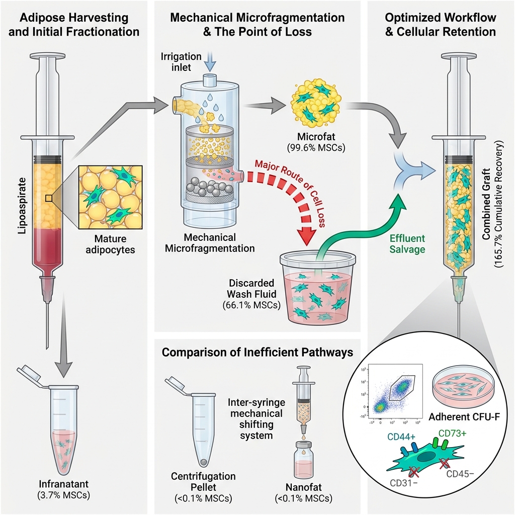
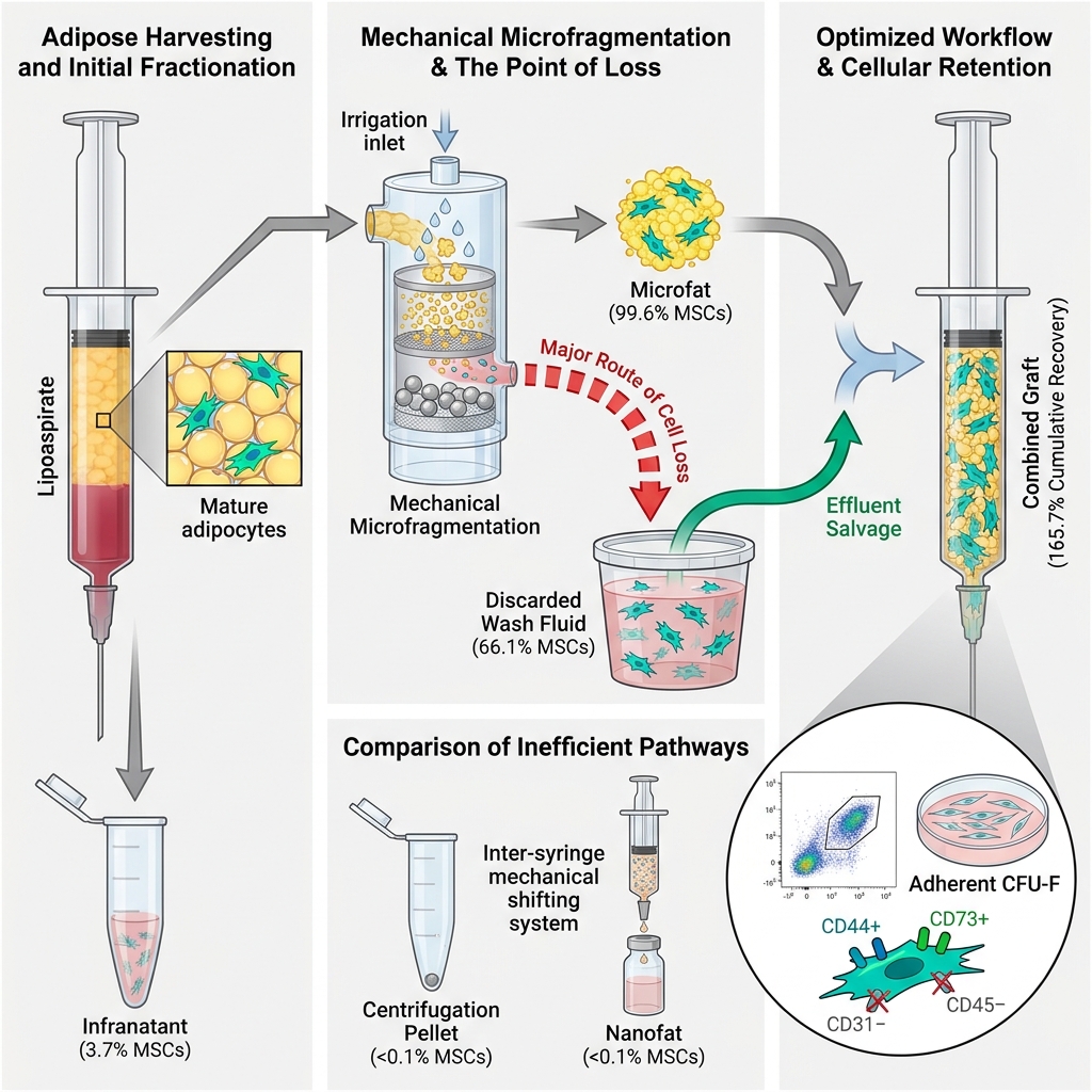
Results: Microfragmented adipose tissue processed using the Lipogems® kit maintained an MSC density comparable to that of standard enzymatically digested fat (99.6% vs. 100%, p = 0.263). Crucially, a substantial proportion of viable MSC-like cells was recovered from the discarded microfat wash liquid (66.1% relative to the standard fat reference), demonstrating marked cellular loss during the intra-device irrigation step. Secondary mechanical dissociation of washed fat into nanofat resulted in an MSC retention of 32.1%, whereas primary centrifugation pellets yielded only 7.1%. Conversely, discarded lipoaspirate infranatant fluid and standard fat wash liquid contained minimal cellularity (<5%). Combining microfat with its discarded wash effluent yielded a cumulative cellularity of 165.7% relative to standard lipoaspirate.
Conclusion: Progenitor cell loss in autologous fat grafting occurs predominantly during post-fragmentation washing and secondary centrifugation steps. Restricting irrigation to the pre-fragmentation phase and avoiding unnecessary secondary centrifugation can substantially improve cellular preservation. Furthermore, intraoperative salvage and concentration of the MSC-rich wash fraction represents an accessible, non-enzymatic strategy to augment graft cellularity and potentially improve clinical outcomes.
Graphical abstract
Introduction
Autologous adipose tissue is an ideal soft-tissue filler offering significant advantages over synthetic biomaterials, including superior biocompatibility, minimal to no risk of foreign-body or allergic reactions, and durable volumetric restoration. Beyond volume augmentation, adipose tissue serves as an abundant reservoir of regenerative cellular components, most notably adipose-derived mesenchymal stem/stromal cells (MSCs)1,2,3,4. These multipotent progenitors exert potent paracrine, immunomodulatory, and pro-angiogenic effects that facilitate tissue remodeling, promote local neo-vascularization, and enhance skin rejuvenation, thereby improving aesthetic and reconstructive outcomes1,2,3,4. Consequently, autologous fat transplantation has become an indispensable clinical procedure in plastic, aesthetic, and reconstructive surgery worldwide.
However, unpredictable and suboptimal graft retention remains a major clinical challenge. Graft survival is governed by multiple variables—including harvested graft volume, recipient-site vascularity, and surgical technique—with reported resorption rates reaching up to 90% within the first postoperative year5. Consequently, repeated surgical procedures are frequently required to achieve and maintain desired aesthetic or reconstructive endpoints, placing an emotional and financial burden on patients and compromising clinical compliance6. To overcome these limitations, several strategies have been developed to enhance graft viability, including multi-planar micro-aliquot grafting, structural micro-fragmentation (microfat grafting), and MSC enrichment.
First, multi-planar, micro-aliquot grafting optimizes graft survival by dispersing minute volumes of adipose tissue across multiple anatomic planes, maximizing contact with vascularized host tissue. Specialized mechanical injection devices, such as the TauTona Adipose Tissue Injector, have been introduced to ensure controlled, uniform extrusion, prevent localized bolus pooling, and reduce central fat necrosis7,8,9.
Second, structural down-sizing via microfat grafting addresses the ischemic vulnerability of large fat lobules, which are prone to core hypoxia, oil cyst formation, and necrosis prior to neo-revascularization. By mechanically fragmenting native adipose clusters (1.0–3.5 mm in diameter) into smaller functional units (approximately 0.2–0.8 mm), this approach produces "microfat" or, with further shear processing, "nanofat". Various commercial closed-system filtration and resizing kits—such as Lipogems®, Tulip Nanofat, Lipocube Nano, and BSL Adinizer—employ distinct filter geometries, mesh apertures, and pass sequences10,11,12. Despite methodological differences, these devices share the common objective of increasing the surface-area-to-volume ratio, facilitating nutrient diffusion from the recipient bed, accelerating revascularization, and minimizing ischemic necrosis10,11,12.
The third strategy, MSC enrichment, harnesses the regenerative potential of adipose tissue, which represents an accessible, abundant source of multipotent progenitor cells13,14. Beyond their trilineage differentiation capacity, adipose-derived MSCs secrete diverse extracellular vesicles and soluble growth factors that modulate inflammation, stimulate local cellular proliferation, and induce robust neovascularization15,16,17,18. According to the "graft replacement theory" proposed by Eto et al., the majority of mature donor adipocytes undergo early ischemic death following transplantation, and long-term graft survival relies predominantly on the survival, activation, and adipogenic differentiation of donor-derived progenitor cells8,19. Consistent with this mechanism, grafts enriched with higher concentrations of stromal cells have repeatedly demonstrated superior volumetric retention20,21.
Clinically, cell enrichment is typically achieved by supplementing lipoaspirates with autologous stromal vascular fraction (SVF), a process termed cell-assisted lipotransfer (CAL)22,23,24,25. Enzymatic digestion (predominantly using bacterial collagenase) has traditionally represented the gold standard for SVF isolation, maximizing cell yield per unit volume. However, enzymatic processing disrupts the native extracellular matrix (ECM) architecture, carries risks of xenogeneic enzyme contamination and immunogenicity, and is classified as "more than minimal manipulation" by regulatory authorities such as the US Food and Drug Administration (FDA) and the European Medicines Agency (EMA), thereby restricting routine chairside clinical adoption26. In response, clinical interest has shifted toward non-enzymatic, mechanical isolation protocols—ranging from automated centrifugation and vortexing to closed mechanical microfragmentation systems like Lipogems®27. Although mechanical methods typically isolate lower absolute cell numbers than enzymatic digestion, they preserve intact microvascular niches and ECM architecture, maintaining pericyte-endothelial interactions and sustaining prolonged regenerative paracrine activity28.
Nevertheless, lipoaspirate processing entails multiple disruptive physical and mechanical steps, including sedimentation, extensive washing, centrifugation, shear fragmentation, and filtration. Each of these manipulation steps risks inadvertent cellular damage, sequestration, or discard. If substantial numbers of progenitor cells are lost during standard preparation, optimizing intraoperative cell retention and salvaging discarded fractions could offer a safer, more cost-effective, and logistically simpler alternative to ex vivo cellular enrichment.
To date, the precise magnitude of progenitor cell loss across specific stages of autologous fat graft processing has not been comprehensively mapped. Therefore, this prospective pilot study aimed to identify and quantify MSC loss throughout sequential washing, centrifugation, microfragmentation, and mechanical dissociation steps, providing evidence-based technical recommendations to maximize regenerative cell retention in clinical practice.
Materials and Methods
Ethical Approval and Donor Eligibility
Human adipose tissue specimens were harvested from adult donors undergoing elective orthopedic, regenerative, or aesthetic surgical procedures involving autologous lipoaspiration at the Image Regenerative Clinic (Milan, Italy). All procedures were performed under sterile conditions in accordance with standard surgical protocols. Clinical demographic reporting adhered to the applicable principles of the Strengthening the Reporting of Observational Studies in Epidemiology (STROBE) guidelines.
The study protocol was approved by the Ethics Committee of the University of Medicine and Pharmacy, Thai Nguyen University (TUMP; approval number 1194/ĐHYD-HĐĐĐ). Written informed consent was obtained from all participants prior to enrollment, permitting the scientific use of residual biological material otherwise destined for disposal, in accordance with Recommendation Rec(2006)4 of the Committee of Ministers of the Council of Europe on research on biological materials of human origin.
Eligible participants included adult men and women aged 37–74 years with no evidence of active systemic disease who had not received chemotherapy, radiotherapy, or systemic immunosuppressive/cytotoxic medications within the preceding two months. Exclusion criteria comprised a body mass index (BMI) > 30 kg/m², diabetes mellitus, uncontrolled hypertension, and active tobacco or alcohol abuse.
Tissue Harvesting Protocol
Subcutaneous adipose tissue was infiltrated with a tumescent solution containing adrenaline (2 μg/mL) as a vasoconstrictor and lidocaine (0.02%) as a local anesthetic in sterile physiological saline. Manual aspiration was performed using a 10-mL Luer-Lok syringe connected to a 19-cm disposable cannula (outer diameter 3 mm) equipped with five oval aspiration ports (1 × 2 mm). A total of 40 lipoaspirate specimens were harvested from 10 consented donors. Harvested samples were sealed in sterile syringes, triple-bagged within certified biohazard transport packaging, maintained at ambient temperature, and processed within 24 hours of collection.
Adipose Tissue Fractionation and Processing
The harvested lipoaspirate was transferred into Falcon tubes (B. Braun, Melsungen, Germany) and held in an upright vertical position for 10 minutes to allow spontaneous gravitational sedimentation, partitioning the specimen into an upper raw adipose layer and a lower aqueous lipoaspirate infranatant.
The raw adipose layer was divided into two experimental arms:
-
One portion was processed through the Lipogems® kit (Lipogems, Milan, Italy) according to the manufacturer's protocol, yielding microfragmented adipose tissue ("microfat") and an associated microfat wash fluid.
-
The remaining raw fat was rinsed with 0.9% (w/v) sodium chloride solution (Sigma-Aldrich, St. Louis, MO, USA) to eliminate residual erythrocytes and cellular debris, yielding washed fat ("fat") and an associated fat wash fluid.
The discarded aqueous fractions—lipoaspirate infranatant, microfat wash fluid, and fat wash fluid—were centrifuged (Eppendorf, Hamburg, Germany) to isolate cell pellets for culture; these were designated samples #4a (aspirate liquid), #2a (microfat wash), and #4b (fat wash), respectively.
Both microfat and washed fat were subjected to an initial primary centrifugation step (Eppendorf, Hamburg, Germany), separating oily supernatants from concentrated adipose tissue and isolating primary centrifugation pellets (designated sample #2c for microfat 1st centrifugation and sample #3a for fat 1st centrifugation).
The concentrated adipose fractions were subsequently processed through either enzymatic digestion or mechanical dissociation:
-
Enzymatic approach: Concentrated fat and microfat were digested with collagenase solution (Sigma-Aldrich) at 37°C for 40 minutes under continuous agitation, neutralized with Dulbecco's Modified Eagle Medium/Nutrient Mixture F-12 (DMEM/F-12) containing 10% fetal bovine serum (FBS; Sigma-Aldrich), and centrifuged (BD Biosciences, Franklin Lakes, NJ, USA) to isolate stromal vascular pellets (designated sample #1 [fat enzyme] and sample #2b [microfat enzyme]).
-
Mechanical approach: Concentrated fat and microfat were mechanically dissociated using a female-to-female three-way stopcock (Thermo Fisher Scientific, Waltham, MA, USA; inner diameter 3.0 mm, outer diameter 4.1 mm) connected to two 5-mL Luer-Lok syringes. Adipose tissue was shifted between syringes for 20 complete passes. Consistent with manual surgical practice, extrusion velocity was standardized by having a single trained investigator execute all procedures using identical equipment and cycle counts. The resulting nanofat was washed with phosphate-buffered saline (PBS; Sigma-Aldrich) and centrifuged (BD Biosciences) to isolate cellular pellets (designated sample #3b [fat-derived nanofat] and sample #2d [microfat-derived nanofat]).
The complete sample allocation and experimental workflow are summarized in Figure 1.
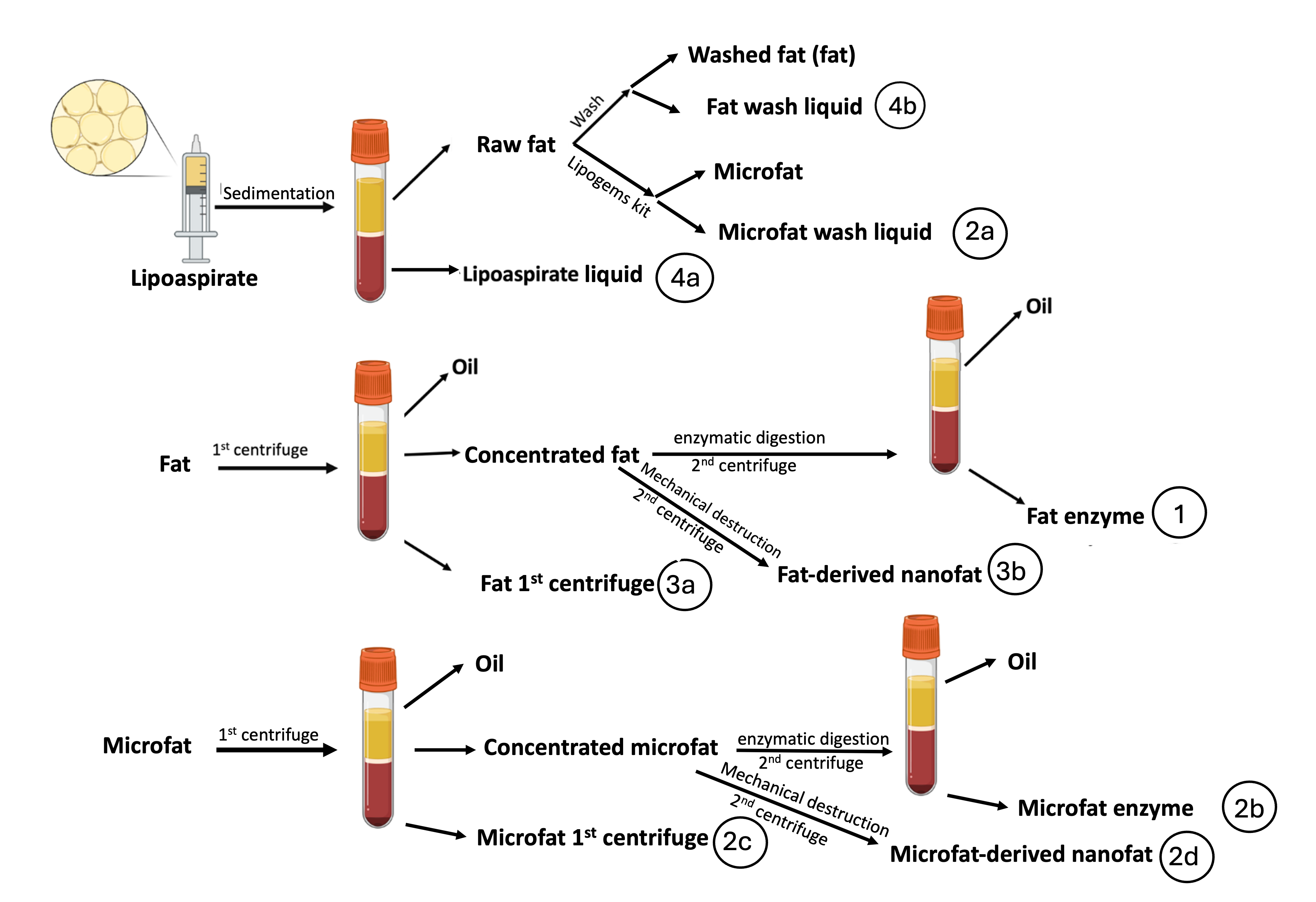
Schematic diagram of adipose tissue harvesting, processing workflows, and sample collection points. Lipoaspirate obtained from elective surgical lipoaspiration was allowed to undergo gravitational sedimentation for 10 minutes, separating raw adipose tissue from the lower aqueous infranatant (sample #4a). Raw fat was partitioned into two preparation pathways: (1) closed mechanical microfragmentation via the Lipogems® kit, yielding microfat and discarded microfat wash fluid (sample #2a), and (2) saline washing (0.9% NaCl), yielding washed fat and fat wash fluid (sample #4b). Microfat and washed fat each underwent primary centrifugation (1st cent) to isolate cellular sediments (samples #2c and #3a, respectively) and concentrated adipose tissue. Concentrated microfat and fat were subsequently processed through either enzymatic digestion (collagenase at 37°C for 40 min followed by secondary centrifugation; samples #1 and #2b) or mechanical shear dissociation (20 inter-syringe passes through a 3.0-mm stopcock followed by secondary centrifugation; samples #2d and #3b). Each isolated fraction was collected for primary culture expansion, cell enumeration, and flow cytometric immunophenotyping.
Primary Cell Culture and Morphological Assessment
All isolated cellular pellets were treated with 0.3% (w/v) hypotonic sodium chloride solution (Sigma-Aldrich) to selectively lyse residual red blood cells. The remaining nucleated cells were seeded into culture dishes and maintained in DMEM/F-12 supplemented with 20% FBS (Sigma-Aldrich) at 37°C in a humidified incubator with 5% CO₂. Fifty percent of the culture medium was replenished every 3–4 days. After 14 days of primary culture, non-adherent cells were removed by gentle washing with PBS. Adherent cell colonies were inspected and photographed under an inverted phase-contrast microscope (Leica Microsystems, Wetzlar, Germany) to evaluate cell morphology and confluence. Adherent cells were detached using 0.05% trypsin-EDTA (Sigma-Aldrich), and total cell counts were determined using a Neubauer hemocytometer (Thermo Fisher Scientific).
Flow Cytometric Immunophenotyping (FACS)
Cultured adherent cells were serially expanded until target cell quantities were achieved. Cell characterization was performed via flow cytometry (fluorescence-activated cell sorting, FACS) using established cluster of differentiation (CD) markers. Cells were incubated with fluorochrome-conjugated monoclonal antibodies (1:50 v/v dilution; Sigma-Aldrich) in the dark at 4°C for 30 minutes, washed, and analyzed using a BD FACSymphony™ flow cytometer running BD FACSDiva™ software (version 9.1; BD Biosciences).
A multi-parameter panel comprising negative lineage markers (anti-CD31 [endothelial] and anti-CD45 [hematopoietic]) and positive mesenchymal markers (anti-CD44 and anti-CD73) was utilized. Events displaying the surface phenotype CD31⁻CD45⁻CD44⁺CD73⁺ were defined as "MSC-like cells" (or "MSC-enriched populations"). Absolute MSC-like cell numbers were calculated by multiplying total viable nucleated cell counts by the percentage of events fulfilling this immunophenotypic profile.
Statistical Analysis
Statistical analyses were performed using GraphPad Prism (version 9.0; GraphPad Software, San Diego, CA, USA). Continuous variables are reported as mean ± standard deviation (SD). Data normality was evaluated using the Shapiro–Wilk test, and homoscedasticity was verified using Levene's test (p > 0.05 for all comparisons). Differences between multiple experimental groups were analyzed using one-way analysis of variance (ANOVA) followed by Tukey's post hoc honestly significant difference (HSD) test. Statistical significance was defined as a two-sided p value < 0.05. As this was an exploratory pilot study designed to establish baseline variance and effect sizes across a patient cohort (n = 9), a priori power calculations were not performed.
Results
Specimen Allocation and Study Cohort
Of the initial 40 lipoaspirate samples collected from 10 patients, four fractions from one donor were excluded due to bacterial contamination during primary culture. Consequently, 36 samples obtained from nine donors (n = 9) successfully completed the experimental protocol and were included in the quantitative analysis.
Morphology of Cultured Nucleated Cells
Phase-contrast microscopy demonstrated that adherent cells isolated from all productive fractions exhibited a characteristic spindle-shaped, fibroblast-like morphology consistent with mesenchymal stem cells (Figure 2). Despite this uniform morphological appearance, marked differences in cell density and culture confluence were observed at day 14, reflecting disparities in initial viable cell seeding numbers. Cultures established from enzymatically digested fat (sample #1), microfat wash liquid (sample #2a), and enzymatically digested microfat (sample #2b) reached high confluence (Figure 2a–c). In contrast, primary cultures derived from the primary centrifugation pellet of microfat (sample #2c) yielded only sparse, isolated colonies (Figure 2d).
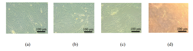
Representative phase-contrast micrographs illustrating the morphological appearance and confluence of cultured adherent nucleated cells across adipose graft fractions. Adherent cells isolated from matched biological donors were cultured for 14 days in DMEM/F-12 supplemented with 20% fetal bovine serum at 37°C in a humidified atmosphere containing 5% CO₂. In all productive fractions, adherent cells displayed a characteristic elongated, spindle-shaped, fibroblast-like morphology consistent with mesenchymal stem cells (MSCs). Panels represent: (a) standard enzymatically digested fat (sample #1); (b) microfat wash liquid (sample #2a); (c) enzymatically digested microfat (sample #2b); and (d) primary centrifugation pellet of microfat (sample #2c). Note the elevated cellular density and high confluence achieved in panels (a–c) relative to the sparse, isolated colony in panel (d), reflecting substantial disparities in initial viable seeding cell numbers. Images were acquired using an inverted phase-contrast microscope at 200× magnification. Scale bars = 100 μm.
Nucleated Cell Count and Distribution
Total nucleated cell concentrations across all experimental fractions are presented in Figure 3. Enzymatic digestion produced the highest yields of nucleated cells, with digested fat (sample #1) and microfat (sample #2b) yielding 353,000 ± 298,000 cells/mL and 350,000 ± 269,000 cells/mL, respectively (p = 0.051). Notably, the microfat wash liquid (sample #2a) contained a substantial nucleated cell concentration (222,000 ± 212,000 cells/mL), confirming that extensive cell shedding occurs during mechanical microfragmentation.
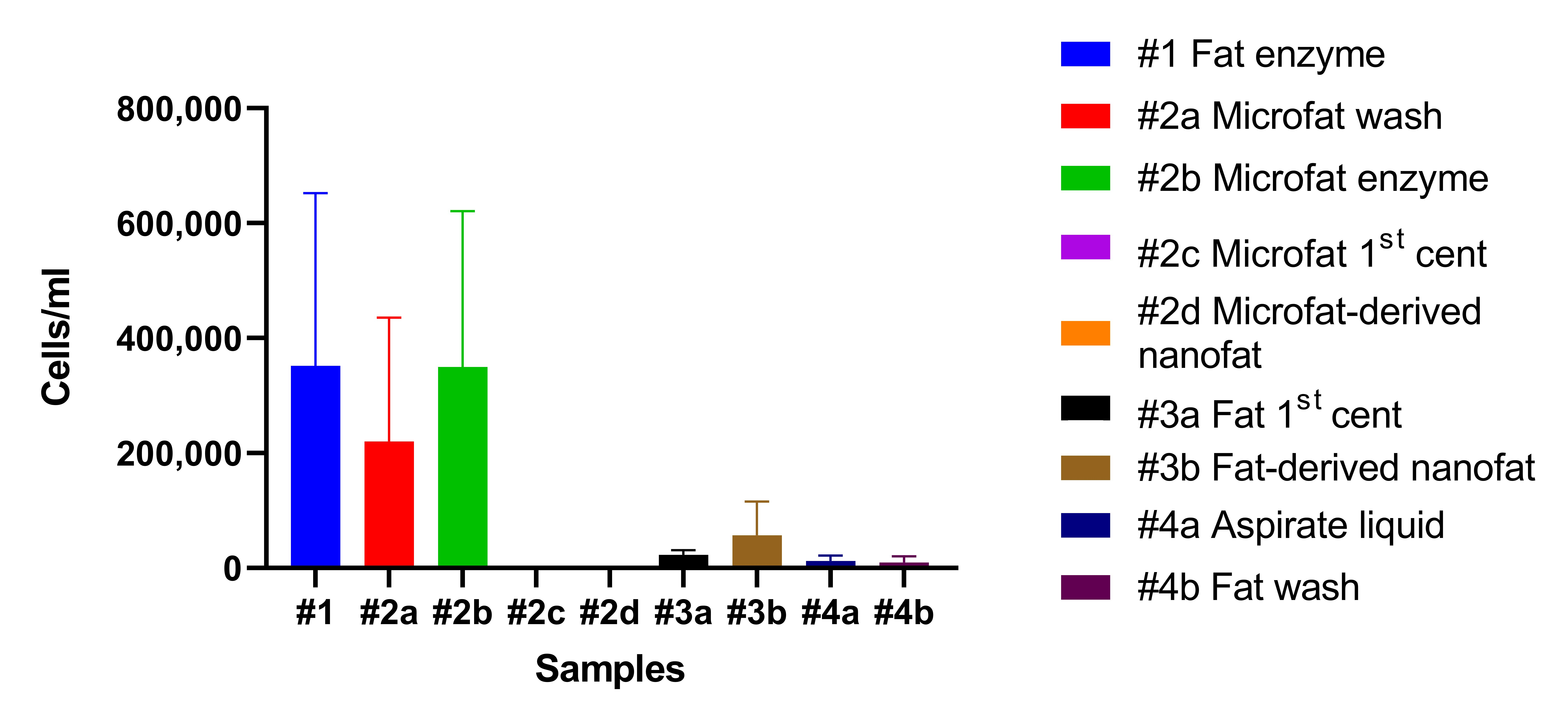
Total nucleated cell concentrations recovered across adipose tissue processing fractions. Adherent nucleated cells were harvested after 14 days of primary culture and quantified using a Neubauer hemocytometer following hypotonic red blood cell lysis. Data are presented as mean cell counts (cells/mL of starting tissue) ± standard deviation (SD) for
Conversely, cellular pellets obtained from primary centrifugation (#2c) and mechanical shear disruption (#2d) of microfat yielded negligible cell counts (mean 178 cells/mL and 30 cells/mL, respectively); these fractions were therefore excluded from subsequent flow cytometric characterization. In contrast, when centrifugation and mechanical dissociation were applied to washed standard fat (samples #3a and #3b), modest cell populations were recovered (24,000 ± 9,000 cells/mL and 58,000 ± 59,000 cells/mL, respectively). Finally, discarded lipoaspirate infranatant fluid (sample #4a) and standard fat wash liquid (sample #4b) contained negligible nucleated cell counts (14,000 ± 8,000 cells/mL and 11,000 ± 9,000 cells/mL, respectively).
MSC-like Cell Count and Distribution
Flow cytometric analysis identified MSC-like cells as CD31⁻CD45⁻CD44⁺CD73⁺ events (Figure 4). The absolute MSC-like cell concentrations per milliliter of starting tissue across all eligible fractions are detailed in Table 1, and normalized relative to the standard enzymatic digestion of fat (sample #1, defined as 100%) in Figure 5.
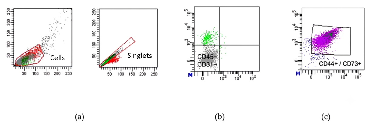
Flow cytometric gating strategy for the immunophenotypic identification of mesenchymal stem cell (MSC)-like cells. Adherent cells harvested from day-14 primary cultures were immunostained with fluorochrome-conjugated monoclonal antibodies and acquired on a BD FACSymphony™ flow cytometer running BD FACSDiva™ software (version 9.1). Gating strategy: (a) Forward scatter area (FSC-A) versus side scatter area (SSC-A) density plot used to delineate intact nucleated cells from subcellular debris, followed by doublet discrimination (FSC-A vs. FSC-H) to gate single cells (singlets). (b) Bivariate contour plot showing exclusion of endothelial (CD31⁺) and hematopoietic (CD45⁺) lineages, isolating the CD31⁻CD45⁻ double-negative subpopulation. (c) Selection of MSC-like cells expressing standard mesenchymal progenitor surface markers, defined as CD44⁺CD73⁺ within the CD31⁻CD45⁻ parent gate. Total MSC-like cell yields were calculated by multiplying total viable nucleated cell counts by the percentage of events satisfying this CD31⁻CD45⁻CD44⁺CD73⁺ immunophenotype.
Quantitative recovery of mesenchymal stem cell (MSC)-like cells across adipose tissue processing fractions.
| Parameter |
#1 Fat enzyme |
#2a Microfat wash |
#2b Microfat enzyme |
#3a Fat 1st cent |
#3b Fat-derived nanofat |
#4a Aspirate liquid |
#4b Fat wash |
|---|---|---|---|---|---|---|---|
| Mean (cells/mL) | 314,982 | 208,106* | 313,665 | 22,436 | 101,190 | 11,720 | 9,020 |
| Standard Deviation | 248,046 | 207,277 | 241,846 | 8,043 | 10,734 | 8,768 | 10,203 |
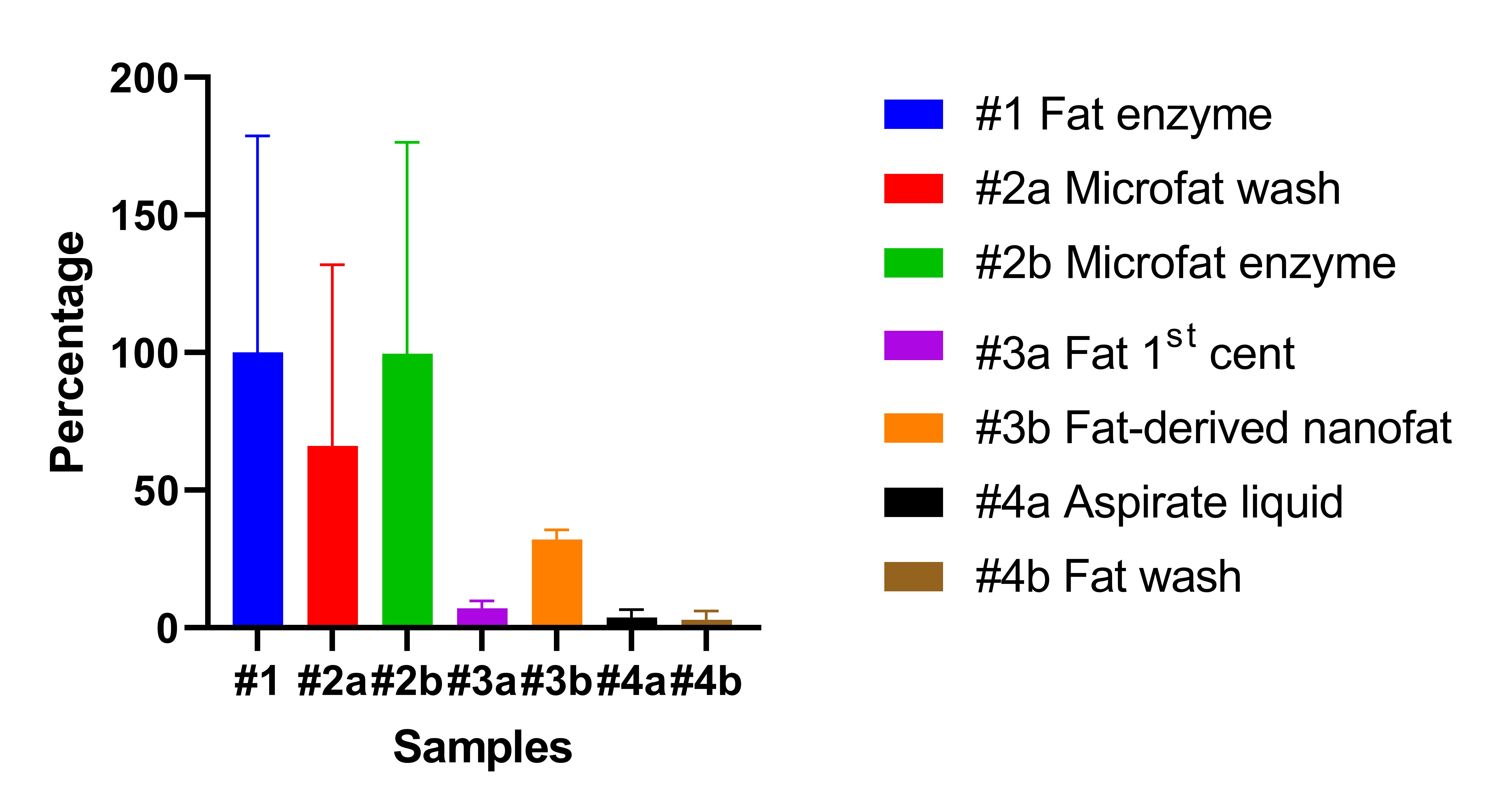
Relative yield of MSC-like cells across processing fractions normalized to standard enzymatically digested fat. Bar graph illustrating the relative recovery of CD31⁻CD45⁻CD44⁺CD73⁺ MSC-like cells across evaluated tissue and fluid fractions, normalized to the yield of standard enzymatically digested fat (sample #1, defined as 100%). Bars represent the mean percentage ± standard deviation (SD) for
In concordance with total nucleated cell yields, enzymatically digested microfat (#2b) retained an MSC-like cell density virtually identical to that of standard digested fat (#1; 313,665 ± 241,846 cells/mL vs. 314,982 ± 248,046 cells/mL; relative yield 99.6%, p = 0.263). Remarkably, the microfat wash liquid (#2a) contained 208,106 ± 207,277 cells/mL, representing 66.1% of the reference MSC yield.
Mechanically dissociated fat-derived nanofat (#3b) retained 101,190 ± 10,734 cells/mL (32.1% relative yield), while the cellular pellet from primary centrifugation of washed fat (#3a) contained 22,436 ± 8,043 cells/mL (7.1% relative yield). Discarded infranatant fluid (#4a) and fat wash liquid (#4b) accounted for only minor fractions of MSC-like cells (11,720 ± 8,768 cells/mL [3.7%] and 9,020 ± 10,203 cells/mL [2.8%], respectively).
Cumulative Cell Recovery in Microfat Processing
When the MSC-like cell population of the microfat graft (#2b) was combined with that recovered from its corresponding wash fluid (#2a), the cumulative cell yield reached 165.7% relative to standard lipoaspirate (Figure 6). This demonstrates that capturing the cell population lost during the Lipogems® washing process could expand total available regenerative cellularity by more than 1.6-fold over standard processing.
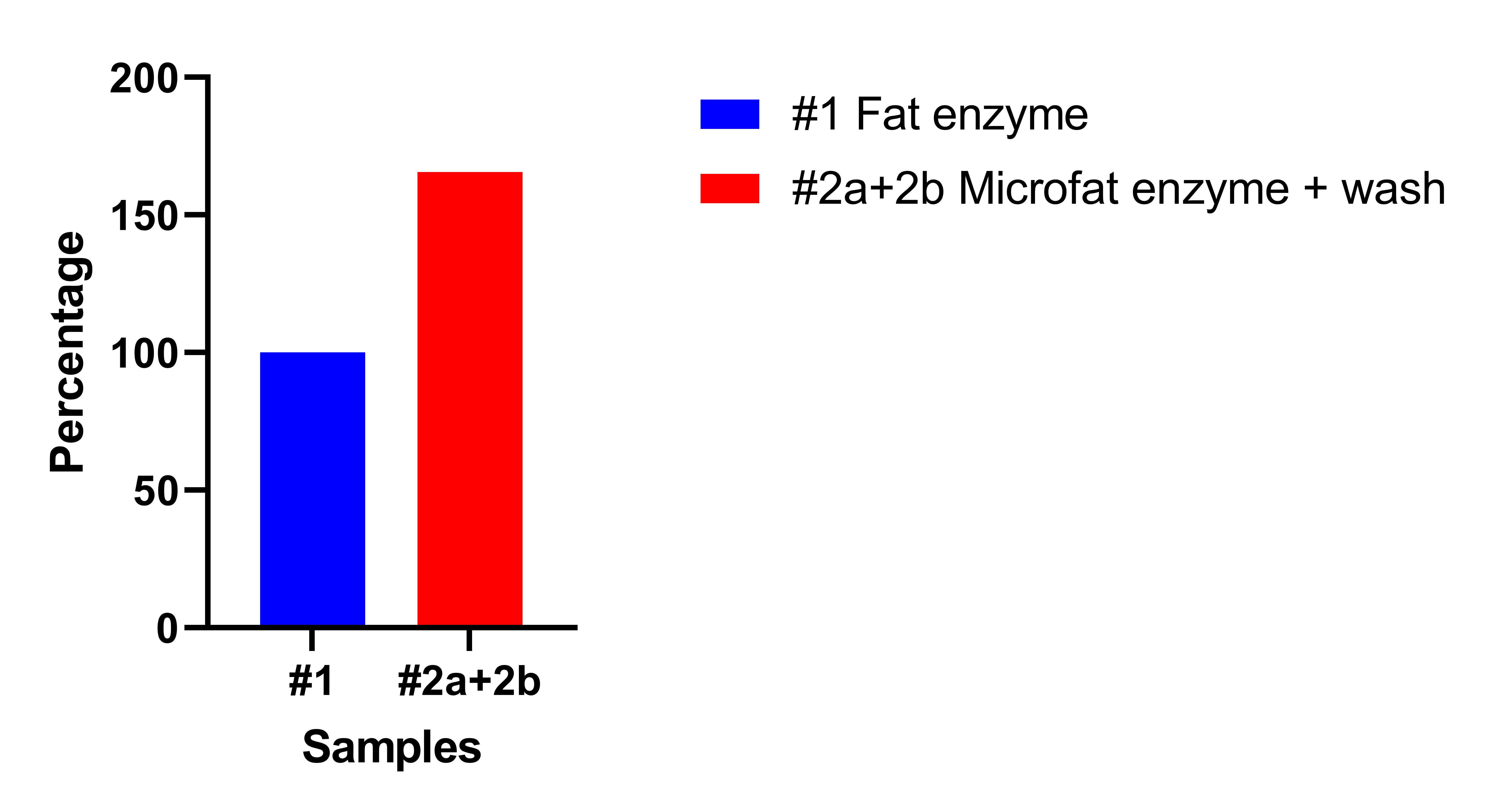
Cumulative recovery potential of MSC-like cells in microfat and its associated wash fluid. Bar chart comparing the MSC-like cell yield of standard enzymatically digested fat (sample #1, 100%) against the cumulative yield obtained by combining enzymatically digested microfat with its corresponding wash liquid (sample #2a + #2b, reaching 165.7% of reference). Values are normalized to the initial volume of processed lipoaspirate. This cumulative recovery illustrates that capturing and salvaging cells inadvertently lost in the discarded microfat wash fraction can substantially augment the total regenerative cellularity of the final autologous fat graft by more than 1.6-fold over standard processing.
Discussion
The primary finding of this investigation is that discarded microfat wash liquid (sample #2a) contains a remarkably high concentration of viable MSC-like cells (66.1% relative to standard enzymatically digested fat). In conventional clinical protocols utilizing the Lipogems® device or analogous microfragmentation systems, this fluid fraction is routinely discarded as biohazard waste. Our findings demonstrate that intense intra-device fluid agitation and mesh filtration liberate a substantial subpopulation of loosely adherent perivascular and stromal cells from the adipose matrix.
Based on these findings, we propose two critical technical modifications for clinical microfragmentation:
First, pre-fragmentation rinsing of the lipoaspirate with saline should be performed to eliminate red blood cells and oil debris, but post-fragmentation washing should be strictly minimized or avoided. Repeated irrigation following tissue morselization dislodges valuable resident MSCs into the effluent, diminishing the regenerative potency of the final graft.
Second, despite shedding substantial numbers of cells into the wash liquid, the concentrated microfat graft (#2b) exhibited an MSC density equivalent to that of standard digested fat (#1; 99.6%). This apparent paradox is explained by volumetric condensation: generating 5–10 mL of microfat typically requires an initial lipoaspirate volume of 10–15 mL. The volumetric compaction of adipose lobules compensates for the absolute cell loss, maintaining high cellular density per unit volume. Crucially, if the discarded wash effluent is collected, concentrated via rapid chairside centrifugation or membrane filtration, and re-introduced into the microfat graft, the net regenerative cellularity could be augmented by 165.7% compared with standard fat.
From a translational perspective, the Lipogems® system effectively serves as a rapid, non-enzymatic extraction platform capable of yielding an MSC-rich fluid fraction containing two-thirds of the cell yield achieved by enzymatic digestion. Unlike enzymatic isolation protocols that require Good Laboratory Practice (GLP) facilities, extensive incubation times, and xenogeneic reagents subject to stringent regulatory oversight (e.g., FDA/EMA "more than minimal manipulation" restrictions)26,27, physical wash fluid collection is rapid, cost-effective, and fully compliant with minimal manipulation standards. This approach holds significant promise for democratizing point-of-care regenerative therapies in outpatient surgical settings.
Our analysis of microfat processed by centrifugation (#2c) or secondary mechanical shearing (#2d) demonstrated negligible cell recovery (178 and 30 cells/mL, respectively). This indicates that the primary microfragmentation process successfully strips the majority of readily mobilizable interstitial cells; therefore, subsequent mechanical passes or spinning of microfat do not yield additional cellular pellets.
Conversely, when applied to standard washed fat, mechanical emulsification into nanofat (#3b) and primary centrifugation (#3a) yielded measurable MSC quantities (32.1% and 7.1% relative to enzymatic control, respectively). Nanofat is widely utilized in aesthetic surgery for intradermal or superficial subdermal injection in delicate anatomical zones, such as the tear trough, lower eyelid, and perioral rhytids, where smooth injectability without visible lobular contour irregularities is required29,30,31. However, our data demonstrate that progressive mechanical shear and subsequent centrifugation cause progressive depletion of viable MSCs. Consequently, two practical recommendations emerge: (1) gentle gravitational sedimentation or low-g physiological washing should be preferred over high-speed centrifugation, and (2) mechanically dissociated nanofat should be injected promptly without excessive secondary centrifugation or rinsing steps.
In contrast to microfat wash effluent, the primary lipoaspirate infranatant (#4a) and standard fat wash liquid (#4b) contained negligible numbers of MSC-like cells (3.7% and 2.8%, respectively). Given that these fractions contain heavy erythrocyte contamination and tumescent debris, attempting to salvage cells from primary lipoaspirate fluid is clinically inefficient and unjustified. Routine disposal of these fractions does not compromise graft regenerative capacity.
Interestingly, the mean MSC yield observed in the microfat wash liquid in this study (66.1%) was higher than that observed in our preliminary investigation (44.1%)32. This discrepancy is attributable to the small cohort (n = 3) of our initial proof-of-concept study. The present 9-patient cohort provides a more robust and statistically reliable estimate of cell recovery, reinforcing the potential of microfat wash fluid as a viable source for cell harvesting.
The clinical impact of graft cellularity is intimately linked to the vascularity and mechanical compliance of the recipient bed. In facial fat grafting, excellent volume maintenance is typically observed even with standard grafts, attributable to the dense facial microvascular plexus and the low-pressure recipient environment33,34. In contrast, large-volume soft-tissue reconstruction—such as post-mastectomy breast reconstruction or gluteal augmentation—faces a hostile, ischemic microenvironment. Introducing large graft volumes into tight anatomic envelopes markedly elevates interstitial fluid pressure, which compresses recipient microvessels, exacerbates tissue hypoxia, and drives extensive central graft necrosis and oil cyst formation, resulting in resorption rates up to 90%5. In these demanding reconstructive scenarios, maximizing MSC density is paramount. High concentrations of MSCs drive robust, early neo-angiogenesis and secrete anti-apoptotic cytoprotective factors that rescue ischemic tissue under elevated interstitial pressures.
Several study limitations must be addressed. First, substantial inter-donor variability (reflected by large standard deviations) was observed across all experimental groups, consistent with established donor-dependent heterogeneity related to age, sex, metabolic status, and harvesting site35,36,37. For instance, Alt et al. and Eckhard et al. demonstrated that adipose tissue from aged donors exhibits markedly diminished proliferation and osteogenic potential compared with younger counterparts36,37. Second, because fresh patient specimens were utilized, primary seeding densities could not be uniformly standardized across all experimental groups, which may have influenced 14-day culture proliferation rates, as lower seeding densities are known to promote higher initial population doublings38,39,40. Third, immunophenotypic identification was performed using a four-marker panel (CD31⁻CD45⁻CD44⁺CD73⁺) rather than the complete ISCT consensus profile (which includes CD90, CD105, and tri-lineage differentiation assays). Consequently, these cells were classified as "MSC-like" or "MSC-enriched" rather than definitive MSCs. Nonetheless, because an identical analytical methodology was systematically applied across all groups, the comparative conclusions regarding relative cellular retention remain internally consistent.
Despite these limitations, this study provides an objective quantitative map of cellular loss during routine fat grafting and establishes a practical foundation for clinical protocols aimed at non-enzymatic cell preservation.
Conclusions
Mechanical processing and washing during autologous fat graft preparation induce substantial, previously unquantified losses of viable mesenchymal progenitor cells. Specifically, extensive post-fragmentation washing discards a potent cell-rich fraction containing two-thirds of the MSC density of standard lipoaspirate. Surgeons utilizing microfragmentation devices such as Lipogems® should restrict washing to the pre-fragmentation phase and avoid excessive centrifugation. Furthermore, intraoperative recovery and concentration of the microfat wash fraction represents an accessible, non-enzymatic strategy to boost graft cellularity and potentially improve clinical retention in soft-tissue reconstruction.
Declarations
Abbreviations
ANOVA: Analysis of variance; ASC: Adipose-derived stem/stromal cell; BMI: Body mass index; CAL: Cell-assisted lipotransfer; CD: Cluster of differentiation; DMEM/F-12: Dulbecco's Modified Eagle Medium/Nutrient Mixture F-12; ECM: Extracellular matrix; EDTA: Ethylenediaminetetraacetic acid; EMA: European Medicines Agency; FACS: Fluorescence-activated cell sorting; FBS: Fetal bovine serum; FDA: Food and Drug Administration; FSC-A: Forward scatter area; FSC-H: Forward scatter height; GLP: Good Laboratory Practice; HSD: Honestly significant difference; ISCT: International Society for Cell & Gene Therapy; MSC: Mesenchymal stem/stromal cell; OD: Outer diameter; PBS: Phosphate-buffered saline; PRIN: Progetti di Rilevante Interesse Nazionale; RBC: Red blood cell; SD: Standard deviation; SSC-A: Side scatter area; STROBE: Strengthening the Reporting of Observational Studies in Epidemiology; SVF: Stromal vascular fraction; TUMP: Thai Nguyen University of Medicine and Pharmacy.
Acknowledgments
The authors thank Prof. Carlo Tremolada (IMAGE Regenerative Clinic, Milan, Italy) for providing biological specimens processed with the Lipogems® system and for insightful clinical discussions. The authors also thank Prof. Michela Bosetti (University of Piemonte Orientale, Novara, Italy) and Prof. Chiara Porta (Next-Gen Flow Cytometry & Sorting Core Facility at Ipazia Center, University of Piemonte Orientale, Novara, Italy) for their expert guidance, technical supervision, and training regarding primary mesenchymal stem cell culture protocols and flow cytometric characterization.
Author’s contributions
None.
Funding
This research was supported by PRIN (Progetti di Rilevante Interesse Nazionale), grant number CUP C53D23003120006 (unit Principal Investigator: Prof. Michela Bosetti, Università degli Studi del Piemonte Orientale). Financial support was also provided in part by Thai Nguyen University of Medicine and Pharmacy (TUMP).
Availability of data and materials
The datasets generated and/or analyzed during the current study are available from the corresponding author upon reasonable request.
Ethics approval and consent to participate
The study protocol was approved by the Ethics Committee of the University of Medicine and Pharmacy, Thai Nguyen University (TUMP; approval number 1194/ĐHYD-HĐĐĐ). Written informed consent was obtained from all participating patients prior to sample acquisition, authorizing the scientific use of discarded biological materials in accordance with Recommendation Rec(2006)4 of the Committee of Ministers of the Council of Europe on research on biological materials of human origin.
Consent for publication
None.
Declaration of generative AI and AI-assisted technologies in the writing process:
None.
Competing interests
The authors declare that they have no competing interests.