
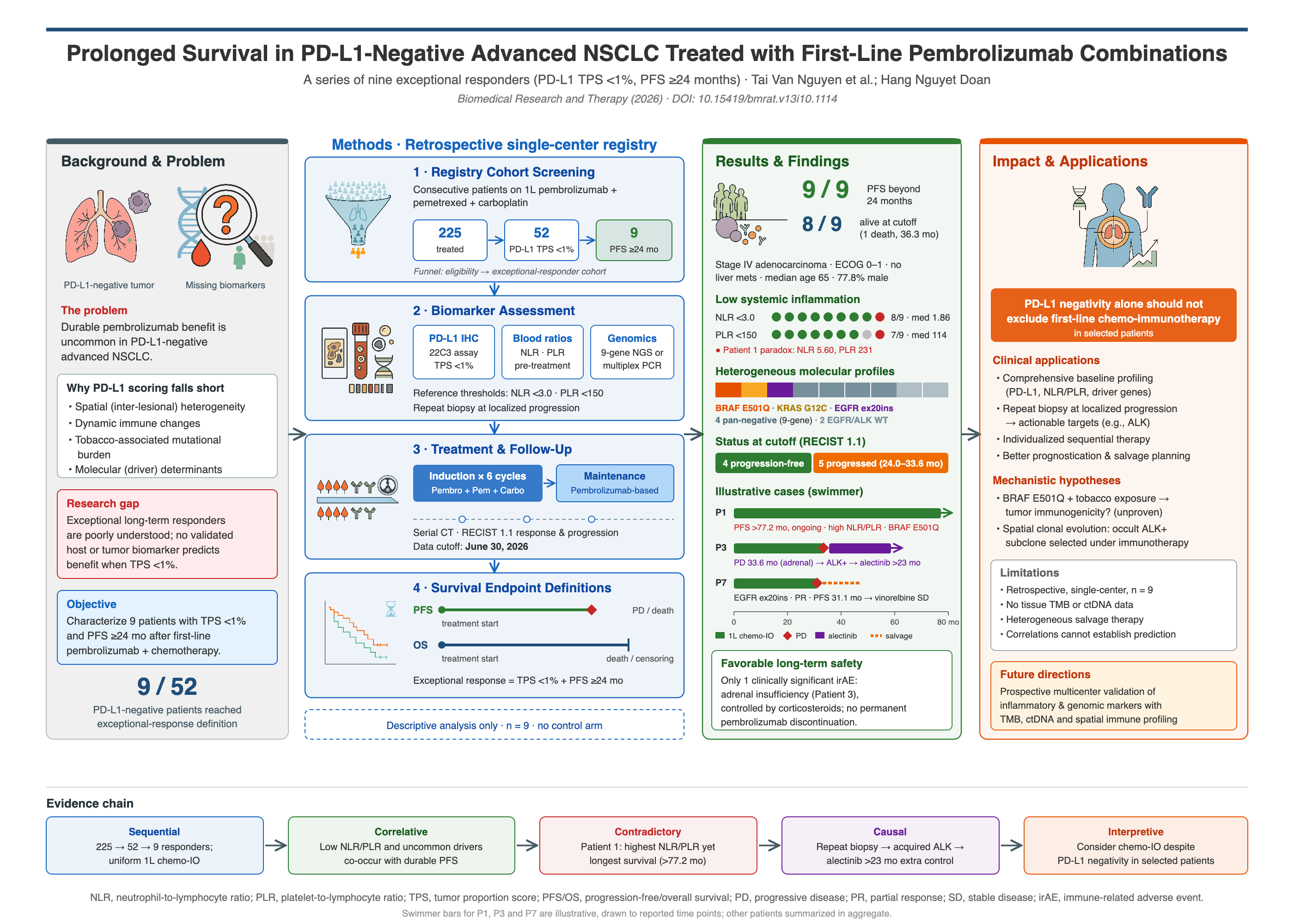


Prolonged Survival in PD-L1-Negative Advanced Non-Small Cell Lung Cancer Treated with First-Line Pembrolizumab Combinations: A Series of Nine Cases
- National Cancer Hospital of Vietnam, 30 Cau Buou, Thanh Liet, Hanoi, Viet Nam
Abstract
Introduction: First-line pembrolizumab-based regimens represent a standard of care for advanced non-small cell lung cancer (NSCLC) lacking actionable driver alterations; however, multi-year progression-free survival (PFS) among patients with programmed death-ligand 1 (PD-L1)-negative tumors remains rare.
Methods: From an institutional registry of 225 consecutive patients with advanced NSCLC treated with first-line pembrolizumab-based chemo-immunotherapy (parent cohort median PFS, 14.25 months; objective response rate, 52.4%), 52 patients (23.1%) had confirmed PD-L1-negative disease (tumor proportion score [TPS] < 1%). We retrospectively evaluated nine consecutive outlier patients (17.3% of the PD-L1-negative subgroup) who achieved exceptional durable clinical benefit, defined as a PFS ≥ 24 months. Baseline systemic inflammatory indices, targeted genomic profiles (via 9-gene next-generation sequencing [NGS] or multiplex polymerase chain reaction [PCR]), and longitudinal clinical-radiographic outcomes were analyzed.
Results: All nine exceptional responders presented with lung adenocarcinoma (median age, 65 years; 77.8% male; 66.7% ever-smokers). Low baseline systemic inflammation was observed in eight patients (cohort median neutrophil-to-lymphocyte ratio [NLR], 1.86; median platelet-to-lymphocyte ratio [PLR], 114). Notably, an inflammatory paradox was observed in Patient 1, who achieved the longest survival (77.2+ months) despite marked baseline systemic inflammation (NLR, 5.60; PLR, 231). Targeted molecular profiling identified non-classical or uncommon driver alterations in three patients (a rare BRAF E501Q variant, a KRAS G12C mutation, and an EGFR exon 20 insertion), whereas four patients were pan-negative by 9-gene NGS and two were confirmed EGFR/ALK wild-type by PCR. In one patient with baseline bone and adrenal metastases (Patient 3), an acquired secondary ALK rearrangement emerged at localized adrenal progression after 33.6 months, which subsequently responded durably to alectinib salvage therapy (overall survival, 56.7+ months).
Conclusion: Selected patients with PD-L1-negative advanced NSCLC can achieve multi-year survival with first-line pembrolizumab plus platinum-doublet chemotherapy. A low baseline systemic inflammatory profile and distinct molecular variants may characterize these exceptional responders, while repeat tissue biopsy and sequential targeted therapy upon localized progression can effectively extend long-term survival.
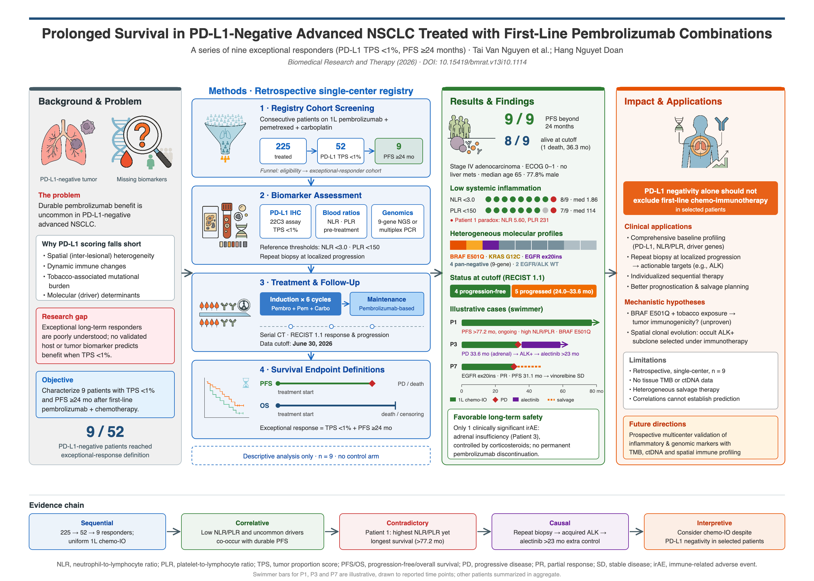
Graphical abstract
INTRODUCTION
First-line pembrolizumab-based regimens represent an established standard of care for patients with advanced non-small cell lung cancer (NSCLC) lacking actionable oncogenic driver mutations1. Clinical treatment algorithms rely heavily on baseline programmed death-ligand 1 (PD-L1) expression2. Whereas pembrolizumab monotherapy is standard for tumors with high PD-L1 expression (tumor proportion score [TPS] ≥ 50%), chemo-immunotherapy combining pembrolizumab with platinum-doublet chemotherapy is recommended for PD-L1-negative disease (TPS < 1%)1. Nevertheless, patients with confirmed tissue PD-L1 negativity who derive multi-year progression-free survival (PFS) benefit remain uncommon. Five-year follow-up data from the pivotal phase 3 KEYNOTE-189 trial demonstrated that although durable benefit is achievable, the 5-year overall survival (OS) rate remains modest at 9.6% among patients with a PD-L1 TPS < 1%, underscoring the rarity of multi-year survival in this subgroup3.
This clinical observation highlights the limitations of relying on a single baseline tissue PD-L1 score as an exclusionary predictor of long-term immunotherapy benefit. Single-site immunohistochemical evaluation frequently fails to capture intratumoral and interlesional spatial heterogeneity, dynamic shifts within the tumor immune microenvironment, or elevated tumor mutational burden (TMB) induced by tobacco carcinogen exposure4. Furthermore, although comprehensive genomic profiling via broad-panel next-generation sequencing (NGS) can elucidate co-occurring molecular determinants of response, its high cost restricts routine upfront implementation in resource-constrained clinical settings5.
Herein, we report the real-world clinical courses, baseline hematological inflammatory indices, and molecular characteristics of nine consecutive patients with advanced PD-L1-negative lung adenocarcinoma who achieved prolonged survival (PFS ≥ 24 months) following first-line pembrolizumab plus pemetrexed-carboplatin therapy.
CASE SERIES PRESENTATION
Cohort Screening and Baseline Clinicopathological Characteristics
From an institutional registry of 225 consecutive patients with advanced NSCLC treated with first-line pembrolizumab-based regimens at the National Cancer Hospital of Vietnam (parent cohort: objective response rate [ORR], 52.4% [118/225]; median PFS, 14.25 months [95% confidence interval (CI), 12.0–16.5]; median OS, 28.8 months [95% CI, 22.3–35.2]), 52 patients (23.1%) had confirmed PD-L1-negative disease (TPS < 1%). We screened this PD-L1-negative subpopulation to identify exceptional responders. Although definitions of prolonged PFS vary across the literature (typically 18 to 24 months), we defined exceptional durable response as a PFS ≥ 24 months, corresponding to the standard 2-year (35-cycle) pembrolizumab treatment duration established in pivotal KEYNOTE trials. All consecutive patients with a confirmed PD-L1 TPS < 1% and PFS ≥ 24 months were included; no patients were excluded due to lost follow-up or incomplete biomarker data, yielding nine exceptional responders (17.3% of the PD-L1-negative cohort).
All nine patients were diagnosed with stage IV lung adenocarcinoma. The median age was 65 years (range, 33–71 years); the series was predominantly male (n = 7, 77.8%), and two-thirds were ever-smokers (n = 6, 66.7%). All patients presented with a favorable baseline Eastern Cooperative Oncology Group (ECOG) performance status of 0–1 and an absence of baseline liver metastases. Individual clinicopathological characteristics and longitudinal survival outcomes are detailed in Table 1, and treatment trajectories with clinical milestones are illustrated in Figure 1.
Individual clinicopathological characteristics, baseline systemic inflammatory ratios, molecular profiles, and longitudinal survival outcomes of the nine patients with PD-L1-negative (TPS < 1%) stage IV lung adenocarcinoma who achieved durable clinical benefit (PFS ≥ 24 months) on first-line pembrolizumab-based chemo-immunotherapy.
| Patient No. | Age (y) / Sex | Smoking Status | Pathology & Genomic Profile (Assay) | Baseline Metastatic Sites | Baseline NLR / PLR | 1L Regimen & Maintenance | PFS (mo) | OS (mo) | Clinical Status & Post-Progression Trajectory |
|---|---|---|---|---|---|---|---|---|---|
| 1 | 65 / M | Ever-smoker | AC, BRAF E501Q (9-gene NGS) | Contralateral lung, pleura | 5.60 / 231 | Pembrolizumab + pemetrexed + carboplatin → pembrolizumab + pemetrexed | 77.2+ | 77.2+ | Alive without progression. Extended follow-up of a previously reported case (PMID: 37151366) |
| 2 | 71 / M | Ever-smoker | AC, EGFR/ALK WT (multiplex PCR) | Contralateral lung, brain | 0.97 / 92 | Brain Gamma Knife radiosurgery; pembrolizumab + pemetrexed + carboplatin → pembrolizumab + vinorelbine | 32.1 | 36.3 | Died following post-pembrolizumab disease progression. |
| 3 | 53 / F | Never-smoker | AC, 9-gene WT (NGS) | Bone, adrenal gland | 2.25 / 200 | Pembrolizumab + pemetrexed + carboplatin → pembrolizumab monotherapy | 33.6 | 56.7+ | Alive. Progressed in left adrenal gland at 33.6 mo; adrenal re-biopsy revealed secondary ALK rearrangement; durable response on 2L alectinib. |
| 4 | 69 / M | Ever-smoker | AC, EGFR/ALK WT (multiplex PCR) | Bone, contralateral lung | 2.11 / 124 | Pembrolizumab + pemetrexed + carboplatin → pembrolizumab + pemetrexed | 24.0 | 48.3+ | Alive. Progressed at 24.0 mo; metachronous secondary bladder cancer diagnosed; stable on 2L paclitaxel. |
| 5 | 70 / M | Ever-smoker | AC, KRAS G12C (9-gene NGS) | Contralateral lung, pleura | 2.03 / 105 | Pembrolizumab + pemetrexed + carboplatin → pembrolizumab + pemetrexed | 45.0+ | 45.0+ | Alive without progression. |
| 6 | 63 / M | Ever-smoker | AC, 9-gene WT (NGS) | Contralateral lung, pleura, bone | 1.86 / 141 | Pembrolizumab + pemetrexed + carboplatin → pembrolizumab + pemetrexed | 39.4+ | 39.4+ | Alive without progression. |
| 7 | 69 / M | Ever-smoker | AC, EGFR exon 20 insertion (9-gene NGS) | Bone, pleura | 1.82 / 114 | Pembrolizumab + pemetrexed + carboplatin → pembrolizumab + pemetrexed | 31.1 | 45.2+ | Alive. Progressed at 31.1 mo (lung, pleura, pelvic bone); currently stable on 2L vinorelbine. |
| 8 | 37 / F | Never-smoker | AC, 9-gene WT (NGS) | Contralateral lung, pleura, bone | 1.63 / 92 | Pembrolizumab + pemetrexed + carboplatin → pembrolizumab + pemetrexed | 34.8+ | 34.8+ | Alive without progression. |
| 9 | 33 / M | Never-smoker | AC, 9-gene WT (NGS) | Adrenal gland, contralateral lung | 1.29 / 102 | Pembrolizumab + pemetrexed + carboplatin → pembrolizumab + pemetrexed | 25.2 | 33.9+ | Alive. Progressed at 25.2 mo; currently receiving 2L ramucirumab + docetaxel. |
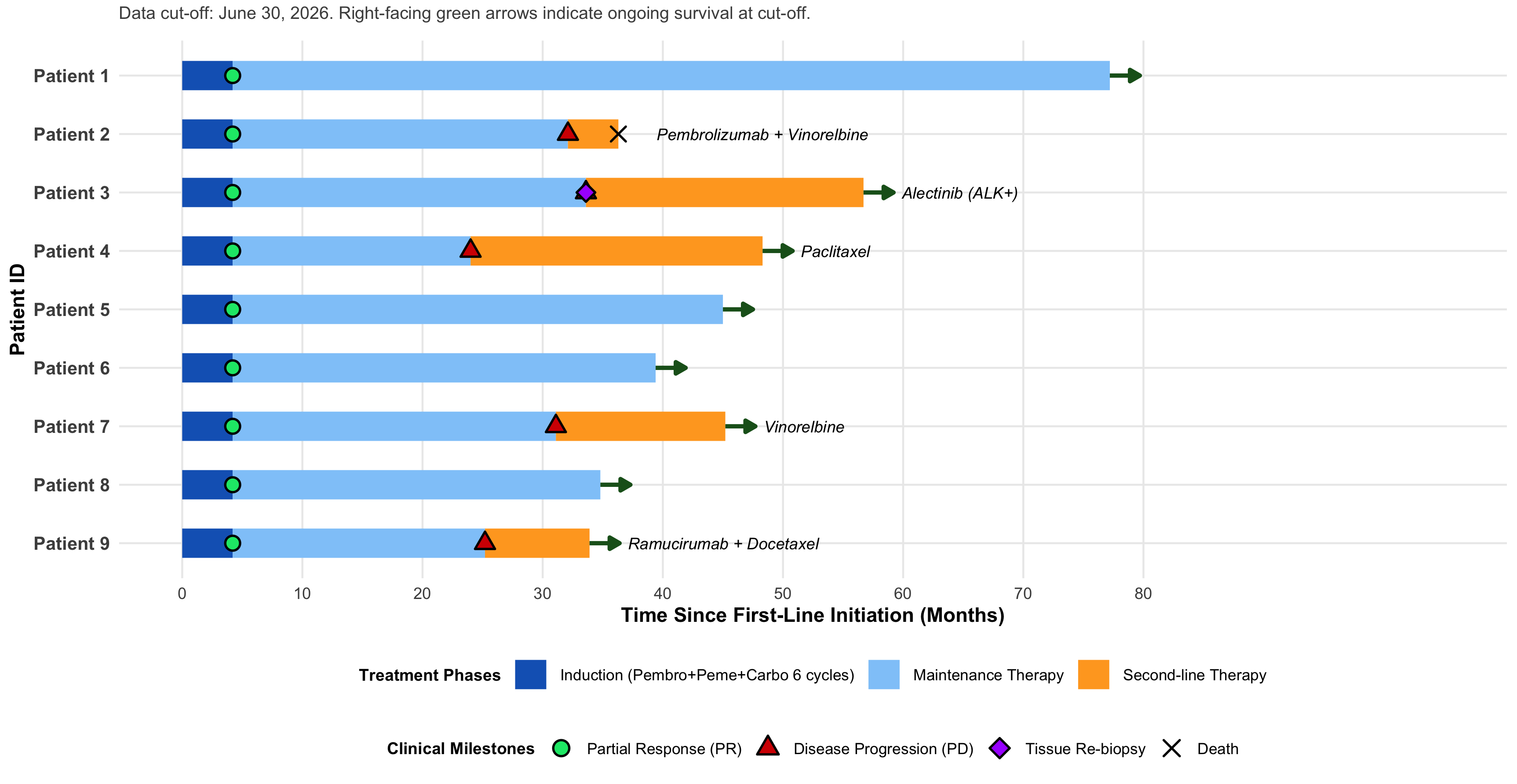
Swimmer plot illustrating individual treatment durations, clinical response milestones, and survival trajectories across the nine PD-L1-negative (TPS < 1%) advanced lung adenocarcinoma outliers (N = 9). Horizontal bars depict longitudinal time (in months) from the initiation of first-line therapy to the study data cut-off date (June 30, 2026), segmented by treatment phase: dark blue bars represent 6-cycle induction therapy with pembrolizumab (200 mg), pemetrexed (500 mg/m2), and carboplatin (AUC 5); light blue bars represent first-line pembrolizumab-based maintenance therapy; and orange bars represent second-line salvage regimens administered after radiographic progression (specific second-line agents and acquired molecular targets are annotated in italics beside each bar). Clinical milestones are denoted by overlaid symbols: green circles indicate initial objective partial response (PR) per RECIST v1.1; red triangles indicate documented radiographic disease progression (PD); the purple diamond (Patient 3, month 33.6) denotes CT-guided adrenal tissue re-biopsy identifying an acquired ALK rearrangement; the black cross (Patient 2, month 36.3) denotes patient death; and right-facing dark green arrows indicate ongoing survival at the June 30, 2026 cut-off. Abbreviations: ALK+, anaplastic lymphoma kinase-positive; AUC, area under the curve; Carbo, carboplatin; CT, computed tomography; PD, progressive disease; PD-L1, programmed death-ligand 1; Pembro, pembrolizumab; Peme, pemetrexed; PR, partial response; RECIST v1.1, Response Evaluation Criteria in Solid Tumors version 1.1; TPS, tumor proportion score.
Biomarker Assessment, Treatment Protocol, and Survival Definitions
Baseline PD-L1 expression was evaluated on diagnostic pre-treatment formalin-fixed, paraffin-embedded (FFPE) tissue specimens using the PD-L1 IHC 22C3 pharmDx assay (Dako, Carpinteria, CA, USA) and scored via TPS, with PD-L1 negativity defined as TPS < 1%. Targeted molecular profiling was performed on diagnostic pre-treatment FFPE tissue. Patients 2 and 4 underwent focused multiplex real-time polymerase chain reaction (PCR) testing and were confirmed as EGFR and ALK wild-type without broad-panel NGS. For the remaining seven patients, targeted NGS was conducted using a 9-gene panel (EGFR, ALK, ROS1, BRAF, KRAS, MET, RET, ERBB2, and NTRK1) on an Illumina platform (mean coverage depth > 500×; minimum nucleic acid input ≥ 20 ng DNA/RNA). Targeted NGS identified specific oncogenic variants in three patients—a rare non-V600E BRAF E501Q variant (Patient 1), a KRAS G12C mutation (Patient 5), and an EGFR exon 20 insertion (Patient 7)—and confirmed driver-negative status across all nine genes in the remaining four cases (Table 1). Baseline peripheral blood counts obtained prior to cycle 1 demonstrated a predominantly low systemic inflammatory profile, with a median neutrophil-to-lymphocyte ratio (NLR) of 1.86 (8 of 9 patients [88.9%] had an NLR < 3.0) and a median platelet-to-lymphocyte ratio (PLR) of 114 (7 of 9 patients [77.8%] had a PLR < 150). Aggregated baseline demographic, inflammatory, and genomic characteristics are summarized in Table 2.
Aggregated baseline demographic, clinicopathological, hematological inflammatory, and genomic characteristics of the exceptional responder cohort (N = 9).
| Baseline Characteristic | Summary Statistic (N = 9) |
|---|---|
| Age (years), median (range) | 65 (33–71) |
| Sex, n (%) | |
| Male | 7 (77.8%) |
| Female | 2 (22.2%) |
| Smoking history, n (%) | |
| Ever-smoker | 6 (66.7%) |
| Never-smoker | 3 (33.3%) |
| ECOG performance status, n (%) | |
| 0–1 | 9 (100.0%) |
| Histology, n (%) | |
| Lung adenocarcinoma | 9 (100.0%) |
| Baseline PD-L1 expression (22C3 pharmDx), n (%) | |
| TPS < 1% (PD-L1-negative) | 9 (100.0%) |
| Baseline systemic inflammatory indices | |
| NLR, median (range) | 1.86 (0.97–5.60) |
| NLR < 3.0, n (%) | 8 (88.9%) |
| PLR, median (range) | 114 (92–231) |
| PLR < 150, n (%) | 7 (77.8%) |
| Baseline molecular profile, n (%) | |
| BRAF E501Q (9-gene NGS) | 1 (11.1%) |
| KRAS G12C (9-gene NGS) | 1 (11.1%) |
| EGFR exon 20 insertion (9-gene NGS) | 1 (11.1%) |
| 9-gene pan-negative wild-type (NGS) | 4 (44.4%) |
| EGFR/ALK wild-type (focused multiplex PCR) | 2 (22.2%) |
| First-line induction regimen, n (%) | |
| Pembrolizumab + pemetrexed + carboplatin | 9 (100.0%) |
All nine patients received first-line induction therapy consisting of pembrolizumab (200 mg intravenously), pemetrexed (500 mg/m2), and carboplatin (area under the curve [AUC] 5) administered every 3 weeks for up to 6 cycles, followed by maintenance pembrolizumab (up to 35 total cycles) plus pemetrexed until disease progression or unacceptable toxicity (Figure 1). The final data cut-off date for survival analysis was June 30, 2026. PFS was calculated from the initiation of first-line therapy to documented radiographic progression per Response Evaluation Criteria in Solid Tumors version 1.1 (RECIST v1.1) or death from any cause; patients alive without progression were right-censored at their last verifiable radiographic assessment. OS was measured from treatment initiation until death from any cause, with surviving patients censored at their last confirmed clinical contact.
Patient 1: Multi-Year Follow-Up of a Rare BRAF E501Q Variant and Baseline Inflammatory Paradox
The initial clinical presentation of Patient 1 was previously described as a preliminary case report6 involving a 65-year-old male heavy smoker with metastatic lung adenocarcinoma harboring a rare non-V600E BRAF E501Q mutation, a PD-L1 TPS < 1%, and marked baseline systemic inflammation (NLR, 5.60; PLR, 231). His inclusion in the present series provides an extended multi-year follow-up. After achieving a partial response following induction chemo-immunotherapy, the patient maintained durable stable disease without radiographic progression or late-onset immune-related toxicities upon completing 35 cycles of pembrolizumab plus pemetrexed maintenance. As of the June 2026 data cut-off, both his PFS and OS have reached 77.2+ months (over 6.4 years), representing one of the longest documented survival durations for this molecular and biomarker subgroup (Table 1, Figure 1).
Patient 3: Acquired Secondary ALK Rearrangement and Clonal Evolution
Patient 3, a 53-year-old female never-smoker, presented with stage IV lung adenocarcinoma involving the contralateral lung, bone, and right adrenal gland. Baseline testing confirmed 9-gene driver-negative, PD-L1-negative (TPS < 1%) status and a low systemic inflammatory profile (NLR, 2.25; PLR, 200). Following 6 cycles of induction pembrolizumab, pemetrexed, and carboplatin, serial computed tomography (CT) demonstrated a marked partial response in the primary left pulmonary lesion and complete radiographic stabilization of the right adrenal metastasis (Figure 2A, 2B). During maintenance therapy, the patient developed immune-related adrenal insufficiency, which was effectively controlled with physiological corticosteroid replacement without requiring pembrolizumab discontinuation. However, owing to persistent chemotherapy-related fatigue and grade 2 anorexia, maintenance pemetrexed was discontinued, and she continued on pembrolizumab monotherapy, achieving a first-line PFS of 33.6 months.
At month 33.6, restaging CT revealed continued control of the primary pulmonary tumor but isolated progression in the left adrenal gland (Figure 2C). A CT-guided core-needle re-biopsy of the progressing adrenal lesion revealed a secondary ALK rearrangement that had not been detected in the baseline primary lung biopsy. Transitioning from pembrolizumab to second-line targeted therapy with alectinib induced a durable secondary partial response (Figure 2D), maintaining disease control for more than 23 additional months and extending her cumulative OS to 56.7+ months as of June 2026.
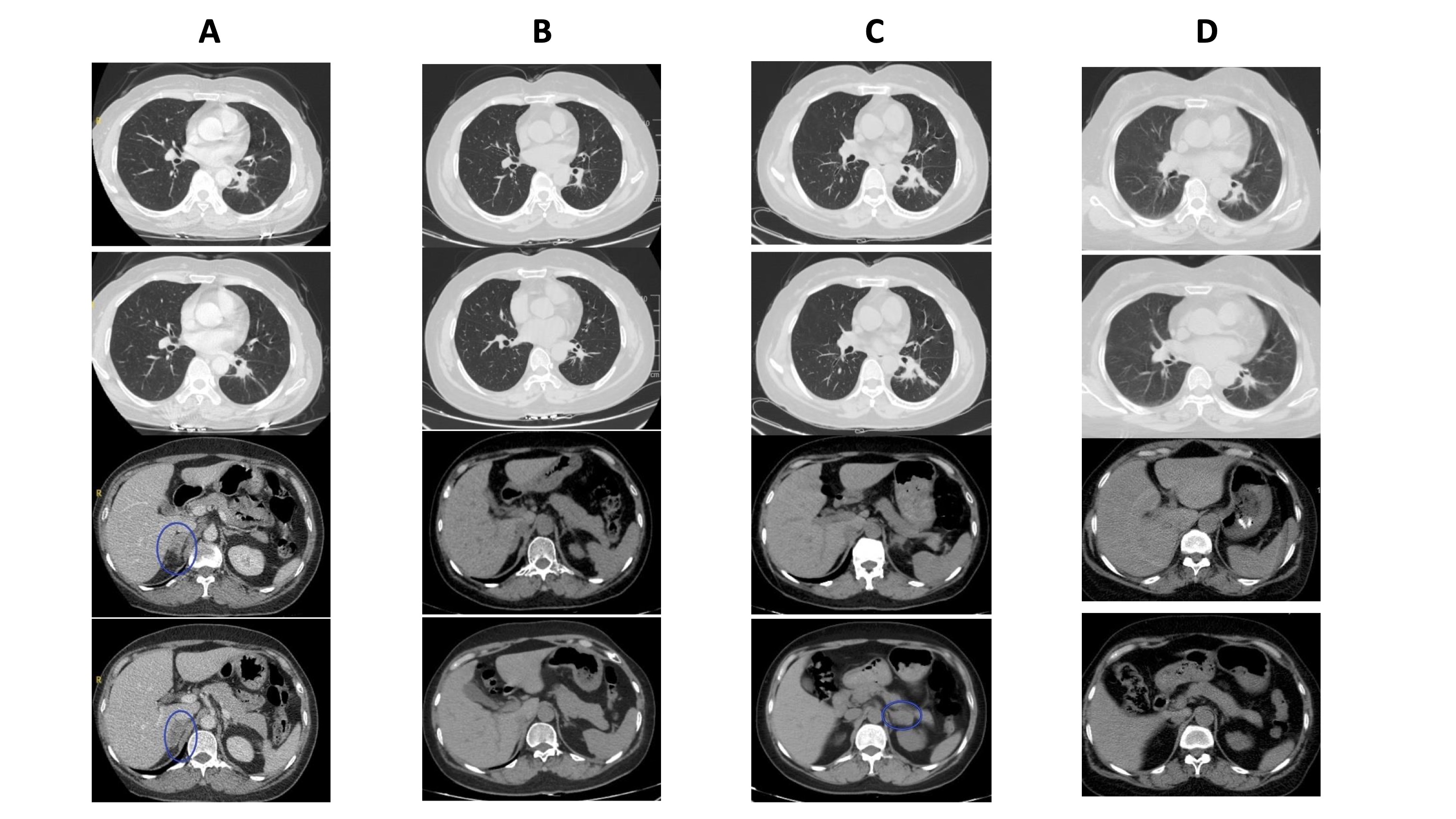
Serial axial computed tomography (CT) scans demonstrating primary pulmonary tumor control, isolated adrenal oligoprogression with acquired ALK rearrangement, and subsequent response to second-line alectinib in Patient 3 (a 53-year-old female never-smoker with baseline 9-gene wild-type, PD-L1-negative lung adenocarcinoma). Within each vertical column, the top two panels display axial chest lung-window views and the bottom two panels display axial abdominal soft-tissue-window views. (A) Baseline pre-treatment CT scans demonstrating the primary left lower lobe pulmonary lesion and a concurrent right adrenal metastasis (encircled in blue). (B) Restaging CT scans following 6 cycles of induction pembrolizumab, pemetrexed, and carboplatin, showing a marked partial response of the left pulmonary mass and radiographic resolution/stabilization of the right adrenal lesion. (C) Restaging CT scans at month 33.6 revealing sustained regression of the primary left lung lesion alongside isolated, localized enlargement of the left adrenal gland (encircled in blue), which prompted a CT-guided core-needle re-biopsy confirming secondary ALK positivity. (D) Follow-up CT scans during second-line targeted alectinib therapy (updated to June 2026), demonstrating a durable secondary partial response in the left adrenal gland and ongoing systemic disease control at 56.7+ months. Abbreviations: ALK, anaplastic lymphoma kinase; CT, computed tomography; PD-L1, programmed death-ligand 1.
Patient 7: Prolonged Frontline Response in EGFR Exon 20 Insertion-Mutated Disease
Patient 7, a 69-year-old male heavy smoker, presented with stage IV lung adenocarcinoma with bone and pleural involvement harboring an EGFR exon 20 insertion and a low baseline inflammatory state (NLR, 1.82; PLR, 114). Although NSCLC tumors carrying EGFR exon 20 insertions are classically refractory to first- and second-generation EGFR tyrosine kinase inhibitors and typically derive limited benefit from single-agent immune checkpoint blockade, this patient achieved a notable partial response after 6 cycles of frontline pembrolizumab, pemetrexed, and carboplatin, reaching a PFS of 31.1 months (Figure 3A, 3B). At month 31.1, radiographic progression occurred, characterized by primary tumor regrowth, increased pleural effusion, and progressive osteolytic pelvic bone metastases (Figure 3C). He was subsequently transitioned to second-line single-agent vinorelbine, achieving disease stabilization (Figure 3D) with an ongoing OS of 45.2+ months at the study cut-off.
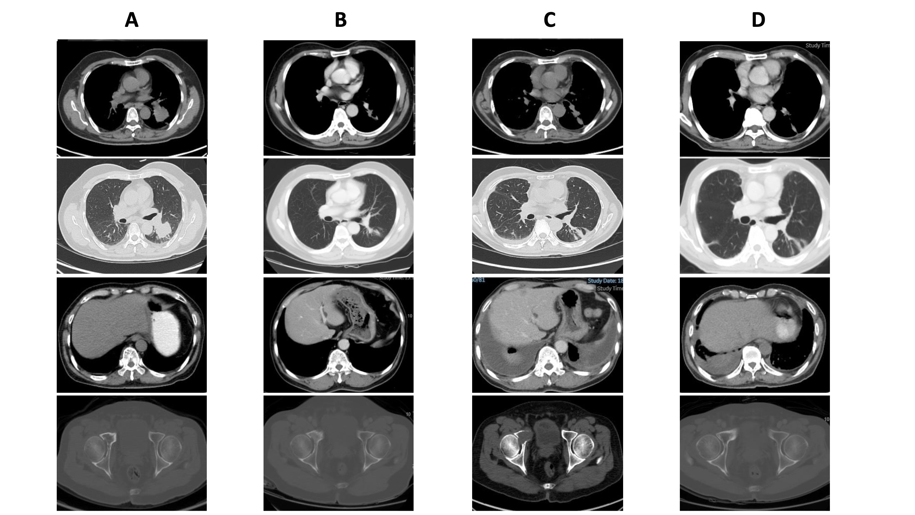
Longitudinal axial computed tomography (CT) evaluation across first-line chemo-immunotherapy and second-line vinorelbine salvage in Patient 7 (a 69-year-old male heavy smoker with PD-L1-negative lung adenocarcinoma harboring a baseline EGFR exon 20 insertion). Within each vertical column, the four panels from top to bottom display axial chest mediastinal-window, chest lung-window, upper abdominal soft-tissue-window, and pelvic bone-window views. (A) Baseline pre-treatment CT scans demonstrating a primary left lower lobe pulmonary mass with ipsilateral pleural thickening, absence of hepatic metastases, and baseline pelvic skeletal integrity. (B) Best partial response following 6 cycles of frontline pembrolizumab, pemetrexed, and carboplatin, showing substantial shrinkage of the primary left lower lobe tumor and resolution of pleural thickening. (C) Documented disease progression at month 31.1 characterized by local regrowth of the primary left pulmonary lesion, recurrent bilateral pleural effusions (abdominal/thoracic base view, third panel), and progressive osteolytic pelvic bone metastases (bottom panel) prior to initiating second-line therapy. (D) Restaging CT scans during second-line single-agent vinorelbine therapy at the June 2026 cut-off (overall survival, 45.2+ months), demonstrating stabilization of the pulmonary lesion, marked reduction of pleural effusion, and radiographic stability of pelvic bone metastases. Abbreviations: CT, computed tomography; EGFR, epidermal growth factor receptor; PD-L1, programmed death-ligand 1.
Post-Progression Management and Long-Term Safety Profile
Across the nine outliers, four patients (Patients 1, 5, 6, and 8) remained alive and progression-free at the June 30, 2026 data cut-off, whereas five patients (Patients 2, 3, 4, 7, and 9) experienced disease progression between 24.0 and 33.6 months and received subsequent lines of systemic therapy tailored to their molecular and clinical profiles (Table 1, Figure 1). Second-line regimens included targeted alectinib for acquired ALK rearrangement (Patient 3), taxane-based therapy with weekly paclitaxel (Patient 4, who also developed a metachronous secondary bladder malignancy) or ramucirumab plus docetaxel (Patient 9), and vinorelbine-based regimens (Patients 2 and 7). Eight of the nine patients remained alive at the final follow-up, with only Patient 2 succumbing to disease progression at 36.3 months.
Overall, the long-term safety profile among these exceptional responders was favorable, with minimal immune-related adverse events (irAEs). Only Patient 3 experienced a clinically significant irAE (immune-related adrenal insufficiency), which was managed with physiological corticosteroid replacement without permanent immunotherapy withdrawal. No late-onset irAEs or cumulative organ toxicities were observed during or after the completion of maintenance therapy.
DISCUSSION
This case series of nine long-term survivors demonstrates that baseline tissue PD-L1 negativity does not preclude multi-year clinical benefit from first-line pembrolizumab plus platinum-doublet chemotherapy in selected patients with advanced lung adenocarcinoma, even though absent PD-L1 expression is broadly associated with lower response rates at the population level1,3. Representing 17.3% (9/52) of the PD-L1-negative subpopulation within our 225-patient institutional cohort7, these outliers all achieved a PFS of at least 24 months. Examining their shared clinicopathological traits alongside their individual molecular trajectories offers clinical insights into host and tumor factors that may support durable anti-tumor immunity despite a negative baseline PD-L1 biomarker.
Although tissue TMB could not be formally quantified in our cohort owing to the use of a focused 9-gene NGS panel or multiplex PCR, a history of heavy smoking was present in 66.7% (6/9) of patients. Tobacco carcinogen exposure is epidemiologically and mechanistically linked to high somatic mutational load and neoantigen generation, which facilitate T-cell recognition and immune-mediated tumor clearance4,8. While elevated TMB remains an unconfirmed hypothesis rather than a directly verified mechanism in our series, future investigations incorporating broad-panel comprehensive genomic profiling are warranted to define the precise mutational signatures driving prolonged responses in PD-L1-negative NSCLC9. Concurrently, a low baseline systemic inflammatory state was a consistent host feature across our series: 88.9% (8/9) of patients exhibited a baseline NLR < 3.0 (median, 1.86), and 77.8% (7/9) had a baseline PLR < 150 (median, 114)10,11. Lower peripheral neutrophil and platelet counts relative to lymphocytes reflect a preserved host adaptive immune reserve and reduced myeloid-driven systemic immunosuppression, enabling patients to sustain treatment sensitivity throughout the 2-year maintenance phase and beyond12.
Despite the predominance of low baseline inflammation across the series, Patient 1 represents a notable biological exception. Presenting with marked baseline systemic inflammation (NLR, 5.60; PLR, 231), this patient nevertheless achieved the longest survival in our cohort, remaining progression-free beyond 77.2 months. This apparent paradox illustrates that peripheral hematological ratios function as probabilistic prognostic indicators rather than absolute mechanistic barriers to immunotherapy efficacy. In selected individuals, potent tumor-intrinsic immunogenicity—potentially driven by tobacco-associated clonal neoantigens or altered mitogen-activated protein kinase (MAPK) signaling linked to the rare non-V600E BRAF E501Q variant—may elicit robust cytotoxic T-lymphocyte responses capable of overcoming peripheral immunosuppressive signaling4,13. Moreover, elevated baseline peripheral neutrophil and platelet counts can occasionally reflect localized tumor necrosis or paraneoplastic cytokine release rather than intrinsic lymphocyte exhaustion14. In such cases, rapid cytoreduction induced by frontline platinum-doublet chemotherapy combined with pembrolizumab may attenuate tumor-derived myeloid mobilization and restore systemic immune competence, permitting sustained multi-year immune surveillance15.
The clinical trajectory of Patient 3 underscores how spatial tumor heterogeneity and clonal evolution shape late resistance following prolonged immunotherapy response. Given that ALK-rearranged NSCLC is characteristically refractory to immune checkpoint inhibitors16, her 33.6-month frontline response strongly suggests that the dominant tumor clone at baseline was genuinely ALK-negative and immune-responsive. Because the progression biopsy was obtained from an enlarging left adrenal metastasis whereas the baseline diagnostic specimen originated from the primary lung mass, the emergence of ALK positivity at month 33.6 likely reflects either interlesional spatial heterogeneity or Darwinian positive selection of an occult, pre-existing ALK-rearranged subclone under prolonged immunotherapeutic selective pressure, rather than de novo genomic rearrangement17. Regardless of whether the clone was spatially segregated at diagnosis or selected over time, it served as an immune-escape pathway. Crucially, performing a repeat tissue biopsy at the site of oligoprogression rather than initiating empiric cytotoxic salvage chemotherapy enabled the identification of an actionable driver and transition to alectinib, securing more than 23 months of additional disease control.
Distinct baseline molecular alterations also contributed to the heterogeneous clinical courses observed in this PD-L1-negative cohort. Patient 5 harbored a KRAS G12C mutation (PFS and OS of 45.0+ months), an alteration frequently associated with tobacco exposure, an inflammatory tumor microenvironment, and heightened sensitivity to PD-1/PD-L1 blockade18. Conversely, the 31.1-month PFS achieved by Patient 7, who harbored an EGFR exon 20 insertion, is clinically noteworthy given the historically poor responsiveness of EGFR-mutant NSCLC to immunotherapy monotherapy16,19. Recent comparative real-world evidence indicates that while single-agent immune checkpoint inhibition yields minimal activity in EGFR exon 20 insertion-positive lung adenocarcinoma, combining platinum-based chemotherapy with immunotherapy can meaningfully extend PFS19. Although definitive mechanistic conclusions cannot be drawn from a single patient, cytotoxic chemotherapy may promote immunogenic cell death and tumor antigen release, sensitizing otherwise immunologically 'cold' EGFR exon 20 insertion tumors to PD-1 blockade20.
Furthermore, while durable first-line disease control was the foundational determinant of survival across our cohort, cumulative OS in the five patients who eventually progressed was substantially reinforced by tailored second-line interventions. Post-progression salvage regimens—including alectinib for acquired ALK rearrangement, ramucirumab plus docetaxel, paclitaxel, and vinorelbine—contributed substantially to post-progression survival. Thus, the multi-year OS observed in this series reflects the synergistic continuum of effective frontline chemo-immunotherapy followed by individualized sequential management.
Several limitations of this study merit consideration. First, our analysis is constrained by its retrospective, single-center design and the small sample size of nine outlier cases, which preclude multivariable statistical modeling. Second, quantitative tissue TMB, programmed death-1 (PD-1)+ tumor-infiltrating lymphocyte density, and serial circulating tumor DNA (ctDNA) dynamics were not assessed due to real-world resource constraints. Finally, long-term OS outcomes were naturally influenced by heterogeneous subsequent therapies administered after first-line progression.
CONCLUSION
Patients with PD-L1-negative advanced lung adenocarcinoma can achieve multi-year PFS and OS when treated with first-line pembrolizumab combined with pemetrexed and carboplatin. Baseline tissue PD-L1 negativity alone should not be viewed as an absolute barrier to durable chemo-immunotherapy benefit. Because long-term survival is shaped by the interplay of host systemic inflammatory status, underlying molecular alterations, and clonal evolution, clinicians should integrate comprehensive clinicopathological profiling at baseline and pursue repeat tissue biopsy upon localized progression to guide precision sequential therapy.
DECLARATIONS
Abbreviations
1L, first-line; AC, adenocarcinoma; ALK, anaplastic lymphoma kinase; AUC, area under the curve; BRAF, v-Raf murine sarcoma viral oncogene homolog B; CI, confidence interval; CT, computed tomography; ctDNA, circulating tumor DNA; DNA, deoxyribonucleic acid; ECOG, Eastern Cooperative Oncology Group; EGFR, epidermal growth factor receptor; ERBB2, Erb-B2 receptor tyrosine kinase 2; FFPE, formalin-fixed, paraffin-embedded; IHC, immunohistochemistry; irAEs, immune-related adverse events; KRAS, Kirsten rat sarcoma viral oncogene homolog; MAPK, mitogen-activated protein kinase; MET, mesenchymal-epithelial transition factor; NGS, next-generation sequencing; NLR, neutrophil-to-lymphocyte ratio; NSCLC, non-small cell lung cancer; NTRK1, neurotrophic receptor tyrosine kinase 1; ORR, objective response rate; OS, overall survival; PCR, polymerase chain reaction; PD, progressive disease; PD-1, programmed cell death protein 1; PD-L1, programmed death-ligand 1; PFS, progression-free survival; PLR, platelet-to-lymphocyte ratio; PR, partial response; RECIST, Response Evaluation Criteria in Solid Tumors; RET, rearranged during transfection; RNA, ribonucleic acid; ROS1, ROS proto-oncogene 1; TMB, tumor mutational burden; TPS, tumor proportion score.
Acknowledgments
None.
Author’s contributions
TVN and KHD conceptualized and designed the study, supervised clinical management, performed formal analysis, and drafted the initial manuscript. PTBN, HTNN, HTT, TMD, TAN, NBH, and HND contributed to patient clinical management, data acquisition, radiographic and molecular data curation, and longitudinal follow-up verification. TVN and KHD critically revised the manuscript for important intellectual content. All authors read and approved the final manuscript.
Funding
None.
Availability of data and materials
The datasets generated and/or analyzed during the current study are available from the corresponding author upon reasonable request, subject to institutional patient privacy regulations.
Ethics approval and consent to participate
This study was approved by the Institutional Ethics Review Board of the National Cancer Hospital of Vietnam (Decision No. 1533/QD-BVK dated April 28, 2020, and Decision No. 566/QD-BVK dated March 18, 2022). Written informed consent to participate was obtained from all patients. All clinical data were anonymized and maintained with strict confidentiality in accordance with the Declaration of Helsinki.
Consent for publication
Written informed consent was obtained from all patients for the publication of this case series and any accompanying anonymized clinical images.
Declaration of generative AI and AI-assisted technologies in the writing process
During the preparation of this work, the authors used Google Gemini to improve language, readability, and grammar. After using this tool, the authors reviewed and edited the content as needed and take full responsibility for the content of the published article.
Competing interests
None. The authors declare that they have no competing interests.