


Serum Parathyroid Hormone-Related Protein Correlates with Clinicopathological Severity in Colorectal Cancer Patients

- Biochemistry Division, Chemistry Department, Faculty of Science, Mansoura University, Mansoura 35516, Egypt
- Biochemistry Department, Faculty of Biotechnology, Sinai University, Kantara 41636, Egypt
- Microbial Biotechnology Department, Biotechnology Research Institute, National Research Centre, Giza 12622, Egypt
- Egypt Centre for Research and Regenerative Medicine (ECRRM), Cairo 4435102, Egypt
- Department of Surgical Oncology, Oncology Center Mansoura University (OCMU), Mansoura University, Mansoura 35516, Egypt
- Research and Development Department, Biotechnology Research Center, New Damietta 34517, Egypt
Abstract
Background: Parathyroid hormone-related protein (PTHrP) is increasingly recognized as a key mediator in colorectal cancer (CRC) biology and a promising circulating biomarker with complementary diagnostic value to carcinoembryonic antigen (CEA). This case–control study provides the first clinical evaluation of serum PTHrP as a diagnostic and disease-severity biomarker in an Egyptian CRC cohort and investigates its relationship with CEA.
Methods: A total of 125 participants were enrolled: 70 patients with histologically confirmed CRC, 25 individuals with benign colorectal polyps, and 30 healthy age- and sex-matched controls. Serum PTHrP and CEA concentrations were quantified using high-sensitivity sandwich enzyme-linked immunosorbent assays (ELISA), alongside the systematic documentation of clinicopathological parameters.
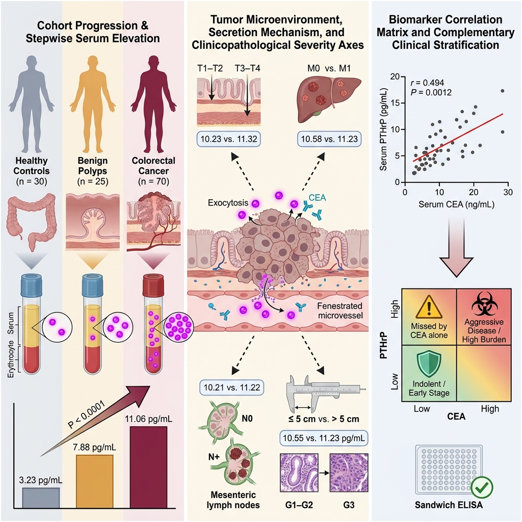
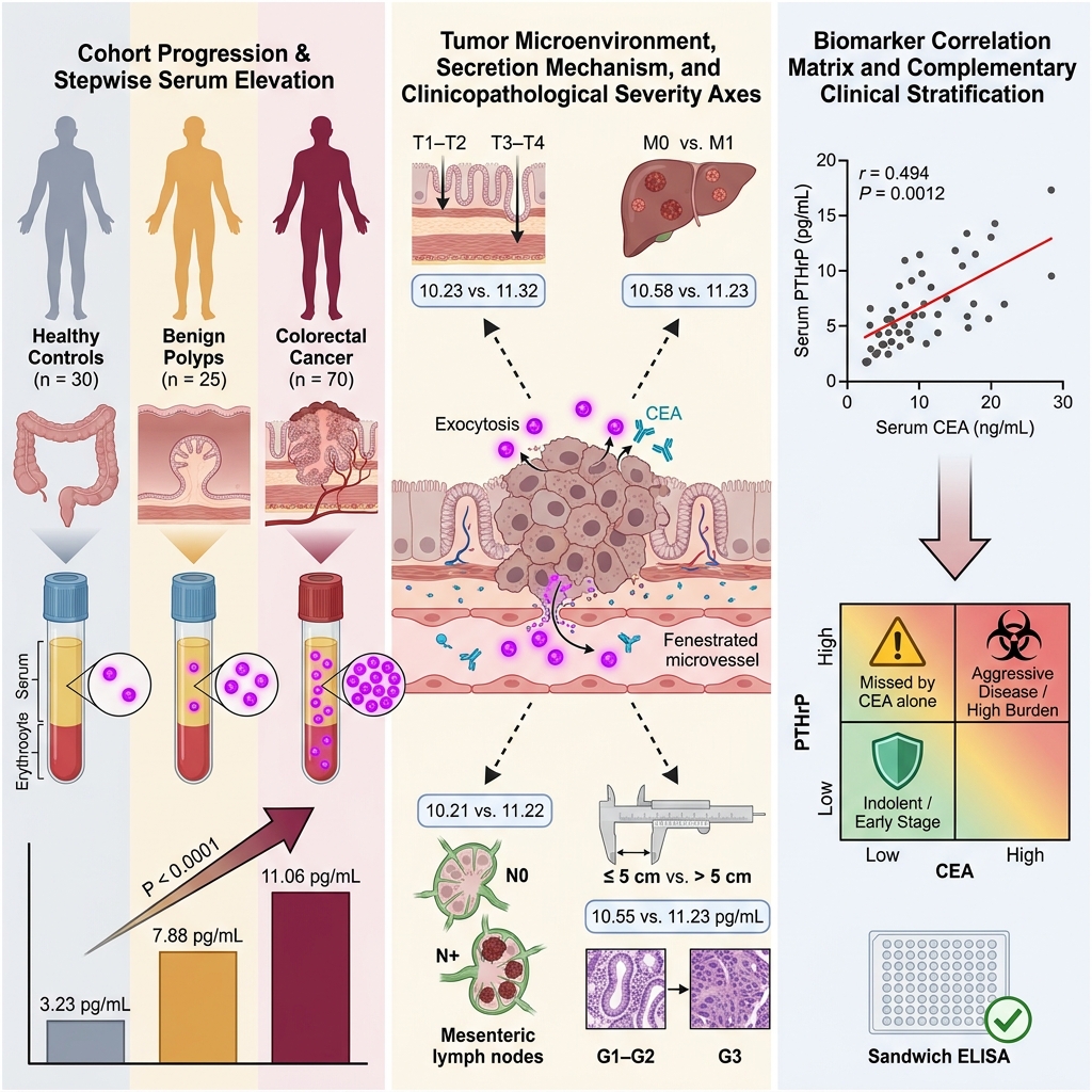
Results: Serum PTHrP concentrations were significantly elevated in CRC patients (median: 11.06 pg/mL; interquartile range [IQR]: 10.11–12.26) compared with patients with benign polyps (median: 7.88 pg/mL; IQR: 6.20–10.06) and healthy controls (median: 3.23 pg/mL; IQR: 2.63–3.97; P < 0.0001). Within the CRC group, higher PTHrP concentrations were significantly associated with adverse clinicopathological features, including advanced primary tumor stage (T3–T4 vs. T1–T2: 11.32 vs. 10.23 pg/mL, P < 0.0001), regional lymph node metastasis (N1 vs. N0: 11.22 vs. 10.21 pg/mL, P = 0.0006), distant metastasis (M1 vs. M0: 11.23 vs. 10.58 pg/mL, P = 0.0020), poorly differentiated histological grade (G3 vs. G1–G2: 11.23 vs. 10.55 pg/mL, P = 0.0021), and larger tumor size (>5 cm vs. ≤5 cm: 11.25 vs. 10.24 pg/mL, P = 0.0010). Furthermore, serum PTHrP demonstrated a moderate positive correlation with CEA (r = 0.494, P = 0.0012), indicating shared yet non-redundant indications of tumor burden.
Conclusions: Elevated circulating PTHrP concentrations correlate with clinicopathological severity and aggressive tumor phenotypes in Egyptian CRC patients. Integrating serum PTHrP measurement with conventional CEA assessment may enhance risk stratification and inform personalized therapeutic approaches, particularly in resource-constrained clinical settings.
Graphical abstract
Introduction
Colorectal cancer (CRC) represents a major global health challenge, ranking as the third most frequently diagnosed malignancy and the second leading cause of cancer-related mortality worldwide. According to recent GLOBOCAN estimates, CRC accounted for more than 1.9 million new cases in 2020, representing approximately 10% of all cancer diagnoses 1, with epidemiological models projecting an increase to over 3 million annual cases by 2040 2. The epidemiological burden is rapidly shifting toward low- and middle-income countries (LMICs), where cancer mortality rates are exacerbated by limited screening infrastructure, diagnostic delays, and restricted access to multimodal therapeutics 3,4. In Egypt, CRC is among the most prevalent malignancies across both sexes, with an age-standardized incidence rate of 9.8 per 100,000 population 5.
Despite improvements in diagnostic and therapeutic techniques, CRC screening uptake remains suboptimal in many LMICs, leading to a predominance of stage III–IV disease at initial presentation 3,6,7. Studies from sub-Saharan Africa and comparable settings indicate that over 60% of patients are diagnosed at advanced stages, significantly compromising curative prospects and overall survival 8,9. Globally, 15% to 30% of patients present with synchronous metastases, and nearly half of those with initially localized tumors eventually develop metachronous distant spread—precipitating a stark decline in five-year overall survival from >85% in stage I to <15% in stage IV disease 10,11. These clinical realities underscore the urgent need for minimally invasive, reliable biomarkers to facilitate early detection, monitor disease progression, and guide therapeutic decision-making—particularly in healthcare systems with constrained endoscopic capacity 12,13,14.
Circulating serum biomarkers offer a minimally invasive, cost-effective modality for CRC detection and longitudinal surveillance. However, conventional biomarkers such as carcinoembryonic antigen (CEA) and carbohydrate antigen 19-9 (CA19-9) demonstrate inconsistent sensitivity and limited prognostic accuracy, particularly in early-stage disease and low-resource settings 15,16,17,18. These diagnostic limitations have spurred growing interest in alternative circulating biomarkers that more faithfully reflect tumor biology and can enhance existing surveillance frameworks 19.
Parathyroid hormone-related protein (PTHrP), initially characterized as the principal humoral mediator of malignancy-associated hypercalcemia, shares substantial N-terminal amino acid homology with parathyroid hormone (PTH), enabling high-affinity binding to the shared type 1 PTH/PTHrP receptor (PTHR1) 20,21. In CRC, PTHrP expression is distinctly upregulated in malignant tissues compared with adjacent normal colonic mucosa and benign adenomatous polyps 22. Furthermore, PTHrP plays a crucial functional role in promoting tumorigenesis through Rac1-driven cell migration, activation of MET and MAPK/PI3K signaling pathways, and integrin-dependent tumor expansion in vivo 23,24,25. Experimental silencing of PTHrP in CRC models markedly attenuates cellular proliferation, induces apoptosis, and restores chemosensitivity, underscoring its pivotal role in tumor cell survival and therapeutic resistance 26. Although tissue-level expression of PTHrP has been documented, circulating PTHrP has not been clinically investigated or validated in Middle Eastern CRC populations. Taken together, these mechanistic insights and translational evidence establish circulating PTHrP as a biologically relevant and clinically tractable candidate to complement existing serum biomarkers for disease staging and risk stratification in CRC.
Therefore, this study aimed to evaluate the association between serum PTHrP levels and clinicopathological severity among Egyptian CRC patients—including advanced primary tumor stage, large tumor size, regional lymph node invasion, high histological grade, and distant metastasis—as well as its correlation with circulating levels of the established biomarker CEA.
Materials and Methods
Study Population and Recruitment
This case–control study was designed and conducted in accordance with the Strengthening the Reporting of Observational Studies in Epidemiology (STROBE) guidelines for case–control studies (a completed STROBE checklist is provided as Supplementary Material). A total of 125 participants were enrolled, comprising 70 patients with histologically confirmed CRC, 25 individuals with benign colorectal conditions (e.g., inflammatory polyps and adenomas without high-grade dysplasia), and 30 healthy, age- and sex-matched control subjects. Participants were recruited from the Oncology Center Mansoura University (OCMU), Mansoura University Hospital, Egypt, between January 2024 and January 2025. All patient blood samples were collected following definitive diagnosis and prior to the initiation of any antineoplastic treatment (surgery, chemotherapy, or radiotherapy).
Inclusion criteria were adult patients (≥18 years of age) with newly diagnosed, treatment-naïve CRC or benign colorectal pathology confirmed via colonoscopic biopsy and histopathological examination. Exclusion criteria encompassed chronic liver or renal disease, active systemic infections, hypercalcemia of any etiology, prior antineoplastic therapy, and any history of malignancy other than CRC. Healthy controls were selected from asymptomatic individuals undergoing routine medical check-ups who had no history of gastrointestinal disorders, neoplastic disease, or chronic systemic illnesses.
The primary variable of interest was serum PTHrP concentration. Secondary variables included serum CEA concentration, primary tumor extent (T stage) and TNM staging according to the American Joint Committee on Cancer (AJCC) classification system 27, histological grade, tumor size, regional lymph node involvement, distant metastasis, demographic parameters, and routine clinical laboratory indices. To minimize potential selection and measurement biases, all CRC cases were histologically verified, blood samples were obtained before treatment initiation, controls were matched for age and sex, and individuals with confounding systemic illnesses that could alter calcium homeostasis or PTHrP levels were strictly excluded.
Ethical Considerations
The study protocol was approved by the Institutional Review Board (IRB) of Mansoura University Hospital (Approval code: R.21.07.1376) and conducted in strict accordance with the ethical principles of the Declaration of Helsinki. Written informed consent was obtained from all participants prior to study procedures. All patient data were anonymized and confidentiality was rigorously maintained throughout the investigation.
Sample Collection and Laboratory Tests
Fasting venous blood samples were collected from each participant into sterile plain tubes (for serum separation) and EDTA-K3 tubes (for hematological analysis). Plain blood tubes were allowed to clot completely at room temperature and then centrifuged at 3,000 rpm (approximately 1,000 × g) for 15 minutes at 4°C using a refrigerated centrifuge (Eppendorf Centrifuge 5430R; Eppendorf, Hamburg, Germany). Serum aliquots were separated immediately and stored at −80°C until batch testing to avoid repeated freeze–thaw cycles.
Routine laboratory testing included complete blood count (CBC), renal function indices (blood urea, serum creatinine), and liver function profiles (alanine aminotransferase [ALT], aspartate aminotransferase [AST], total bilirubin, serum albumin), all quantified using automated clinical analyzers (Sysmex Corporation, Kobe, Japan; Hitachi High-Tech Corporation, Tokyo, Japan). Serum CEA and PTHrP concentrations were measured using commercial enzyme-linked immunosorbent assay (ELISA) kits strictly according to the manufacturers' instructions. Serum CEA was quantified using a solid-phase sandwich ELISA kit (MyBioSource, San Diego, CA, USA) with a linear analytical range of 0.625–40.00 ng/mL. Serum PTHrP concentrations were measured using a high-sensitivity sandwich ELISA kit (Innova Biotech Co., Ltd., Beijing, China) with an analytical sensitivity of 0.69 pg/mL and a dynamic detection range of 3.00–200.00 pg/mL. All assay measurements were performed in duplicate.
Statistical Analysis
Statistical analyses were performed using IBM SPSS Statistics for Windows (version 26.0; IBM Corp., Armonk, NY, USA) and GraphPad Prism (version 6.0; GraphPad Software, San Diego, CA, USA). Data normality was evaluated using the Shapiro–Wilk test. Continuous variables are presented as mean ± standard deviation (SD) for normally distributed parameters or as median with interquartile range (IQR; 25th–75th percentiles) for non-normally distributed parameters. Categorical variables are expressed as absolute frequencies and percentages.
Inter-group comparisons among CRC patients, individuals with benign colorectal polyps, and healthy controls were evaluated using one-way analysis of variance (ANOVA) followed by Tukey's post-hoc test for normally distributed continuous variables, or the Kruskal–Wallis test followed by Dunn's post-hoc multiple comparison test for non-normally distributed variables. Categorical variables were compared using the Chi-square test. Within the CRC cohort, differences in serum PTHrP concentrations across dichotomized clinicopathological subgroups were analyzed using the two-sided Mann–Whitney U test. The correlation between serum PTHrP and CEA levels was examined using Spearman's rank correlation coefficient (r). Complete-case analysis was applied, and no data imputation was performed. A two-sided P value < 0.05 was considered statistically significant.
Results
Patient Characteristics
No statistically significant differences in age distribution were observed across the three study cohorts (P = 0.333), demonstrating demographic comparability between CRC patients (mean ± SD: 51.50 ± 12.10 years), individuals with benign conditions (49.00 ± 10.30 years), and healthy controls (47.90 ± 5.10 years). Similarly, the distribution of sex did not differ significantly among the cohorts (P = 0.324). Furthermore, hepatic enzyme activities, renal parameters, and hematological indices did not differ significantly between CRC patients and control subjects, consistent with the strict exclusion of individuals with chronic systemic comorbidities.
Serum CEA concentrations were significantly elevated in CRC patients (median: 3.65 ng/mL; IQR: 2.00–23.00) compared with both the benign polyp cohort (median: 2.00 ng/mL; IQR: 1.10–4.50) and healthy controls (median: 2.00 ng/mL; IQR: 1.00–3.50; P = 0.015; Table 1). The clinicopathological classification of CRC patients by AJCC TNM stage, tumor grade, regional lymph node invasion, distant metastasis, and tumor size is summarized in Table 2.
Demographic, clinical, and laboratory characteristics of the study population
| Variables | Colorectal Cancer (n = 70) | Benign Polyps (n = 25) | Healthy Controls (n = 30) | |
|---|---|---|---|---|
| Gender (Male/Female) | 49 / 21 | 18 / 7 | 20 / 10 | 0.324a |
| Age (years) | 51.50 ± 12.10 | 49.00 ± 10.30 | 47.90 ± 5.10 | 0.333b |
| Hemoglobin (g/dL) | 11.50 ± 1.90 | 11.60 ± 2.14 | 11.62 ± 1.82 | 0.241b |
| RBCs (1012/L) | 4.15 ± 0.71 | 4.30 ± 0.55 | 4.50 ± 0.60 | 0.632b |
| WBCs (109/L) | 7.80 ± 3.10 | 7.90 ± 1.90 | 6.99 ± 1.50 | 0.109b |
| Platelet count (109/L) | 249.20 ± 59.10 | 255.00 ± 65.20 | 269.00 ± 61.70 | 0.191b |
| ALT (U/L) | 26.70 ± 10.10 | 23.50 ± 4.20 | 21.20 ± 8.10 | 0.425b |
| AST (U/L) | 46.15 ± 10.10 | 41.11 ± 8.50 | 39.13 ± 7.40 | 0.095b |
| Total bilirubin (mg/dL) | 0.80 ± 0.21 | 0.71 ± 0.15 | 0.66 ± 0.19 | 0.591b |
| Albumin (g/dL) | 3.75 ± 0.42 | 3.92 ± 0.44 | 4.10 ± 0.33 | 0.122b |
| Creatinine (mg/dL) | 0.88 ± 0.29 | 0.78 ± 0.14 | 0.75 ± 0.16 | 0.399b |
| Urea (mg/dL) | 26.12 ± 5.31 | 24.80 ± 5.90 | 23.60 ± 4.80 | 0.610b |
| CEA (ng/mL) | 3.65 (2.00–23.00) | 2.00 (1.10–4.50) | 2.00 (1.00–3.50) | 0.015c* |
Clinicopathological classification of colorectal cancer patients (n = 70)
| Clinicopathological Features | Number of Patients, n (%) |
|---|---|
| Primary Tumor Stage (T Stage) | |
| Early stage (T1–T2) | 30 (42.9%) |
| Late/advanced stage (T3–T4) | 40 (57.1%) |
| Regional Lymph Node Invasion (N Stage) | |
| Negative (N0) | 21 (30.0%) |
| Positive / present (N1) | 49 (70.0%) |
| Distant Metastasis (M Stage) | |
| Negative / absent (M0) | 51 (72.9%) |
| Positive / present (M1) | 19 (27.1%) |
| Histological Grade | |
| Low grade (G1–G2; well-to-moderately differentiated) | 29 (41.4%) |
| High grade (G3; poorly differentiated) | 41 (58.6%) |
| Tumor Size | |
| Small (≤ 5.0 cm) | 30 (42.9%) |
| Large (> 5.0 cm) | 40 (57.1%) |
Elevated Serum PTHrP Levels in CRC Development and Disease Severity
Serum PTHrP concentrations were markedly elevated in CRC patients (median: 11.06 pg/mL; IQR: 10.11–12.26) compared with patients with benign colorectal polyps (median: 7.88 pg/mL; IQR: 6.20–10.06) and healthy controls (median: 3.23 pg/mL; IQR: 2.63–3.97; P < 0.0001; Figure 1).
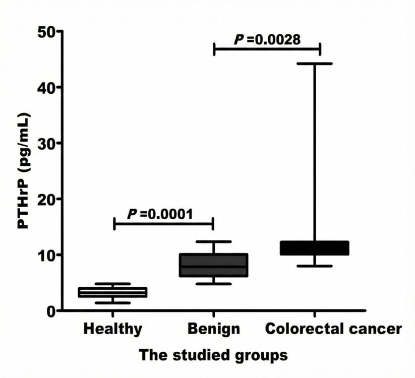
Circulating parathyroid hormone-related protein (PTHrP) concentrations across study cohorts. Quantification of serum PTHrP levels was performed using a high-sensitivity sandwich enzyme-linked immunosorbent assay (ELISA; detection range: 3.00–200.00 pg/mL) in healthy control subjects (n = 30), patients with benign colorectal polyps (n = 25), and patients with histologically confirmed colorectal cancer (CRC; n = 70). Box-and-whisker plots display the median (solid horizontal line within the box), interquartile range (IQR, 25th to 75th percentiles; lower and upper box boundaries), and minimum-to-maximum range (error bars). Serum PTHrP concentrations were significantly elevated in patients with CRC (median: 11.06 pg/mL; IQR: 10.11–12.26) compared with patients with benign polyps (median: 7.88 pg/mL; IQR: 6.20–10.06) and healthy controls (median: 3.23 pg/mL; IQR: 2.63–3.97). Global differences among the three study groups were evaluated using the non-parametric Kruskal–Wallis test (
Within the CRC cohort, elevated serum PTHrP concentrations were consistently associated with aggressive clinicopathological features. Specifically, patients presenting with advanced primary tumor stage (T3–T4) exhibited significantly higher PTHrP levels than those with early-stage disease (T1–T2: 11.32 vs. 10.23 pg/mL, P < 0.0001). Significant elevations were likewise observed in patients with regional lymph node metastasis (N1 vs. N0: 11.22 vs. 10.21 pg/mL, P = 0.0006) and distant metastasis (M1 vs. M0: 11.23 vs. 10.58 pg/mL, P = 0.0020). Similarly, serum PTHrP was significantly higher in poorly differentiated tumors (G3 vs. G1–G2: 11.23 vs. 10.55 pg/mL, P = 0.0021) and in tumors larger than 5 cm (>5 cm vs. ≤5 cm: 11.25 vs. 10.24 pg/mL, P = 0.0010; Table 3).
Association between serum PTHrP levels and clinicopathological features in colorectal cancer patients
| Clinicopathological Parameter | Serum PTHrP (pg/mL), Median (IQR) | |
|---|---|---|
| Primary Tumor Stage | ||
| Early stage (T1–T2; n = 30) | 10.23 (9.10–11.20) | < 0.0001* |
| Late stage (T3–T4; n = 40) | 11.32 (10.90–22.30) | |
| Regional Lymph Node Invasion | ||
| Negative (N0; n = 21) | 10.21 (9.10–11.24) | 0.0006* |
| Present / positive (N1; n = 49) | 11.22 (10.72–18.19) | |
| Distant Metastasis | ||
| Negative (M0; n = 51) | 10.58 (9.54–11.45) | 0.0020* |
| Present / positive (M1; n = 19) | 11.23 (10.75–24.08) | |
| Histological Grade | ||
| Low grade (G1–G2; n = 29) | 10.55 (9.24–11.40) | 0.0021* |
| High grade (G3; n = 41) | 11.23 (10.82–31.20) | |
| Tumor Size | ||
| Small (≤ 5.0 cm; n = 30) | 10.24 (9.20–11.26) | 0.0010* |
| Large (> 5.0 cm; n = 40) | 11.25 (10.57–15.79) | |
Correlation with Established Biomarkers
A statistically significant, moderate positive correlation was identified between serum PTHrP and CEA levels in CRC patients (Spearman's r = 0.494, P = 0.0012; Figure 2). This correlation suggests an additive and complementary role for circulating PTHrP in clinicopathological profiling and disease burden assessment.
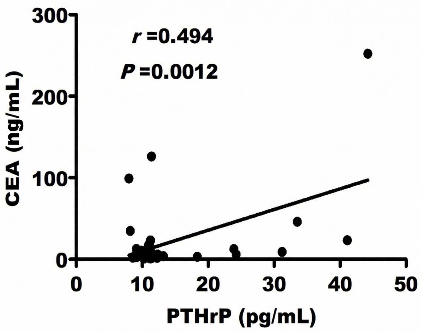
Correlation between serum parathyroid hormone-related protein (PTHrP) and carcinoembryonic antigen (CEA) levels in patients with colorectal cancer. Bivariate correlation between serum concentrations of PTHrP (pg/mL) and CEA (ng/mL) in patients with histologically confirmed CRC (n = 70). Both biomarkers were quantified from pre-treatment fasting serum samples using commercial ELISA kits. Scatter plot representing individual patient values. The solid linear line indicates the best-fit linear regression, and dashed lines demarcate the 95% confidence intervals. Non-parametric correlation analysis revealed a statistically significant, moderate positive association between circulating PTHrP and CEA levels (Spearman's rank correlation coefficient r = 0.494,
Discussion
Among emerging oncological biomarkers, PTHrP has attracted increasing attention in CRC due to its multifaceted role in tumor biology. Initially identified as the primary endocrine mediator of humoral hypercalcemia of malignancy, PTHrP regulates key cellular processes—including proliferation, differentiation, and survival—through interaction with the common type 1 PTH/PTHrP receptor 21. Beyond its classical endocrine actions, PTHrP functions through autocrine, paracrine, and intracrine signaling pathways, activating oncogenic cascades that support primary tumor growth, local invasion, and distant dissemination 28. In vitro investigations have consistently demonstrated that PTHrP stimulates CRC cell proliferation, suppresses programmed apoptotic death, and enhances cell motility and invasion, highlighting its pro-metastatic significance in experimental models 29,30.
To our knowledge, no previous studies have systematically evaluated the clinical relevance of circulating PTHrP in relation to CRC progression in Egyptian cohorts, nor more broadly within Middle Eastern populations, despite its established oncogenic functions 30. This evidence gap is especially pertinent because Egyptian CRC patients present disproportionately with late-stage and early-onset disease, reflecting limited population screening and diagnostic delays 31,32. In this study, we quantified serum PTHrP in an Egyptian CRC cohort and analyzed its correlation with key clinicopathological markers of tumor aggressiveness. Notably, cases and controls were well balanced in terms of age and sex, and individuals with severe hepatic, renal, or systemic inflammatory conditions were excluded. This rigorous matching strategy minimized confounding from age-related endocrine fluctuations or organ dysfunction, thereby strengthening the validity of the observed association between tumor biology and circulating PTHrP concentrations.
In agreement with preclinical findings, Egyptian CRC patients exhibited significantly higher circulating PTHrP levels (median: 11.06 pg/mL; IQR: 10.11–12.26) than individuals with benign colorectal polyps (7.88 pg/mL) and healthy controls (3.23 pg/mL; P < 0.0001). Similar elevations have been described in sporadic case series of CRC-associated hypercalcemia, in which elevated circulating PTHrP levels tracked with extensive hepatic metastases and poor clinical outcomes 20. Mechanistically, PTHrP triggers protein kinase A (PKA) and protein kinase C (PKC) downstream pathways, thereby accelerating primary tumor expansion and facilitating metastatic colonization 28,33.
In our cohort, serum PTHrP concentrations increased stepwise in parallel with established indicators of clinicopathological severity. Patients with large primary tumors (>5 cm), advanced primary stage (T3–T4), or poorly differentiated histology (G3) exhibited significantly higher median PTHrP levels (11.25, 11.32, and 11.23 pg/mL, respectively) than those with smaller, early-stage, or well-to-moderately differentiated lesions (10.24, 10.23, and 10.55 pg/mL, respectively; all P < 0.005). Consistent gradients were observed in relation to dissemination: node-positive patients exhibited significantly higher PTHrP concentrations than node-negative individuals (11.22 vs. 10.21 pg/mL, P = 0.0006), and patients with distant metastases (M1) had higher concentrations than non-metastatic patients (11.23 vs. 10.58 pg/mL, P = 0.0020). These clinical findings corroborate the histopathological observations of Nishihara et al., who reported that elevated intratumoral PTHrP expression correlated with mural invasion, lymphatic permeation, and liver metastasis in resected colorectal tumors 34. These associations also align with translational studies linking tumor PTHrP expression to epithelial–mesenchymal transition (EMT) induction and MET receptor activation, which collectively orchestrate the establishment of a pro-metastatic niche 26. Although a PTHrP-driven EMT program mediated via the RANKL/NF-κB axis has been described in breast cancer, the operation of this specific pathway in CRC requires further elucidation 35,36.
At the cellular and molecular level, PTHrP orchestrates several cooperative oncogenic cascades in CRC. It upregulates integrin α6β4 expression, which in turn engages PI3K/Akt signaling to promote cell survival, motility, and anchorage-independent growth 23. In LoVo CRC cells, PTHrP-induced α6β4 signaling stimulates Akt phosphorylation and inhibits glycogen synthase kinase-3 (GSK-3), fostering enhanced cell proliferation and invasion 29. Additional downstream pathways further substantiate its pro-tumorigenic role. PTHrP activates the extracellular signal-regulated kinase 1/2 (ERK1/2) and p38 MAPK cascades, resulting in downstream RSK phosphorylation, cyclin D1 and CDK6 upregulation, and p53 transcriptional repression—molecular hallmarks of accelerated cell-cycle progression 37. Concurrently, PTHrP increases focal adhesion kinase (FAK) phosphorylation and enhances Rac1 GTPase activity via Tiam1 and α6β4 integrin, directly promoting cytoskeletal remodeling and metastatic cell migration 38. Furthermore, PTHrP stimulates tumor angiogenesis by inducing VEGF, HIF-1α, and MMP-9 expression in neoplastic cells; conditioned media from PTHrP-stimulated CRC cells significantly promote endothelial cell proliferation and capillary tube formation in a VEGF-dependent manner 39. Collectively, these findings delineate a comprehensive molecular network through which PTHrP accelerates colorectal tumor progression, vascularization, and dissemination.
Although serum CEA remains the standard circulating biomarker for CRC surveillance, its limited diagnostic sensitivity in early-stage disease and in molecularly distinct tumor subsets constrains its clinical utility 40. To address this limitation, our study provides the first systematic evidence that circulating PTHrP may serve as a valuable complementary biomarker to CEA. In this cohort, serum PTHrP demonstrated a moderate positive correlation with CEA (Spearman's r = 0.494, P = 0.0012), indicating that both markers increase concurrently with advancing tumor burden and metastatic potential. This observation is supported by previous case reports of metastatic CRC wherein elevated PTHrP and CEA occurred simultaneously in the setting of malignancy-associated hypercalcemia, pointing toward a shared biological trajectory of advanced disease 41.
Multi-marker panel strategies are well established in gastrointestinal oncology. For example, concurrent elevation of CEA and CA19-9 is associated with significantly poorer five-year overall survival compared with elevation of either marker alone 42,43. By analogy, dual quantification of PTHrP and CEA may capture complementary facets of CRC biology: CEA primarily reflects glycoprotein burden and secretory shedding, whereas circulating PTHrP reflects active pro-invasive, angiogenic, and pro-metastatic signaling. Nonetheless, several limitations of the present study should be noted. This was a single-center investigation with a relatively modest sample size, which may limit the immediate generalizability of our findings. However, the rigorous inclusion criteria and inclusion of well-matched control groups reinforce the internal validity of our observations. Prospective, multicenter longitudinal studies in larger patient cohorts are warranted to validate these findings and to establish clinically actionable diagnostic cutoff values for PTHrP-inclusive biomarker panels, particularly in low-resource screening settings.
Conclusion
In conclusion, this study provides compelling clinical evidence that elevated circulating PTHrP concentrations correlate significantly with clinicopathological severity and aggressive tumor phenotypes in Egyptian CRC patients. The marked elevation of PTHrP in advanced primary tumors, nodal involvement, and distant metastasis, combined with its moderate positive concordance with CEA levels, highlights its potential clinical utility as an informative biomarker of tumor burden. Because it is measurable via standardized, cost-effective immunoassays, serum PTHrP represents a promising candidate to complement existing surveillance protocols and improve risk stratification. Incorporating PTHrP into routine clinical assessment could facilitate more timely interventions and support personalized therapeutic management, addressing an urgent clinical need in resource-limited healthcare environments.
Abbreviations
AJCC: American Joint Committee on Cancer; ALT: Alanine aminotransferase; ANOVA: Analysis of variance; AST: Aspartate aminotransferase; CA19-9: Carbohydrate antigen 19-9; CBC: Complete blood count; CDK6: Cyclin-dependent kinase 6; CEA: Carcinoembryonic antigen; CRC: Colorectal cancer; EDTA: Ethylenediaminetetraacetic acid; ELISA: Enzyme-linked immunosorbent assay; EMT: Epithelial–mesenchymal transition; ERK: Extracellular signal-regulated kinase; FAK: Focal adhesion kinase; GSK-3: Glycogen synthase kinase-3; HIF-1α: Hypoxia-inducible factor 1-alpha; IQR: Interquartile range; IRB: Institutional Review Board; LMICs: Low- and middle-income countries; MAPK: Mitogen-activated protein kinase; MMP-9: Matrix metalloproteinase-9; NF-κB: Nuclear factor kappa B; OCMU: Oncology Center Mansoura University; PI3K: Phosphoinositide 3-kinase; PKA: Protein kinase A; PKC: Protein kinase C; PTH: Parathyroid hormone; PTHR1: Parathyroid hormone/parathyroid hormone-related peptide receptor type 1; PTHrP: Parathyroid hormone-related protein; RBC: Red blood cell; RSK: Ribosomal S6 kinase; SD: Standard deviation; STROBE: Strengthening the Reporting of Observational Studies in Epidemiology; TNM: Tumor–node–metastasis; VEGF: Vascular endothelial growth factor; WBC: White blood cell
Acknowledgments
The authors express their sincere gratitude to the medical, nursing, and clinical laboratory staff at the Oncology Center Mansoura University (OCMU) for their invaluable assistance in patient recruitment, clinical data coordination, and biological sample collection.
Author’s contributions
MEF and MAA conceptualized, designed, and supervised the study. HG, AAT, and AA contributed to clinical participant recruitment, biological sample acquisition, and experimental assays. MEF, MAA, and AAT performed data curation, statistical analyses, and data interpretation. MEF and MAA drafted the initial manuscript. All authors critically reviewed, revised, and approved the final version of the manuscript.
Funding
This research received no external funding.
Availability of data and materials
The datasets generated and/or analyzed during the current study are available from the corresponding authors upon reasonable request.
Ethics approval and consent to participate
This study was conducted in accordance with the ethical principles of the Declaration of Helsinki and was approved by the Institutional Review Board (IRB) of Mansoura University Hospital (Approval Code: R.21.07.1376). Written informed consent was obtained from all participants prior to study enrolment.
Consent for publication
Not applicable.
Declaration of generative AI and AI-assisted technologies in the writing process
None. The authors did not use generative AI or AI-assisted technologies in the writing of this manuscript.
Competing interests
The authors declare that they have no competing interests.