


Antimicrobial Resistance, Biofilm Formation, and Genetic Diversity of Clinical Pseudomonas aeruginosa Isolates Revealed by tDNA-Polymerase Chain Reaction
- Department of Pathological Analysis, Kufa Technical Institute, Al-Furat Al-Awsat Technical University, 31001 Kufa, Al-Najaf, Iraq
Abstract
Background: Pseudomonas aeruginosa is a major opportunistic pathogen associated with severe healthcare-associated infections. Managing infections caused by this organism presents substantial clinical challenges due to multidrug resistance and biofilm-mediated persistence. Therefore, investigating the antimicrobial resistance profiles, virulence determinants, and genetic diversity of clinical isolates is critical for epidemiological surveillance and therapeutic decision-making.
Methods: This cross-sectional, laboratory-based study was conducted between February and October 2025. A total of 260 non-duplicate clinical specimens were collected from patients presenting with diverse infections at Al-Hakeem Teaching Hospital in Najaf, Iraq. Bacterial isolation, identification, and antimicrobial susceptibility testing of P. aeruginosa were performed using standard microbiological methods and the automated VITEK® 2 Compact system. Biofilm formation was assessed semi-quantitatively using the tube adherence method. Polymerase chain reaction (PCR) was performed to detect resistance- and biofilm-associated genes (mexD, oprD, pelA, and blaNDM-1). Clonal relatedness and molecular epidemiology were evaluated by transfer RNA intergenic spacer PCR (tDNA-PCR), followed by calculation of the clustering rate, Simpson’s Diversity Index (SDI), and Hunter–Gaston Discriminatory Index (HGDI).
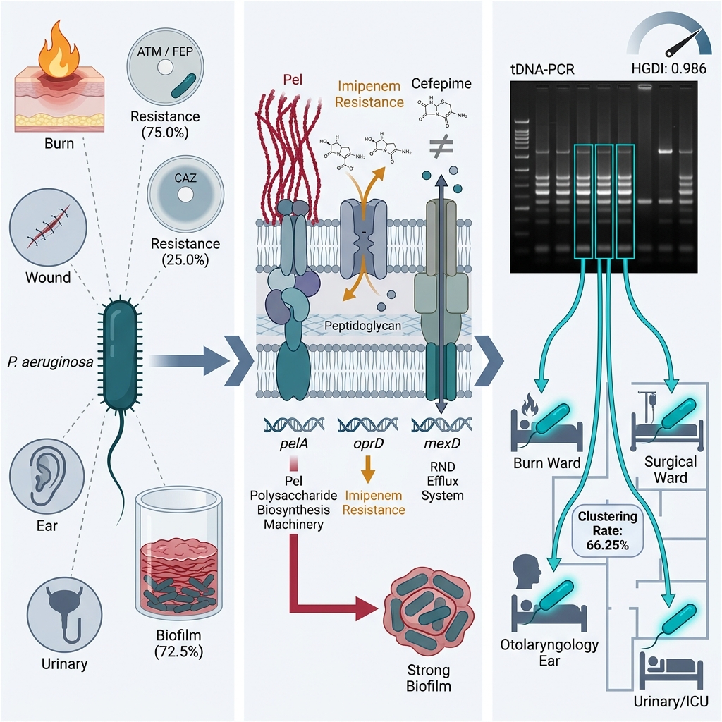
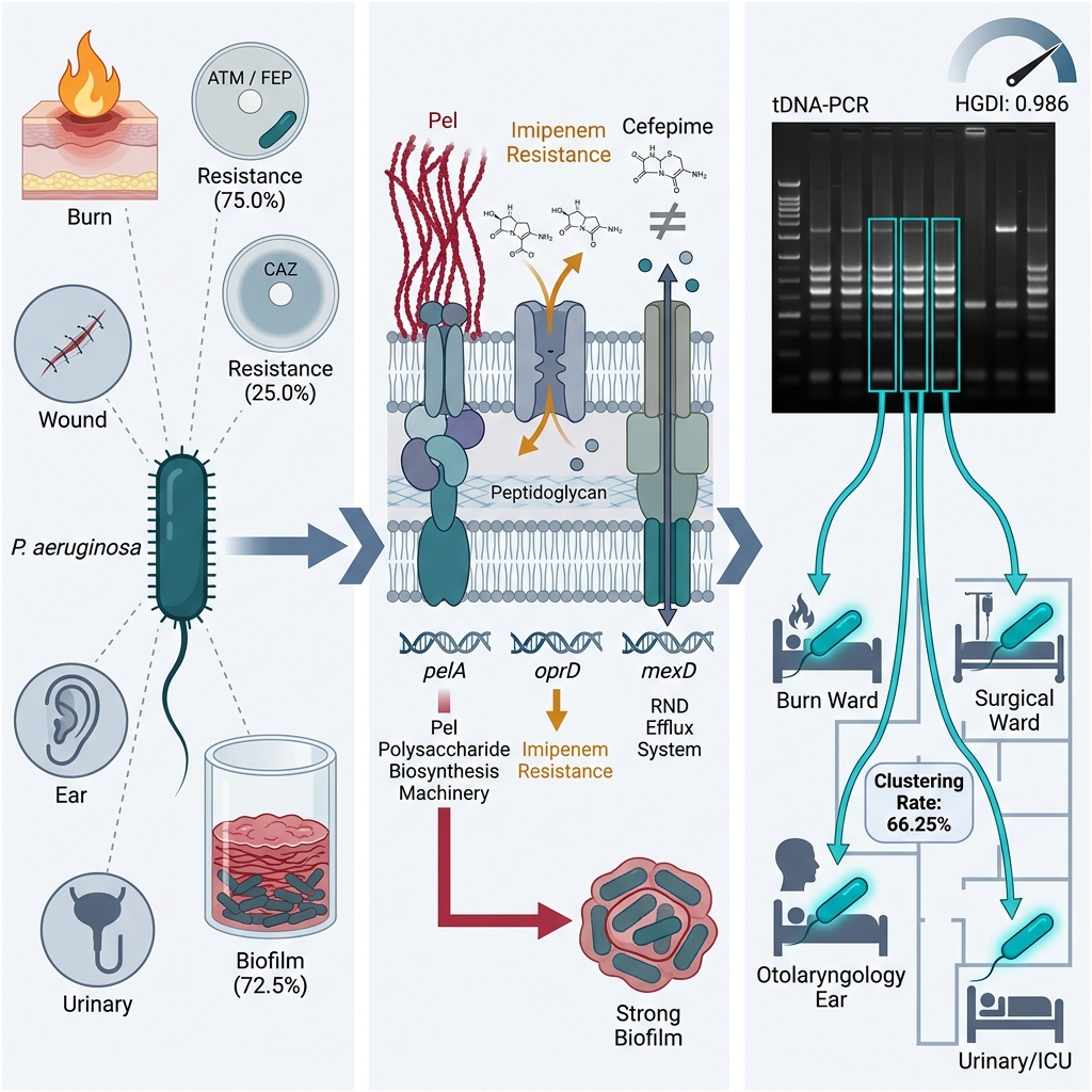
Results: P. aeruginosa was identified in 30.7% (80/260) of clinical specimens, with the highest recovery frequency observed in burn wound infections (32.5%), followed by ear infections (27.5%), urinary tract infections (22.5%), and wound infections (17.5%). Antimicrobial susceptibility testing showed the highest resistance rates against aztreonam and cefepime (75.0% each), followed by ticarcillin–clavulanate (66.3%), imipenem (50.0%), ciprofloxacin (50.0%), and meropenem (41.3%), whereas ceftazidime exhibited the lowest resistance rate (25.0%). Biofilm formation was demonstrated in 72.5% of isolates (42.5% strong and 30.0% moderate producers). Distribution of biofilm categories across specimen sources was not statistically significant (P = 0.263). PCR analysis detected mexD in 68.7% of isolates, oprD in 42.5%, and pelA in 37.5%; blaNDM-1 was not detected. PCR positivity for pelA was significantly associated with strong biofilm production (P = 0.0035), and oprD amplicon detection was significantly associated with imipenem resistance (P < 0.001). tDNA-PCR fingerprinting resolved 49 distinct genotypes among the 80 isolates (27 unique patterns and 22 shared clusters comprising 53 isolates), yielding an overall clustering rate of 66.25%. Both SDI and HGDI reached 0.986, indicating high genetic heterogeneity.
Conclusion: Clinical P. aeruginosa isolates demonstrated high rates of antimicrobial resistance and biofilm-forming capacity. tDNA-PCR proved to be a practical, cost-effective, and highly discriminatory molecular typing technique for elucidating genetic diversity and detecting clonal dissemination in clinical microbiology laboratories with limited resources.
Graphical abstract
Introduction
The remarkable metabolic adaptability and physiological plasticity of Pseudomonas aeruginosa underpin its persistence and success as a prominent opportunistic pathogen in clinical settings. This bacterium possesses an array of intrinsic and acquired virulence determinants that substantially complicate the management of healthcare-associated infections, including a functional type III secretion system, alginate capsular polysaccharide, type IV pili, and a potent capacity for biofilm development1. According to estimates by the National Institutes of Health (NIH), microbial biofilms contribute to approximately 65% of all acute microbial infections and up to 80% of chronic infectious diseases1,2. Within biofilm architectures, bacterial cells are encased in a self-produced extracellular polymeric matrix that impedes antimicrobial penetration, protects against host immune defenses, and fosters physiological tolerance2.
P. aeruginosa employs multiple multifaceted mechanisms that confer resistance to clinically vital antimicrobial classes. These mechanisms include the inducible production of chromosomally encoded AmpC β-lactamase, an enzyme that hydrolyzes penicillins and cephalosporins3; transcriptional downregulation, mutational disruption, or total loss of the OprD outer membrane porin, which selectively mediates carbapenem entry into the bacterial cell4,5; and the active extrusion of antimicrobial agents via multidrug resistance-nodulation-division (RND) efflux pumps, predominantly MexAB–OprM and MexCD–OprJ (the latter partially encoded by mexD)4,5. Furthermore, mobile genetic elements frequently mediate the acquisition of metallo-β-lactamases (MBLs), such as New Delhi metallo-β-lactamase (NDM), conferring broad resistance against almost all β-lactams, including carbapenems5.
Molecular typing methods are essential for elucidating the transmission dynamics, genetic diversity, and clonal relatedness of clinical pathogens. Reference approaches in molecular epidemiology include multilocus sequence typing (MLST), whole-genome sequencing (WGS), and pulsed-field gel electrophoresis (PFGE)6. While MLST and WGS represent sequence-based modalities and PFGE is a macrorestriction band-based technique, each provides high discriminatory resolution for tracking epidemic lineages6. However, routine clinical implementation of these advanced tools remains severely constrained in many resource-limited hospital laboratories due to high instrumentation costs, specialized bioinformatic requirements, and prolonged turnaround times7.
Transfer RNA intergenic spacer PCR (tDNA-PCR) has emerged as a rapid, accessible, and reproducible PCR-based fingerprinting method8,11. This technique utilizes consensus primers complementary to conserved flanking tRNA gene sequences to amplify the variable spacer regions located between adjacent tRNA loci. Because bacterial lineages exhibit pronounced structural variations in both the length and nucleotide sequence of these intergenic spacers, electrophoretic resolution of the resulting amplicons generates distinct, strain-specific banding patterns8,11. Multiple studies have demonstrated that tDNA-PCR delivers robust discriminatory performance, making it a viable preliminary surveillance tool when sequencing-based technologies are unavailable8. Accordingly, this study aimed to investigate the antimicrobial resistance patterns, biofilm-forming capacity, distribution of key resistance and virulence genes (mexD, oprD, pelA, and bla), and genetic diversity of clinical P. aeruginosa isolates using tDNA-PCR fingerprinting.
Materials and Methods
Study Design and Clinical Setting
A cross-sectional, laboratory-based investigation was conducted from February to October 2025 at Al-Hakeem Teaching Hospital in Najaf Governorate, Iraq. The study was structured and reported in accordance with the Strengthening the Reporting of Observational Studies in Epidemiology (STROBE) guidelines. A total of 260 consecutive, non-duplicate clinical specimens were collected from inpatients and outpatients presenting with symptoms of urinary tract, wound, burn, and ear infections. Only one specimen was collected per eligible patient who visited the hospital during the designated sampling interval. A formal a priori sample size calculation was not conducted, and all presenting patients meeting inclusion criteria were enrolled. Exclusion criteria comprised patients with known chronic pre-existing medical conditions, pregnant women, and individuals undergoing ongoing antimicrobial therapy or systemic chemotherapy at the time of specimen collection.
Specimen Collection and Bacterial Identification
Clinical specimens—including wound swabs, midstream urine samples, burn exudate swabs, and ear discharge swabs—were obtained under strict aseptic precautions and transported immediately to the diagnostic microbiology laboratory. Specimens were inoculated onto nutrient agar, MacConkey agar, and cetrimide agar (selective medium for Pseudomonas), followed by aerobic incubation at 37 °C for 24 hours. Presumptive identification of P. aeruginosa was established based on colony morphology, characteristic pigmentation, grape-like odor, Gram-negative bacillary morphology, and conventional biochemical reactions (including oxidase and catalase positivity). Final species confirmation was performed using the automated VITEK® 2 Compact system (bioMérieux, Marcy-l'Étoile, France) with GN identification cards in accordance with the manufacturer's operational instructions.
Antimicrobial Susceptibility Testing
Antimicrobial susceptibility testing of all confirmed P. aeruginosa isolates was executed on the automated VITEK® 2 Compact system utilizing AST-N222 cards. Susceptibility interpretations were categorized into resistant, intermediate/susceptible-dose dependent (SDD), and susceptible adhering to the Clinical and Laboratory Standards Institute (CLSI) M100 guidelines (35th edition, 2025). The antimicrobial panel evaluated eight agents: meropenem (MEM), imipenem (IPM), aztreonam (ATM), ticarcillin–clavulanate (TIM), piperacillin/tazobactam (TZP), ceftazidime (CAZ), cefepime (FEP), and ciprofloxacin (CIP).
Detection of Biofilm Production
Phenotypic biofilm production was evaluated using the semi-quantitative tube adherence method originally described by Christensen et al.10, with reference strain P. aeruginosa PAO1 (ATCC 15692) serving as the positive control. Briefly, pure bacterial colonies were inoculated into glass culture tubes containing 10 mL of trypticase soy broth (TSB) supplemented with 1% D-glucose and incubated statically at 37 °C for 24 hours. Following incubation, the contents were decanted, and the tubes were rinsed four times with phosphate-buffered saline (PBS, pH 7.2) to eliminate non-adherent, planktonic cells. After air-drying at room temperature, adherent biofilms were fixed and stained with 0.1% (w/v) crystal violet solution for 15 minutes. Excess stain was decanted, and the tubes were washed thoroughly with deionized water and inverted to dry. Biofilm production was scored visually: the presence of an adherent dye film lining both the lower tube walls and bottom was classified as positive (categorized as strong or moderate), whereas the absence of staining or the presence of a faint ring restricted exclusively to the liquid–air interface was classified as negative (weak or non-producer)9,10. Although the quantitative microtiter plate assay represents the reference standard, the semi-quantitative tube adherence method was selected due to its operational simplicity, low cost, and proven utility for screening clinical isolates in resource-limited diagnostic settings9,10.
DNA Extraction and PCR Detection of Resistance and Biofilm Genes
Bacterial genomic DNA was extracted from single overnight colonies cultured on Luria–Bertani (LB) agar using the Geneaid DNA Mini Kit (Geneaid Biotech Ltd., New Taipei City, Taiwan) according to the manufacturer’s protocol. Extracted DNA purity and concentration were verified spectrophotometrically. Conventional uniplex PCR assays were conducted to detect the presence of mexD, oprD, pelA, and bla using 2× Promega PCR Master Mix (Promega Corp., Madison, WI, USA). Specific oligonucleotide primer sequences and their expected amplicon sizes are provided in Supplementary Table S1. Detailed thermal cycling parameters are summarized in Supplementary Table S2. Amplified products were resolved alongside a 100-bp to 3,000-bp molecular weight DNA ladder on a 1.5% (w/v) agarose gel stained with ethidium bromide in 1× TAE buffer at 80 V for 60 minutes. Band visualization and digital image acquisition were carried out using an ultraviolet (UV) transilluminator documentation system.
Molecular Typing by tDNA-PCR
Genotypic fingerprinting was performed using transfer RNA intergenic spacer PCR (tDNA-PCR) with the consensus primer pair T5A (5′-AGT CCG GTG CTC TAA CCA ACT GAC-3′) and T3B (5′-AGG TCG CGG GTT CGA ATC C-3′), which target conserved tRNA gene margins8,11. PCR amplifications were assembled in a final reaction volume of 25 µL containing 50 ng of template genomic DNA, 20 pmol of each primer, 0.16 mM of each dNTP, 1.5 mM MgCl, 1 U of Taq DNA polymerase, and 1× reaction buffer8,11. Amplification was executed under the following thermal cycling parameters: initial denaturation at 94 °C for 2 minutes; 30 cycles of denaturation at 94 °C for 30 seconds, primer annealing at 55 °C for 30 seconds, and extension at 72 °C for 2 minutes; followed by a final extension cycle at 72 °C for 10 minutes. Amplified products were separated by horizontal electrophoresis on a 1.6% (w/v) agarose gel in 0.5× TBE buffer at a field strength of 10 V/cm and stained with ethidium bromide. Banding patterns were visualized under UV transillumination.
Statistical and Epidemiological Analysis
Data entry and statistical analyses were performed using IBM SPSS Statistics version 23.0 (IBM Corp., Armonk, NY, USA). Descriptive variables were summarized as counts and percentages. Categorical associations were examined using Pearson’s Chi-square test (χ²) or Fisher’s exact test when expected cell frequencies were less than 5. A two-tailed P-value < 0.05 was defined as statistically significant. Clonal relatedness from tDNA-PCR profiles was established by cluster analysis using the unweighted pair group method with arithmetic mean (UPGMA). Clustered patterns were defined as two or more isolates displaying identical electrophoretic fingerprints. The clustering rate was computed using the standard formula:
Clustering rate (%) = (N / N) × 100
where N represents the number of clustered isolates and N represents the total number of isolates. The discriminatory capability of tDNA-PCR was quantified using Simpson’s Diversity Index (SDI) and the Hunter–Gaston Discriminatory Index (HGDI). Complete datasets were available for all 80 confirmed P. aeruginosa isolates, with no missing values.
Ethics Approval and Consent to Participate
The study protocol was reviewed and approved by the Institutional Review Board and Ethics Committee of Al-Hakeem Teaching Hospital, Najaf Health Directorate (IRB Approval No. 3668, approved on January 30, 2025). The requirement for individual patient informed consent was formally waived by the ethics committee as clinical specimens were collected during standard diagnostic procedures and all patient-identifying information was fully anonymized prior to analysis.
Results
Prevalence and Clinical Distribution of P. aeruginosa
A total of 260 clinical specimens collected from distinct infection sites were processed. Through conventional microbiological examination, selective culture, biochemical assays, and automated VITEK® 2 identification, P. aeruginosa was isolated from 30.7% (80/260) of the specimens. Stratification of the 80 P. aeruginosa isolates across anatomical infection sources showed that burn wound infections accounted for the largest proportion (32.5%, 26/80), followed by ear discharge infections (27.5%, 22/80), urinary tract infections (22.5%, 18/80), and general wound infections (17.5%, 14/80). A statistically significant association was observed between the clinical specimen source and bacterial category (P. aeruginosa versus non-P. aeruginosa pathogens; χ² = 45.12, df = 3, P < 0.001; Table 1).
Distribution of
| Specimen Source | Other Bacteria n (%) | Total Specimens n (%) | |
|---|---|---|---|
| Wound infection | 14 (17.5) | 83 (46.1) | 97 (37.3) |
| Burn infection | 26 (32.5) | 14 (7.8) | 40 (15.4) |
| Urinary tract infection (UTI) | 18 (22.5) | 62 (34.4) | 80 (30.8) |
| Ear infection | 22 (27.5) | 21 (11.7) | 43 (16.5) |
| Total | 80 (100.0) | 180 (100.0) | 260 (100.0) |
Antimicrobial Susceptibility Profiles
Susceptibility testing against eight antipseudomonal agents revealed marked resistance among the 80 confirmed clinical isolates (Table 2). The highest resistance rates were observed for aztreonam (75.0%, 60/80) and cefepime (75.0%, 60/80), followed by ticarcillin–clavulanate (66.3%, 53/80), imipenem (50.0%, 40/80), and ciprofloxacin (50.0%, 40/80). Intermediate levels of resistance were recorded for meropenem (41.3%, 33/80) and piperacillin/tazobactam (33.8%, 27/80; an additional 10.0% [8/80] were categorized as susceptible-dose dependent [SDD]). Conversely, the lowest resistance rate was documented for ceftazidime (25.0%, 20/80; 75.0% susceptible).
Antimicrobial susceptibility and resistance profiles of clinical
| Antimicrobial Agent | Resistant n (%) | SDD / Intermediate n (%) | Susceptible n (%) |
|---|---|---|---|
| Meropenem (MEM) | 33 (41.3) | 0 (0.0) | 47 (58.8) |
| Imipenem (IPM) | 40 (50.0) | 0 (0.0) | 40 (50.0) |
| Aztreonam (ATM) | 60 (75.0) | 0 (0.0) | 20 (25.0) |
| Ticarcillin–clavulanate (TIM) | 53 (66.3) | 0 (0.0) | 27 (33.8) |
| Piperacillin/tazobactam (TZP) | 27 (33.8) | 8 SDD (10.0) | 45 (56.3) |
| Ceftazidime (CAZ) | 20 (25.0) | 0 (0.0) | 60 (75.0) |
| Cefepime (FEP) | 60 (75.0) | 0 (0.0) | 20 (25.0) |
| Ciprofloxacin (CIP) | 40 (50.0) | 0 (0.0) | 40 (50.0) |
Biofilm Formation Phenotypes
Assessment of biofilm-forming capacity revealed that 72.5% (58/80) of P. aeruginosa isolates were biofilm formers, consisting of 42.5% (34/80) strong and 30.0% (24/80) moderate producers. Only 17.5% (14/80) produced weak biofilms, and 10.0% (8/80) were classified as non-biofilm formers (Table 3). Isolates recovered from ear infections displayed the highest proportion of strong biofilm producers (63.6%, 14/22), followed by wound infections (50.0%, 7/14), burn infections (34.6%, 9/26), and urinary tract infections (22.2%, 4/18). Moderate biofilm production predominated among burn (42.3%, 11/26) and urinary tract isolates (38.9%, 7/18). Cross-tabulation of biofilm categories by anatomical infection source demonstrated no statistically significant difference (χ² = 11.185, df = 9, P = 0.263; Table 3).
Phenotypic biofilm formation capacity of clinical
| Infection Source | Strong n (%) | Moderate n (%) | Weak n (%) | Non-producer n (%) | Total n |
|---|---|---|---|---|---|
| Burn infections | 9 (34.6) | 11 (42.3) | 4 (15.4) | 2 (7.7) | 26 |
| Ear infections | 14 (63.6) | 4 (18.2) | 2 (9.1) | 2 (9.1) | 22 |
| Urinary tract infections (UTIs) | 4 (22.2) | 7 (38.9) | 5 (27.8) | 2 (11.1) | 18 |
| Wound infections | 7 (50.0) | 2 (14.3) | 3 (21.4) | 2 (14.3) | 14 |
| Total | 34 (42.5) | 24 (30.0) | 14 (17.5) | 8 (10.0) | 80 |
Prevalence of Resistance and Biofilm Virulence Genes
PCR screening of target loci demonstrated divergent distribution across the isolates (Table 4). The multidrug efflux pump gene mexD was detected in 68.7% (55/80) of isolates. The carbapenem porin-encoding gene oprD was amplified in 42.5% (34/80) of isolates, whereas the biofilm exopolysaccharide biosynthesis gene pelA was present in 37.5% (30/80). In contrast, the acquired metallo-β-lactamase gene bla was not detected in any isolate (0.0%, 0/80).
Detection frequency of antimicrobial resistance and biofilm-associated virulence genes in clinical
| Gene Target | Encoded Function / Target | Positive (n) | Positive (%) |
|---|---|---|---|
| Outer membrane porin D (carbapenem entry channel) | 34 | 42.5 | |
| MexCD–OprJ multidrug efflux pump component | 55 | 68.7 | |
| Pel exopolysaccharide biosynthesis deacetylase | 30 | 37.5 | |
| New Delhi metallo-β-lactamase 1 (carbapenemase) | 0 | 0.0 |
Genotype–Phenotype Correlation Analysis
Statistical correlation revealed a significant association between PCR detection of the pelA gene and the strong biofilm-forming phenotype (χ² = 8.525, P = 0.0035; Supplementary Table S3). Similarly, a highly significant association was identified between oprD PCR amplicon detection and imipenem resistance (χ² = 46.036, P < 0.001). Conversely, no statistically significant association was observed between mexD PCR positivity and cefepime resistance (χ² = 1.571, P = 0.2101), nor between oprD PCR positivity and meropenem resistance (χ² = 0.201, P = 0.6542; Supplementary Table S3).
Molecular Fingerprinting and Genetic Diversity by tDNA-PCR
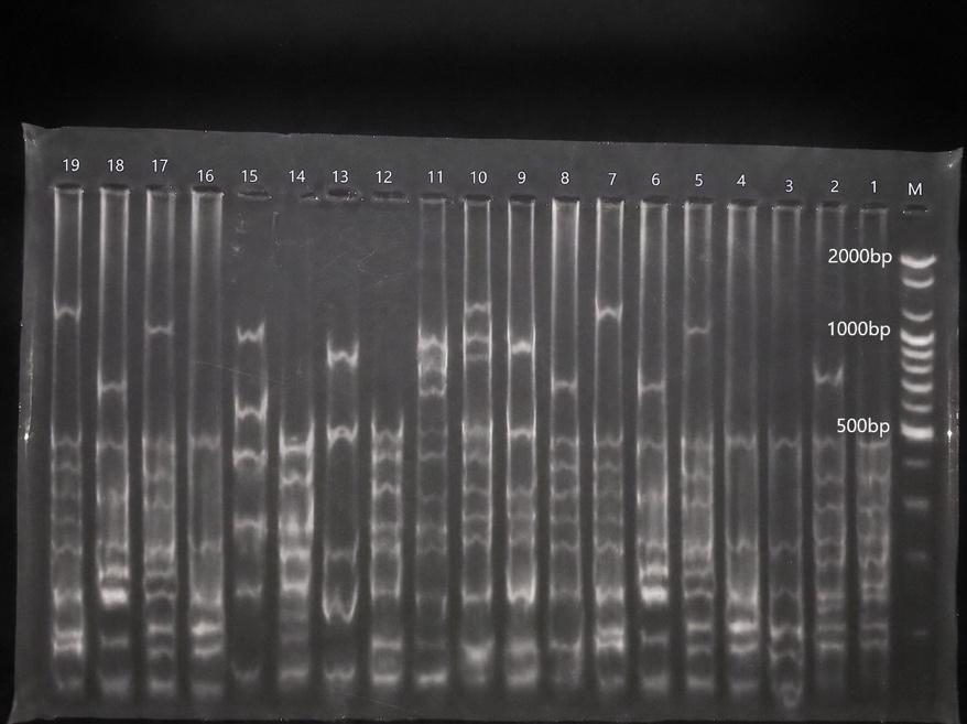
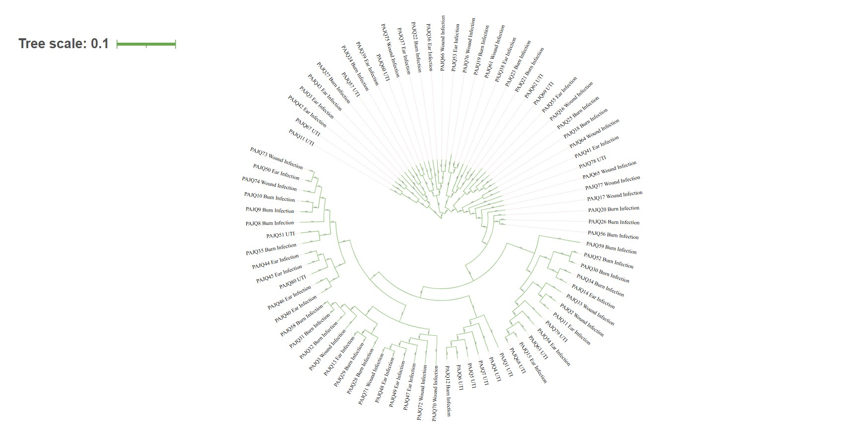
Cluster analysis of tDNA-PCR fingerprints resolved the 80 clinical P. aeruginosa isolates into two major clusters, designated Cluster A and Cluster B, at a 50% pattern similarity threshold. At a 70% similarity cutoff, each major cluster was further subdivided into two subclusters: Subcluster A1 (n = 26 isolates), Subcluster A2 (n = 18 isolates), Subcluster B1 (n = 30 isolates), and Subcluster B2 (n = 6 isolates) (Figure 1, Figure 2). Each subcluster contained isolates originating from disparate anatomical infection sources. Furthermore, multiple isolates obtained from distinct patient infection types exhibited identical tDNA-PCR electrophoretic profiles. In total, the 80 isolates were classified into 49 distinct genotypes, comprising 27 unique profiles and 22 shared profiles encompassing 53 clustered isolates. The overall clustering rate was 66.25% (Table 5). Both Simpson’s Diversity Index (SDI) and the Hunter–Gaston Discriminatory Index (HGDI) yielded an identical value of 0.986, indicating high discriminatory capacity and extensive genetic polymorphism among circulating isolates.
Dendrogram of genetic relatedness and clonal distribution among 80 clinical
Representative agarose gel electrophoresis of tDNA-PCR fingerprint profiles of clinical
Molecular epidemiological typing metrics and discriminatory indices of tDNA-PCR fingerprinting for clinical
| Typing Parameter | Observed Value |
|---|---|
| Total isolates analyzed ( | 80 |
| Distinct genotypes identified | 49 |
| Unique genotypes (single-isolate patterns) | 27 |
| Clustered genotypes (shared patterns) | 22 |
| Total number of clustered isolates ( | 53 |
| Clustering rate (%) | 66.25% |
| Simpson’s Diversity Index (SDI) | 0.986 |
| Hunter–Gaston Discriminatory Index (HGDI) | 0.986 |
Discussion
In this hospital-based investigation, P. aeruginosa accounted for 30.7% of 260 clinical specimens, corroborating its standing as a major opportunistic pathogen in healthcare settings. This recovery rate aligns closely with surveillance studies conducted in Duhok, Iraq, which reported a 29.3% isolation frequency12, and in Baghdad, Iraq, where P. aeruginosa constituted 36.0% of clinical isolates13. In contrast, a lower prevalence (19.3%) was reported from specialized hospital settings in Ethiopia14. The clinical ubiquity and persistence of P. aeruginosa stem from its minimal nutritional requirements, tolerance to physical desiccation, intrinsic antimicrobial resistance, and propensity to colonize medical devices and moist hospital reservoirs15.
Analysis by infection site revealed that burn wounds were the primary clinical source (32.5%), consistent with findings from northern Iran reporting a 36.1% prevalence in burn units16. Severe thermal injuries disrupt the cutaneous epithelial barrier, impair local cellular immunity, and necessitate prolonged hospitalization and extensive therapeutic intervention, thereby establishing an optimal niche for P. aeruginosa colonization and invasive disease. Conversely, studies in Addis Ababa, Ethiopia, documented a lower frequency (12.8%) among burn patients17, reflecting regional differences in infection control protocols and patient populations.
The antimicrobial susceptibility profile revealed pronounced resistance to frontline antipseudomonal β-lactams, particularly aztreonam (75.0%), cefepime (75.0%), and ticarcillin–clavulanate (66.3%), alongside elevated resistance to imipenem (50.0%) and ciprofloxacin (50.0%). These findings mirror high resistance rates reported across the Middle East and North Africa; for instance, an Egyptian surveillance study recorded resistance exceeding 80% to multiple β-lactams and aminoglycosides18. In contrast, Said et al.12 observed that 78.9% of isolates remained imipenem-susceptible, while Asamenew et al.14 reported lower resistance to cefepime (51.0%), ceftazidime (50.0%), imipenem (28.4%), and ciprofloxacin (14.9%). The elevated resistance observed in our center reflects intense selective pressures driven by empirical broad-spectrum antibiotic usage.
A notable observation in this study was the higher resistance rate to cefepime (75.0%) compared to ceftazidime (25.0%). In typical AmpC β-lactamase hyperproducers, cross-resistance between both extended-spectrum cephalosporins is customary. However, preferential resistance to cefepime while retaining ceftazidime susceptibility has been documented previously19,20. This phenotypic discordance is frequently governed by active multidrug efflux mechanisms, specifically the upregulation of MexCD–OprJ or MexXY–OprM efflux systems, combined with mutations in regulatory genes (e.g., nfxB) that selectively diminish intracellular cefepime accumulation without conferring concomitant high-level ceftazidime resistance19,20.
Biofilm formation was confirmed in 72.5% of isolates, with 42.5% displaying a strong biofilm phenotype. These rates are congruent with reports by Farhan et al.21 (80.0% biofilm formers) and Abou Elez et al.18 (100% biofilm formers, 73.1% strong). Biofilm development serves as a formidable survival adaptation that thwarts antimicrobial efficacy by creating a physical barrier to antibiotic diffusion, establishing oxygen- and nutrient-depleted microenvironments that promote metabolic dormancy, and shielding bacteria from host complement and phagocytic clearance22.
Molecular analysis identified the mexD efflux gene in 68.7% of isolates. Overexpression of the MexCD–OprJ multidrug efflux pump contributes significantly to elevated minimum inhibitory concentrations (MICs) against fluoroquinolones, zwitterionic cephalosporins (cefepime), and macrolides, often acting synergistically with target mutations or enzymatic degradation23. The porin gene oprD was detected in 42.5% of isolates, while pelA was detected in 37.5%. We identified a statistically significant association between pelA PCR positivity and the strong biofilm phenotype (P = 0.0035), corroborating the structural necessity of the Pel exopolysaccharide in maintaining biofilm matrix integrity and cell–cell cohesion24,25. Nevertheless, the overall prevalence of pelA (37.5%) was lower than the total biofilm-forming rate (72.5%). This discrepancy underscores that biofilm biogenesis in P. aeruginosa is a multifactorial process coordinated by alternative redundant pathways, including the Psl polysaccharide operon, alginate biosynthesis machinery, type IV pili, flagella, and bis-(3′-5′)-cyclic dimeric guanosine monophosphate (c-di-GMP) signaling circuits26. Consequently, biofilm development cannot be ascribed solely to a single genetic locus.
The metallo-β-lactamase gene bla was absent across all 80 isolates. This finding indicates that carbapenem resistance in this cohort was mediated by non-NDM mechanisms. Resistance to carbapenems in P. aeruginosa frequently arises from mutational inactivation of OprD, stable derepression of chromosomal AmpC, or overexpression of MexAB–OprM, as well as the potential presence of other transferable carbapenemase genes such as bla, bla, or bla variants26,27. Interestingly, oprD PCR amplicons were significantly more frequent among imipenem-resistant isolates (P < 0.001). This phenomenon is consistent with published evidence demonstrating that positive PCR amplification of an oprD fragment does not guarantee the synthesis of an intact, functional porin channel28,29. Previous molecular investigations have demonstrated that PCR-positive oprD alleles in carbapenem-resistant isolates frequently harbor disruptive point mutations, frame-shifts, premature stop codons, or insertion elements that truncate or abrogate OprD protein expression28,29. Similarly, the association between mexD detection and cefepime resistance was not statistically significant (P = 0.2101). Standard PCR demonstrates gene presence rather than transcriptional expression or functional pump assembly; hence, PCR detection alone is insufficient to predict phenotypic resistance30,31.
Genotypic fingerprinting by tDNA-PCR revealed 49 distinct patterns among the 80 isolates, including 27 unique genotypes and 22 clustered genotypes encompassing 53 strains (clustering rate = 66.25%). Both Simpson’s Diversity Index and the Hunter–Gaston Discriminatory Index reached 0.986. These results demonstrate extensive genetic polymorphism among clinical strains while simultaneously confirming the existence of clonal dissemination across the institution. Crucially, identical fingerprint profiles were identified in isolates recovered from different anatomical sources (burns, wounds, urine, ear). This observation points to cross-transmission or the circulation of shared environmental clones across distinct hospital departments. Similar molecular diversity patterns have been documented in Brazil11 (eight genotypes among clinical isolates) and Egypt8 (21 genotypes, seven clusters, Simpson's index 0.921). The higher diversity index observed here (0.986) underscores the polyclonal composition of clinical P. aeruginosa in this healthcare center.
The high propensity for biofilm development identified herein emphasizes the urgent clinical necessity for non-conventional therapeutic modalities to overcome chronic recalcitrant infections. Novel adjunctive strategies under active investigation include bacteriophage therapy, synthetic antimicrobial peptides, and quorum-sensing inhibitors32,33. Several recent studies have shown that bacteriophage–antibiotic combinations exert potent synergistic effects, degrading extracellular matrix architecture, reducing biofilm biomass, and clearing established P. aeruginosa infections34,35. Incorporating these biological and molecular strategies into conventional antimicrobial regimens represents a promising avenue for mitigating persistent infections.
Study Limitations
This study has several limitations that should be acknowledged. First, biofilm production was assessed using the semi-quantitative tube adherence assay rather than the quantitative microtiter plate assay or confocal laser scanning microscopy. Although the tube method is cost-effective and suited for screening, its visual scoring involves potential subjectivity and inter-observer variability. Second, conventional PCR assays only established the presence or absence of target gene regions, without providing nucleotide sequence integrity, mutational analysis, or quantitative transcriptional expression levels (e.g., via RT-qPCR). Third, although tDNA-PCR demonstrated high discriminatory capacity, it offers lower resolution and inter-laboratory standardization than whole-genome sequencing or multilocus sequence typing. Finally, the study was conducted at a single tertiary hospital, which may limit the generalizability of the findings to broader geographical regions.
Conclusion
This study demonstrates a high prevalence of multidrug resistance and biofilm-forming capability among clinical P. aeruginosa isolates recovered from diverse hospital infections. Resistance to aztreonam and cefepime was particularly prominent. Biofilm formation was documented in 72.5% of isolates and was significantly associated with the presence of the pelA gene. The absence of bla, coupled with the detection of mexD and oprD, indicates that chromosomal mechanisms and non-NDM pathways drive carbapenem and β-lactam resistance in this setting. Molecular fingerprinting by tDNA-PCR revealed extensive genetic diversity (HGDI = 0.986) while identifying shared clonal clusters circulating across different clinical services. tDNA-PCR represents a rapid, economical, and discriminative molecular epidemiological tool for tracking clinical P. aeruginosa in resource-constrained environments.
Abbreviations
ATM: Aztreonam; CAZ: Ceftazidime; c-di-GMP: Bis-(3′-5′)-cyclic dimeric guanosine monophosphate; CIP: Ciprofloxacin; CLSI: Clinical and Laboratory Standards Institute; df: Degrees of freedom; DNA: Deoxyribonucleic acid; dNTP: Deoxynucleotide triphosphate; FEP: Cefepime; HGDI: Hunter–Gaston Discriminatory Index; IPM: Imipenem; IRB: Institutional Review Board; LB: Luria–Bertani / Lysogeny broth; MBL: Metallo-β-lactamase; MEM: Meropenem; MIC: Minimum inhibitory concentration; MLST: Multilocus sequence typing; NDM-1: New Delhi metallo-β-lactamase-1; NIH: National Institutes of Health; PBS: Phosphate-buffered saline; PCR: Polymerase chain reaction; PFGE: Pulsed-field gel electrophoresis; RND: Resistance-nodulation-division; SDD: Susceptible-dose dependent; SDI: Simpson’s Diversity Index; SPSS: Statistical Package for the Social Sciences; STROBE: Strengthening the Reporting of Observational Studies in Epidemiology; TAE: Tris-acetate-EDTA; TBE: Tris-borate-EDTA; tDNA-PCR: Transfer RNA intergenic spacer polymerase chain reaction; TIM: Ticarcillin–clavulanate; TSB: Trypticase soy broth; TZP: Piperacillin/tazobactam; UTI: Urinary tract infection; UV: Ultraviolet; WGS: Whole-genome sequencing
Acknowledgments
The authors express their sincere gratitude and appreciation to the administration, medical staff, and laboratory personnel of Al-Hakeem Teaching Hospital (Najaf, Iraq) for their generous assistance, technical support, and facilitation of clinical specimen collection throughout this study.
Author’s Contributions
All authors contributed significantly to the conception, study design, collection of clinical specimens, laboratory investigations, data analysis, and manuscript preparation. All authors critically reviewed, revised, and approved the final manuscript.
Funding
None.
Availability of Data and Materials
The datasets generated and/or analyzed during the current study are available from the corresponding author upon reasonable request.
Ethics Approval and Consent to Participate
The study protocol was approved by the Institutional Review Board and Ethics Committee of Al-Hakeem Teaching Hospital, Najaf Health Directorate, Iraq (IRB Approval No. 3668, approved on January 30, 2025). The requirement for informed consent was waived by the committee because the research involved routine diagnostic clinical specimens and did not disclose any personal patient-identifying information.
Consent for Publication
Not applicable.
Declaration of Generative AI and AI-Assisted Technologies in the Writing Process
During the preparation of this manuscript, the authors used artificial intelligence assistance solely for language editing, grammatical correction, and scientific style refinement to improve the readability and clarity of the paper. The authors thoroughly reviewed and edited the resulting text and take full responsibility for the integrity and accuracy of the content of the published article.
Competing Interests
The authors declare that they have no competing interests.