


Biomolecular Condensates: Molecular Mechanisms, Advanced Technologies, and Translational Therapeutic Strategies for Prion Diseases
- Department of Biological Sciences, Fourah Bay College, University of Sierra Leone, Freetown, Sierra Leone
- Key Laboratory of Luminescence Analysis and Molecular Sensing (Ministry of Education), College of Pharmaceutical Sciences and Chinese Medicine, Southwest University, Chongqing 400715, China
- Department of Biochemistry, College of Science, Evangel University, Akaeze, Ebonyi State, Nigeria
Abstract
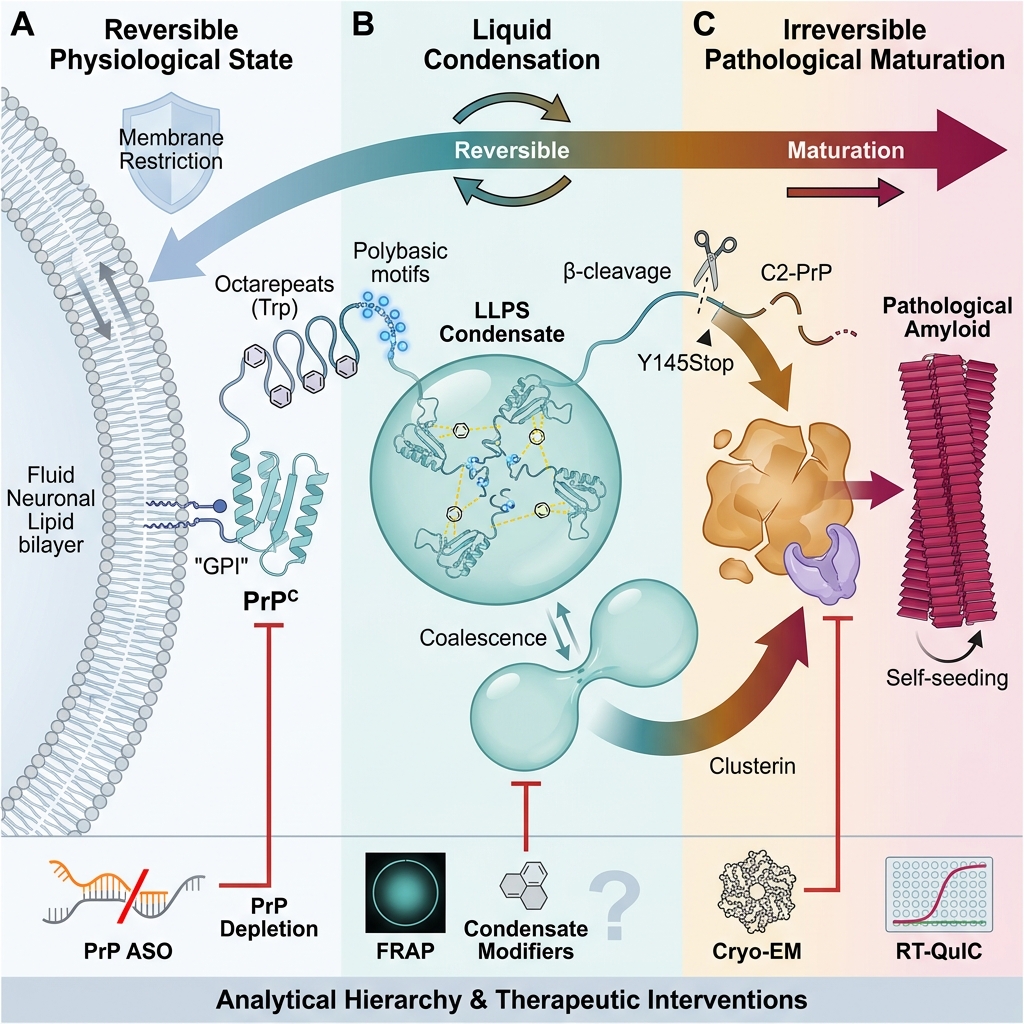
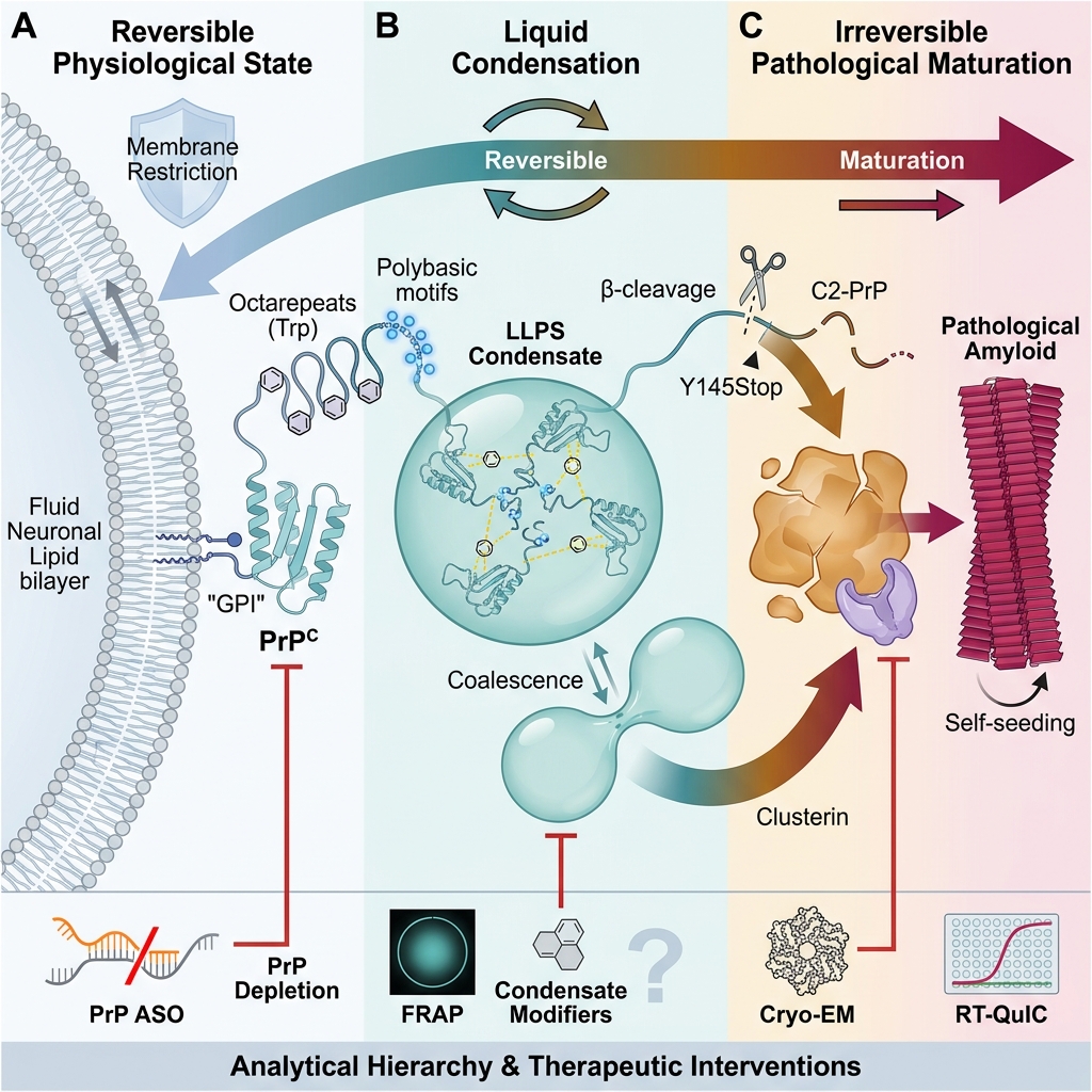
Prion diseases are fatal neurodegenerative disorders driven by conformational conversion of cellular prion protein (PrPC) into self-propagating pathogenic PrP assemblies. No approved therapy has yet demonstrated disease modification. Biomolecular condensation has emerged as a mechanistically relevant framework because recombinant full-length PrP, its N-terminal intrinsically disordered region, and disease-associated truncation variants can undergo liquid-liquid phase separation (LLPS) and, under defined conditions, mature into less dynamic or amyloid-like states. This review critically evaluates evidence linking PrP domain architecture, membrane topology, heterotypic interactions, regulated proteolysis, and condensate ageing with pathological conversion. PubMed/MEDLINE, PubMed Central, Crossref-indexed metadata, and publisher databases were searched through 5 August 2026, and the literature was assessed using SANRA principles. We distinguish direct measurements of PrP phase behavior, including confocal microscopy, droplet fusion, fluorescence recovery after photobleaching, fluorescence correlation spectroscopy, Raman spectroscopy, and atomic force/electron microscopy, from orthogonal measurements of amyloid structure or prion seeding, including cryo-electron microscopy and real-time quaking-induced conversion (RT-QuIC). Therapeutic evidence is classified according to whether interventions directly modify PrP condensates or indirectly reduce PrP substrate abundance or prion propagation. The evidence hierarchy spans chaperone-mediated heterotypic condensation, Clusterin, nucleic-acid ligands, PrP-lowering antisense oligonucleotides, and anti-PrP antibodies. Current support is strongest at the biochemical and preclinical levels; direct evidence that endogenous pathological PrP condensates constitute an obligatory intermediate in the mammalian brain remains lacking. LLPS should therefore be viewed as a testable mechanistic and potentially therapeutic framework rather than a universal pathway for all prion strains.
Graphical abstract
Introduction
Prion diseases are a rare group of neurodegenerative and transmissible disorders that affect both humans and animals1,2. Human prion diseases include sporadic Creutzfeldt-Jakob disease (sCJD), genetic CJD, Gerstmann-Sträussler-Scheinker syndrome, fatal familial insomnia, variant CJD, and kuru. Animal prion diseases include scrapie in sheep and goats, bovine spongiform encephalopathy in cattle, and chronic wasting disease in cervids. Their defining molecular event is the conformational conversion of host-encoded PrP into assemblies that template further conversion and can acquire infectivity.
No disease-modifying treatment is currently approved or curative, despite evaluation of antibodies, small molecules, and gene-silencing strategies. Genetic and animal-model evidence strongly supports PrP lowering, whereas many aggregation-directed compounds have failed because of inadequate central nervous system exposure, toxicity, suboptimal timing, or insufficient target engagement3,4. Symptomatic prion disease often progresses rapidly, reinforcing the need for intervention at an early molecular stage.
Biomolecular condensates are non-stoichiometric assemblies that can concentrate proteins, nucleic acids, and metabolites without a delimiting membrane. They are typically formed through multivalent weak interactions and can exhibit liquid-like fusion, deformation, and molecular exchange5,6. Because condensates can also mature into gels, glasses, or amyloid-enriched solids, phase behavior provides a plausible mechanism linking reversible protein organization with irreversible aggregation7. This framework is particularly relevant to PrP: its N-terminal region is intrinsically disordered and contains polybasic and aromatic motifs, whereas its C-terminal region is glycosylated and tethered to the outer leaflet of the plasma membrane by a glycosylphosphatidylinositol (GPI) anchor8,9.
Notably, evidence does not support the claim that PrP LLPS is the sole or obligatory source of PrP in vivo. Most direct observations derive from recombinant proteins, engineered truncations, or experimental cell-topology models. Accordingly, this review distinguishes established phase behavior from mechanistic inference and from demonstrated prion infectivity. It aims to define the molecular grammar of PrP condensation, characterize the transition from dynamic droplets to amyloid-competent states, map analytical methods to the readouts they actually provide, and evaluate therapeutic strategies within an explicit evidence hierarchy8,9.
Search Strategy and Study Selection
This structured narrative review was prepared in accordance with the quality domains emphasized by the Scale for the Assessment of Narrative Review Articles (SANRA), including justification of importance, statement of aims, description of the search, appropriate referencing, scientific reasoning, and presentation of relevant data10. PubMed/MEDLINE, PubMed Central, Crossref-indexed bibliographic metadata, and publisher databases were searched from January 2000 to 5 August 2026; seminal earlier work on prion biology and immunotherapy was retained when directly relevant. The core Boolean string was: ("prion protein" OR PrP OR PRNP) AND ("liquid-liquid phase separation" OR LLPS OR condensate* OR coacerv* OR "liquid-to-solid" OR "phase transition"). This was combined with method terms (FRAP, fluorescence correlation spectroscopy, Raman, AFM, electron microscopy, cryo-EM, RT-QuIC, PMCA) and intervention terms (chaperone, Clusterin, Ydj1, aptamer, antibody, antisense oligonucleotide, RNA interference, membrane anchor).
Eligible evidence included peer-reviewed primary studies of mammalian PrP phase behavior, aggregation, or prion propagation; mechanistic studies of PrP domains, membrane topology, ligands, or proteolysis; preclinical therapeutic studies; and authoritative reviews. Studies of artificial prion-like proteins were excluded unless they provided a method or conceptual principle explicitly identified as non-PrP evidence. When a peer-reviewed article was available, corresponding preprints were excluded. Each source cited in Tables 1 and 2 was checked against the methods and findings of the original paper; references were removed when the cited paper did not actually evaluate the stated technology, therapeutic category, or outcome. Additional primary studies were identified by reference-list screening and forward citation chaining. Because the literature was heterogeneous and predominantly mechanistic, no meta-analysis was performed; findings were synthesized narratively.
Molecular and Biophysical Principles of Biomolecular Condensates
LLPS, material states, and evidentiary criteria
LLPS occurs when a mixture demixes into a biomolecule-rich dense phase and a dilute phase. The threshold is governed by protein concentration, temperature, ionic strength, pH, molecular crowding, post-translational modification, and the concentration and valence of interacting partners. In sticker-spacer terms, transiently interacting residues or motifs act as stickers, while intervening sequence regions tune accessibility, solvation, and chain dynamics. Electrostatic attraction, cation-pi and pi-pi interactions, hydrophobic contacts, and hydrogen bonding collectively determine whether a dense phase remains fluid or progresses toward a kinetically arrested state5,7.
Operational evidence for a liquid condensate should combine morphology with dynamics; spherical appearance alone is insufficient. Stronger evidence includes concentration dependence, droplet fusion, wetting behavior, reversibility, and rapid fluorescence recovery after photobleaching (FRAP). In contrast, declining FRAP recovery, irregular morphology, increased beta-sheet/Thioflavin-T signal, and fibrillar ultrastructure support maturation toward a gel-like or amyloid-enriched solid. FRAP (mobility), spectroscopy (conformation), microscopy (morphology), and seeding assays (templated amplification) provide complementary rather than interchangeable readouts5,7.
Structural Basis and Pathological Progression of PrP Phase Behavior
Domain architecture and molecular interaction grammar
The human PrP is synthesized as a 253-residue precursor. Residues 1-22 form the N-terminal signal peptide that directs entry into the secretory pathway, while the C-terminal GPI-signal sequence (approximately residues 232-253) is removed during maturation. Mature PrP comprises residues 23-231 and is displayed on the extracellular surface of the plasma membrane. Residues 23-120 are predominantly disordered, whereas residues 121-231 form a globular domain containing three alpha-helices, a short antiparallel beta-sheet, a Cys179-Cys214 disulfide bond, and two N-linked glycosylation sites at Asn181 and Asn1979,11.
The disordered N terminus contains two lysine-rich polybasic clusters (residues 23-31 and approximately 100-110), an octarepeat region enriched in regularly spaced tryptophan residues, and a hydrophobic segment extending toward the folded domain. Deletion and substitution studies showed that the N-terminal region is required and sufficient for PrP LLPS under the tested conditions12. More recent work refined this interaction grammar by showing that conserved polybasic motifs and octarepeat tryptophans promote phase separation, consistent with electrostatic and cation-pi contacts. Importantly, the octarepeat contribution was independent of its histidines and of copper binding in that experimental system13. Thus, the octarepeat should not be described as a copper-dependent LLPS switch.
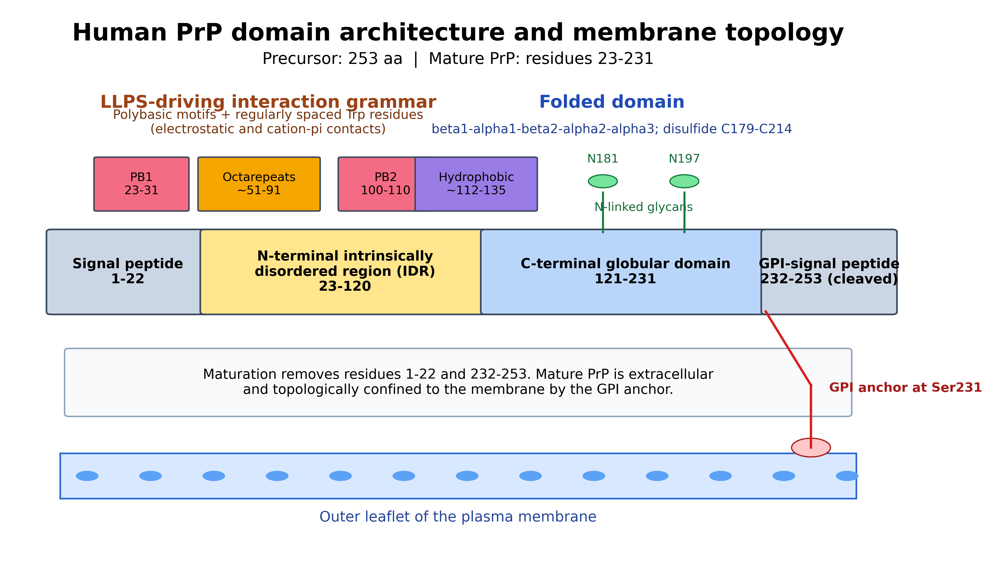
Structural domain layout of human PrP and its extracellular membrane localization. Residue boundaries are approximate for the mature human protein. PB, polybasic motif; GPI, glycosylphosphatidylinositol.
The folded C-terminal domain influences the conformational consequences of condensation. In vitro, LLPS can precede the emergence of protease-resistant, beta-rich conformations in full-length recombinant PrP14. In Y145Stop, loss of most of the globular domain yields a highly charged intrinsically disordered chain that forms dynamic droplets and subsequently self-seeding amyloid fibrils15. These systems show that phase separation can locally concentrate PrP and create an environment conducive to nucleation; they do not, by themselves, establish that the resulting material is equivalent to all infectious PrP strains.
Membrane topology as a suppressor of aberrant phase behavior
Membrane topology provides another layer of regulation. GPI anchoring confines PrP to two-dimensional diffusion at the membrane surface and restricts the orientational and translational freedom available for bulk three-dimensional condensation. Cell-based and biochemical experiments show that this topological confinement stabilizes native PrP and suppresses spontaneous LLPS and seeding-competent aggregation, whereas release from the membrane promotes aggregation16. These findings refine the earlier simplification that the GPI anchor merely prevents cytoplasmic condensation: mature PrP is extracellular, and the relevant control is dimensional confinement within the membrane context rather than direct cytoplasmic sequestration.
From dynamic condensates to arrested and amyloid-rich assemblies
PrP can progress from reversible condensation to arrested assemblies through several routes. First, disease-associated truncation can remove structural elements that restrain aggregation. Y145Stop provides direct evidence that an intrinsically disordered PrP fragment can transition from LLPS to self-seeding amyloid15. Second, regulated beta-cleavage can generate C2-PrP. In a reconstituted system, beta-cleavage produced immobile C2-PrP aggregates only when full-length PrP had undergone LLPS before proteolysis; C2-PrP remained soluble when cleavage occurred in non-phase-separated PrP17. This context dependence indicates that dense-phase concentration or molecular organization can expose latent aggregation pathways.
Third, nucleic acids and heterotypic protein partners can stabilize or destabilize condensates. A high-affinity DNA aptamer altered PrP phase separation and fibrillation in a sequence- and stoichiometry-dependent manner18. Electrostatic complementarity between the positively charged PrP N terminus and the acidic alpha-synuclein C terminus enables heterotypic condensate formation; RNA can reorganize these assemblies into multiphasic condensates and alter amyloid conversion19. Such findings argue against labeling all condensates as uniformly protective or toxic. Outcome depends on material state, composition, stoichiometry, residence time, and the order of molecular events.
Fourth, cellular stressors can alter PrP concentration, redox state, membrane trafficking, proteolysis, and ligand availability. Oxidative stress, pH shifts, metal dyshomeostasis, and proteostasis failure are therefore plausible modifiers of phase boundaries, although their causal role in endogenous neuronal PrP condensation is less directly established than the recombinant-protein evidence8,9. A rigorous model should therefore distinguish three evidence levels: direct measurements of PrP condensates, orthogonal evidence of amyloid or seeded conversion, and inference from broader condensate biology.
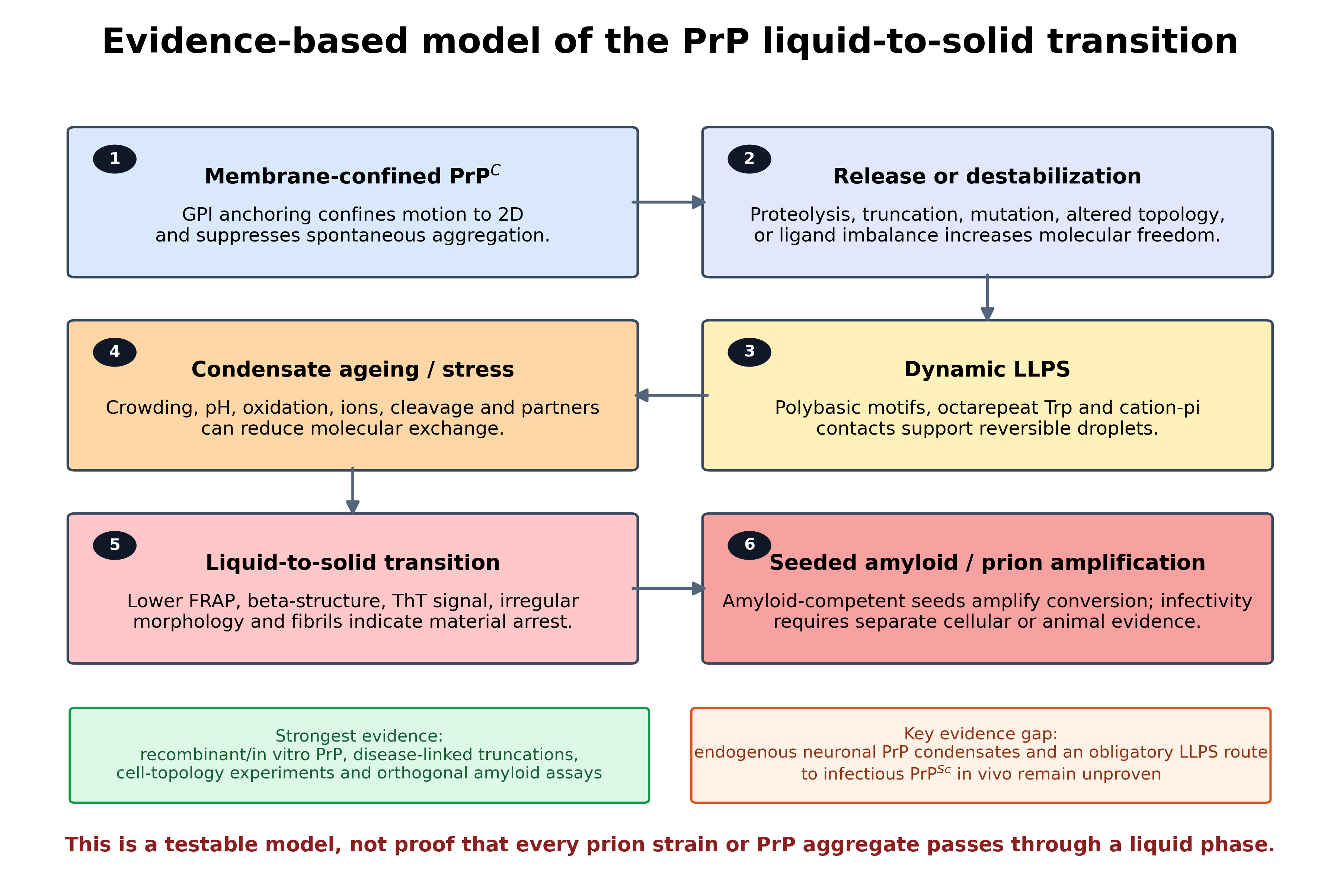
Stepwise biophysical model for the physiological-to-pathological PrP transition. The figure separates direct phase-behavior evidence from the unresolved question of whether endogenous neuronal condensates are an obligatory precursor of infectious PrPSc.
Advanced Techniques for Studying PrP Condensates and Prion Conversion
No single technique simultaneously establishes the liquid-to-solid transition, amyloid structure, and disease-relevant infectivity. Table 1 cross-tabulates each method against the PrP readout that has actually been demonstrated and its principal limitation. This evidence-matched approach avoids attributing methods such as X-ray photon correlation spectroscopy, optogenetic control, proximity labeling, or cryo-electron tomography to studies that did not use those techniques.
Evidence-matched analytical techniques for PrP phase behavior, amyloid structure, and seeded conversion.
| Method | Readout/principle | Verified application to PrP | Principal limitation | Evidence-matched references |
|---|---|---|---|---|
| Confocal microscopy, concentration series, and droplet-fusion analysis | Visualizes condensate formation, coalescence, morphology, and component partitioning. | Used directly for full-length PrP, N-terminal fragments, Y145Stop, PrP/alpha-synuclein mixtures, and chaperone-containing condensates. | Spherical droplets alone do not establish equilibrium LLPS or liquid material properties. | |
| FRAP | Quantifies exchange and mobility inside assemblies; declining recovery supports ageing or arrest. | Directly distinguishes dynamic full-length PrP droplets from immobile C2-PrP aggregates and characterizes Y145Stop/chaperone condensates. | Recovery depends on bleach geometry, binding, diffusion, and photophysics; it is not a direct structural assay. | |
| Fluorescence correlation spectroscopy and time-resolved anisotropy | Measures diffusion, local viscosity, and binding/reorientation dynamics at high sensitivity. | Applied to Y145Stop and Ydj1 heterotypic condensates to resolve altered molecular mobility and interaction networks. | Requires fluorescent labeling and careful model selection; most applications remain in vitro. | |
| Raman/infrared spectroscopy, circular dichroism, and Thioflavin-T kinetics | Reports secondary-structure change and amyloid-associated beta-sheet formation. | Links condensate maturation to conformational conversion in full-length PrP, Y145Stop, and ligand-modulated systems. | Thioflavin-T is not specific for infectivity and may miss noncanonical aggregates. | |
| AFM and transmission electron microscopy | Resolves nanoscale morphology and fibril development after condensate ageing. | Validates emergence of fibrillar assemblies from phase-separated Y145Stop or ligand-modulated PrP. | Usually end-point and ex situ; sample preparation can alter morphology. | |
| Super-resolution or membrane-topology imaging | Tests nanoscale clustering and the effect of two-dimensional membrane confinement. | Supports domain mapping and the conclusion that GPI anchoring suppresses spontaneous PrP aggregation. | Does not by itself establish a thermodynamic phase transition. | |
| Cryo-electron microscopy | Determines near-atomic architecture of mature, brain-derived prion fibrils. | Defines the structural endpoint and strain-specific fibril features. | It does not measure condensate dynamics or prove that a fibril passed through LLPS. | |
| RT-QuIC and related seeded-conversion assays | Measures the capacity of a sample to seed recombinant PrP amyloid amplification. | Used as an orthogonal test showing that Clusterin inhibits seeded aggregate formation from CJD cerebrospinal fluid. | A positive signal reflects seeding activity, not direct observation of LLPS; clinical infectivity is not measured directly. |
Therapeutic Strategies: Direct Condensate Modulation versus Indirect Prion Control
A therapeutically relevant condensate-directed strategy should preserve essential PrP biology, reach the appropriate extracellular or endocytic compartment in the brain, and act before irreversible fibrils predominate. The evidence is therefore most clearly organized by proximity to the proposed mechanism. Direct condensate modifiers have experimentally altered PrP phase behavior, whereas indirect strategies change PrP abundance or prion propagation without demonstrating condensate-specific target engagement (Table 2).
Therapeutic strategies classified by whether the cited evidence directly demonstrates modification of PrP phase behavior.
| Strategy | Mechanistic rationale | Relationship to PrP condensates | Evidence/development stage | Verified references |
|---|---|---|---|---|
| Hsp40/Ydj1-mediated heterotypic condensation | Ydj1 partitions with Y145Stop and reorganizes the dense phase into a dynamic, non-amyloid state. | Direct: multicolor imaging, FRAP/FCS, anisotropy, Raman, AFM. | In vitro mechanistic study; no mammalian efficacy or CNS-delivery data. | |
| Clusterin | Does not prevent full-length PrP LLPS but prevents beta-cleavage-induced C2-PrP aggregation when present before arrest; also inhibits RT-QuIC amplification from CJD CSF. | Direct for PrP phase-transition context; orthogonal seeded-conversion evidence. | In vitro; could not dissolve preformed C2-PrP aggregates. | |
| Nucleic-acid ligands/aptamers | Alter PrP partitioning, droplet material state, and fibrillation in a sequence- and stoichiometry-dependent manner. | Direct in vitro phase-behavior and fibrillation evidence. | Delivery, nuclease stability, off-target binding, and dose-window remain unresolved. | |
| PrP-lowering antisense oligonucleotides | Reduce synthesis of PrPC, the required substrate for prion replication and any PrP condensation pathway. | Indirect with respect to LLPS; strong disease-modifying evidence in prion-infected mice across stages and strains. | CNS preclinical; intrathecal delivery and early treatment are key translational issues. | |
| Anti-PrP monoclonal antibodies | Bind accessible PrP epitopes, inhibit replication, and promote clearance or degradation of pathological species. | Indirect: no condensate-specific mechanism established in the cited studies. | Cell and animal evidence; neurotoxicity, epitope selection, and BBB exposure require careful control. | |
| Membrane-topology stabilization | Maintain GPI-mediated two-dimensional confinement or prevent pathological release of PrP. | Mechanistic target concept supported by topology experiments, not yet a validated drug class. | Target discovery stage; physiological trafficking and cleavage must be preserved. |
Chaperone-mediated control of condensate maturation
The strongest evidence that a pathological phase trajectory can be redirected comes from chaperone studies that do not require elimination of condensation itself. Ydj1 undergoes heterotypic condensation with Y145Stop and prevents amyloid formation, whereas Clusterin acts after full-length PrP has condensed but before C2-PrP becomes irreversibly arrested17,20. These findings define a time-dependent therapeutic window in which an intervention may need to enter condensates, preserve molecular exchange, or shield aggregation-prone surfaces before solidification.
Nucleic-acid ligands
Nucleic-acid ligands are similarly context dependent. The same broad class of polyanions may promote, reshape, or inhibit conversion depending on sequence, structure, concentration ratio, and ionic conditions. Aptamers therefore should not be treated as generic LLPS inhibitors. Their translational development requires quantitative phase diagrams, selectivity against physiological nucleic-acid interactions, stability in serum and cerebrospinal fluid, and evidence of benefit in infected animals9,18.
PrP lowering by antisense oligonucleotides
PrP-lowering ASOs currently provide the strongest in vivo therapeutic evidence among the strategies discussed here. Reducing PrP increased survival and improved disease-relevant endpoints in prion-infected mice across treatment paradigms and prion strains3,4. Their relationship to condensates is mechanistically indirect: lowering PrP should move the system below the concentration required to sustain homotypic condensation while also reducing substrate available for seeded conversion, but the cited animal studies did not quantify LLPS. Maintaining this distinction is essential for citation integrity.
Immunotherapy
Anti-PrP antibodies can inhibit prion propagation or promote degradation in cellular and animal models, although the cited studies did not demonstrate dissolution of PrP condensates22,23. Antibodies could nevertheless complement PrP lowering or direct condensate modifiers when their epitopes remain accessible in early assemblies. Safety requires particular attention because some PrP-targeting antibodies may perturb normal PrP signaling or induce cross-linking, while systemically administered biologics generally achieve limited brain exposure.
Translational Challenges and Validation Framework
Five interconnected challenges shape translation. First, target engagement must be demonstrated in the relevant material state: a compound that dissolves recombinant droplets may not act on membrane-associated, glycosylated, or seeded assemblies in vivo. Second, selectivity is essential because physiological PrP interactions and proteolytic processing cannot be indiscriminately disrupted. Third, cellular compartmentalization and blood-brain barrier exposure differ substantially among small molecules, antibodies, aptamers, and ASOs. Fourth, biomarkers are needed both for presymptomatic detection and for distinguishing reduced PrP abundance from altered condensate dynamics or reduced seeding activity. Fifth, prion strain and PRNP genotype may influence structural routes to aggregation, making a universally effective phase modifier unlikely3,4,16,18,22,23.
These risks can be addressed through a staged validation pipeline. Candidate interventions should first be assessed with orthogonal phase, mobility, and structural assays, including target-engagement studies using full-length glycosylated or membrane-tethered PrP and disease-linked PrP variants. Promising candidates should then be tested in prion-infected cells and brain-derived seeding assays, followed by pharmacokinetic and efficacy studies in at least two prion models. Biomarkers should include PrP concentration, RT-QuIC lag time or seeding dose, markers of neurodegeneration, and, where feasible, imaging or biochemical indicators of assembly state. This sequence helps prevent a favorable condensation phenotype from being mistaken for genuine anti-prion activity.
Future Directions
Priority experiments should establish whether endogenous PrP forms reversible mesoscale assemblies in neurons, where such assemblies occur, and whether they precede pathological PrP accumulation in infected brain. Artefacts caused by overexpression could be reduced through endogenous tagging, single-particle tracking, super-resolution imaging, spatial proteomics, and rapid fixation or cryogenic methods. Phase diagrams should incorporate membrane attachment, native glycans, lipid composition, pH, redox conditions, and physiologically relevant RNA and protein partners. Finally, causal perturbations, not descriptive colocalization alone, should demonstrate that altering condensate material properties changes prion seeding, neurotoxicity, and survival.
Combination strategies should also be considered during therapeutic development. PrP lowering can reduce substrate; an extracellular chaperone or small molecule may stabilize early assemblies; and an antibody may clear residual pathological species. Such combinations will require careful sequencing and pharmacodynamic biomarkers. The objective is not to relabel all aggregates as condensates, but to identify a reversible, measurable state that lies before irreversible conversion and can be therapeutically targeted.
Conclusion
PrP has an established capacity to undergo LLPS, encoded largely by its N-terminal disordered region and regulated by polybasic motifs, octarepeat tryptophans, heterotypic ligands, proteolysis, and membrane topology. Under defined experimental conditions, condensates can mature into immobile or amyloid-rich assemblies, and chaperones can redirect this trajectory. These findings make phase behavior a plausible mechanistic and therapeutic framework for prion disease. However, condensate formation has not been demonstrated to be an obligatory intermediate for every infectious prion in vivo. An evidence-graded approach therefore remains the most rigorous path forward: dynamic and structural assays should define phase state, seeded-conversion and infection models should establish prion relevance, and methods or therapeutic effects should not be attributed to citations that did not test them. In this framework, condensate biology complements rather than replaces established approaches such as PrP lowering and immunotherapy.
Declarations
Conflict of Interest
The authors declare no potential conflicts of interest, financial or otherwise, related to this work.
Ethics Approval
Not applicable.
Author Contributions
JV conceptualized the review. PCA, JV, and SX conducted the literature search, verified the evidence tables, and drafted the manuscript. All authors critically revised and approved the final manuscript.
Funding
The authors declare no external funding for this study.
Availability of Data and Materials
Not applicable. This article is a narrative review of publicly available literature.