


Boosting Thioredoxin-1 Protects Against Acetaminophen-Induced Cardiotoxicity

- Molecular Cell Biology Unit, Division of Biochemistry, Faculty of Science, Tanta University, Tanta 31527, Egypt
- Molecular Carcinogenesis Laboratory, Department of Zoology, Faculty of Science, Tanta University, Tanta 31527, Egypt
- Department of Histology and Cell Biology, Faculty of Medicine, Alexandria University, Alexandria, Egypt
Abstract
Background: Although overdose-induced acute hepatic failure following acetaminophen (APAP) administration is well documented, clinical studies have also reported myocardial infarction, cardiac dysfunction, arrhythmias, pericarditis, and cardiac myocyte necrosis in APAP poisoning. As APAP overdose is associated with excessive reactive oxygen species (ROS) production and metabolism-mediated glutathione depletion, the cytosolic thioredoxin system (Trx-1/TrxR1/NADPH) may offer an alternative mechanism for cardioprotection. Although transgenic mice overexpressing thioredoxin-1 (Trx-1) are protected against doxorubicin/Adriamycin-induced cardiotoxicity, whether pharmacological enhancement of the cytosolic Trx system can mitigate APAP-induced cardiotoxicity and improve the therapeutic efficacy of chlorpromazine (CPZ) remains unclear.
Methods: Cardiotoxicity was induced in wild-type C57BL/6 mice using APAP (400 mg/kg, i.p.). Expression of the Trx system was upregulated via chronic daily administration of low-dose epinephrine (EP, 0.2 mg/kg, s.c. for 14 days), and mice were subsequently treated with a subtoxic dose of CPZ (6 mg/kg, i.p.).
Results: EP significantly upregulated the expression of Trx-1, thioredoxin-like protein 1 (Trx-L1), and thioredoxin reductase 1 (TrxR1) in cardiac tissue. Furthermore, EP attenuated APAP-induced acute cardiac injury, and its combination with CPZ additively amplified these cardioprotective effects. These findings were supported by the normalization of serum cardiac biomarkers (aspartate aminotransferase [AST], lactate dehydrogenase [LDH], and cardiac troponin I [cTnI]), reduction of oxidative stress (restoration of reduced glutathione [GSH], superoxide dismutase [SOD], and nuclear factor erythroid 2-related factor 2 [Nrf2]) and inflammatory markers (tumor necrosis factor-alpha [TNF-α]) in cardiac tissue, and preservation of normal myocardial histological architecture. Mechanistically, activation of the Trx system was accompanied by upregulation of β-arrestin-1 and cAMP response element-binding protein 1 (CREB1), indicating their involvement in Trx-mediated cardioprotection.
Conclusion: Pharmacological enhancement of the cytosolic thioredoxin system protects against APAP-induced acute cardiac injury, and combining EP with CPZ exerts an additive cardioprotective effect.
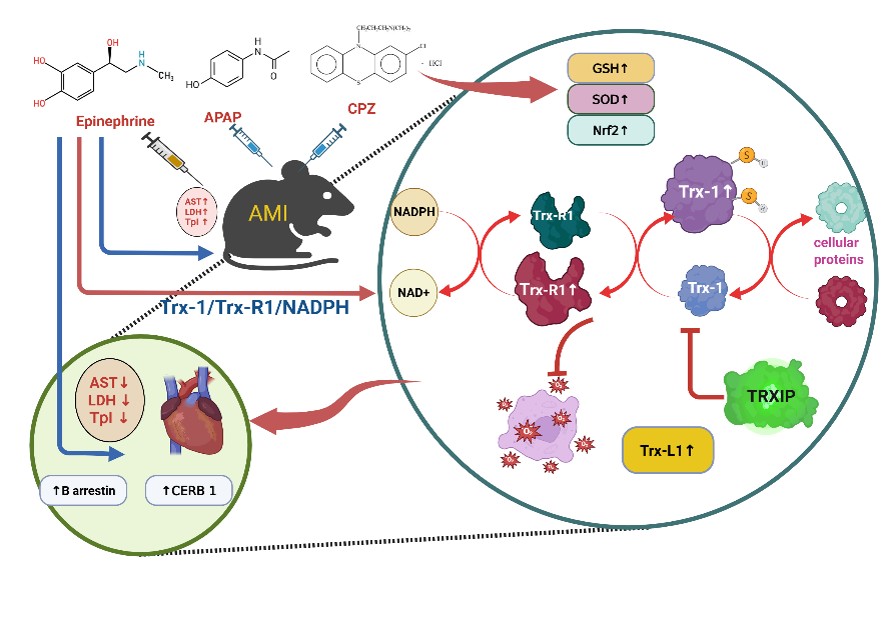
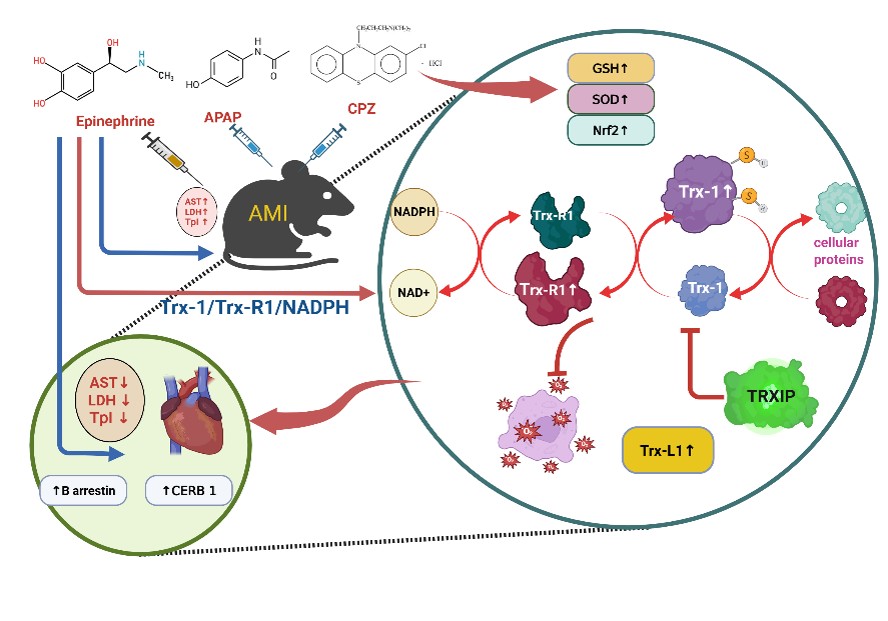
Graphical Abstract
Introduction
Acute liver injury is the primary clinical manifestation of acetaminophen (APAP) overdose; however, cardiac involvement has been increasingly recognized in APAP and other drug-related poisonings 1,2,3. Although relatively uncommon, APAP overdose can lead to myocardial infarction, cardiac dysfunction, arrhythmias, pericarditis, myocyte necrosis, and sudden cardiac death 4. These severe adverse events are primarily attributed to excessive oxidative stress, overproduction of reactive oxygen species (ROS), and mitochondrial dysfunction. Other proposed mechanisms include: (i) depletion of cellular reduced glutathione (GSH) 5 following its conjugation with the toxic APAP metabolite N-acetyl-p-benzoquinone imine (NAPQI) during phase II reactions 6; (ii) cardiac arrhythmias secondary to myocardial ischemia 7; (iii) altered gene expression related to oxidative stress, DNA damage, and apoptosis 8; and (iv) secondary cardiac complications arising from drug-induced liver injury (DILI) 9.
These pathophysiological pathways highlight the critical role of the GSH-independent thioredoxin system (thioredoxin [Trx-1/2]/thioredoxin reductase [TrxR1/2]/NADPH) as an alternative cytosolic and mitochondrial antioxidant defense system. Overexpression of cytosolic Trx-1 has consistently conferred cardioprotection in experimental animal models and cell-based acute cardiac injury models. Early studies demonstrated that transgenic mice overexpressing Trx-1 (Trx-1) were protected against focal ischemic brain damage 10. Similarly, Trx-1 mice exhibited resistance to chronic myocardial infarction (MI) 11 and hindlimb ischemia 12. Furthermore, adenovirus-mediated Trx-1 gene therapy mitigated myocardial damage from ischemia-reperfusion injury 13 and improved outcomes in ischemic myocardium in diabetic rats 3. In a murine model of sepsis-induced cardiomyopathy, Trx-1 overexpression markedly preserved myocardial function by counteracting oxidative stress and inhibiting pro-apoptotic signaling, underscoring its anti-apoptotic and redox-regulatory roles in safeguarding the myocardium against inflammatory and oxidative damage 14,15,16. In vitro studies have also demonstrated that elevated Trx-1 levels in natural killer (NK) cells enhance tolerance to HO-induced oxidative stress 17. By scavenging ROS, Trx-1 prevents the activation of pro-apoptotic cascades, including caspase activation and mitochondrial cytochrome c release 18. Additionally, Trx-1 directly interacts with apoptosis-related proteins to modulate their activity and prevent cell death 19.
Conversely, some studies have reported upregulation of Trx-1 following myocardial infarction 2. These differing findings have been attributed to variations in the hepatic microenvironment where drugs undergo extensive biotransformation, as well as complex interactions between Trx-1 and cellular targets that differentially regulate Trx system activity. Moreover, Trx-1 activity is negatively regulated by thioredoxin-interacting protein (TXNIP) 20,21, an arrestin family member that controls clathrin-mediated endocytosis (CME), membrane receptor trafficking, and downstream cell signaling. Consequently, pharmacological strategies that inhibit CME—such as chlorpromazine (CPZ) treatment 22—or enhance the Trx system 23 hold therapeutic promise for cardioprotection. However, pharmacological and natural product-based strategies to upregulate the Trx system remain insufficiently explored. Given these considerations, this study was designed to investigate whether pharmacological enhancement of the cytosolic thioredoxin system (Trx-1/TrxR1) combined with a subtoxic dose of CPZ confers protection against APAP-induced acute cardiac injury.
Methods
Key chemicals and reagents
Acetaminophen (APAP; CAS No. 103-90-2) was purchased from Sigma-Aldrich (St. Louis, MO, USA). Chlorpromazine (CPZ; Cat. No. C6410) was obtained from ABExBIO (Houston, TX, USA). Epinephrine (EP) was obtained from Misr Company for Pharmaceuticals (Kalubia, Egypt). Enzyme-linked immunosorbent assay (ELISA) kits for Mouse Tumor Necrosis Factor-alpha (TNF-α; Cat. No. CSB-E04741m) and Mouse Nuclear Factor Erythroid 2-Related Factor 2 (Nrf2; Cat. No. CSB-E16188m) were purchased from CUSABIO Technology LLC (Houston, TX, USA). The Mouse Troponin I ELISA Kit (Cat. No. ab285235) was obtained from Abcam (Cambridge, UK). Assessment of reduced glutathione (GSH) and superoxide dismutase (SOD) activities was performed using the GSH Colorimetric Assay Kit (Cat. No. E-BC-K030-M; Elabscience, Bethesda, MD, USA) and SOD Colorimetric Assay Kit (Cat. No. 13485; RayBiotech, Norcross, GA, USA), respectively. Protein concentrations in heart tissue homogenates were determined using the Pierce™ BCA Protein Assay Kit (Cat. No. 23227; Thermo Fisher Scientific, Waltham, MA, USA). Primary antibodies against caspase-3 (E-8; sc-7272), caspase-8 (8CSP03; sc-56070), β-arrestin-1 (25-G10; sc-53780), Trx-1 (sc-13536), and TrxR1 (30F1; BSA-free NBP2-59489) were supplied by Santa Cruz Biotechnology, Inc. (Santa Cruz, CA, USA).
In vivo treatment protocols
Eight- to nine-week-old wild-type male C57BL/6 mice (n = 42) were obtained from Misr University for Science and Technology (Giza, Egypt). All animal handling and experimental procedures complied with the National Institutes of Health (NIH) Guide for the Care and Use of Laboratory Animals and were approved by the Institutional Animal Care and Use Committee (IACUC-SCI-TU-0243) of the Faculty of Science, Tanta University, Egypt. Mice were acclimatized for 1 week under a standard 12 h light/12 h dark cycle with free access to food and water. Animals were randomly divided into seven groups (n = 6 per group; Figure 1):
-
Group I (Control): Received normal saline vehicle.
-
Group II (APAP): Intraperitoneally (i.p.) injected with a single toxic dose of APAP (400 mg/kg) 24,25.
-
Group III (CPZ): Received a single subtoxic dose of CPZ (6 mg/kg, i.p.) 26.
-
Group IV (APAP + CPZ): Received a single dose of APAP (400 mg/kg, i.p.) followed by CPZ (6 mg/kg, i.p.).
-
Group V (EP): Subcutaneously (s.c.) injected daily with a low dose of epinephrine (0.2 mg/kg/day) 27 for 14 consecutive days.
-
Group VI (EP + APAP): Received daily EP (0.2 mg/kg, s.c.) for 14 days prior to a single injection of APAP (400 mg/kg, i.p.).
-
Group VII (EP + APAP + CPZ): Received daily EP (0.2 mg/kg, s.c.) for 14 days, followed by APAP (400 mg/kg, i.p.) and CPZ (6 mg/kg, i.p.).
Figure 1 outlines the complete treatment timeline and order of drug administration. At the end of the experimental period, mice were euthanized, and blood and heart tissue samples were immediately collected for biochemical and histological analyses.
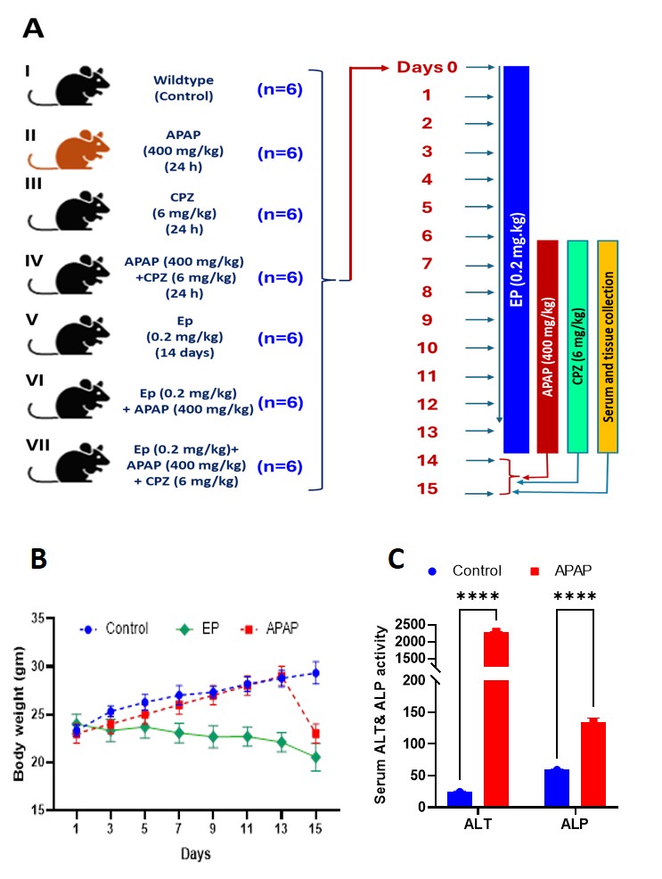
Experimental design, treatment schedule, and serum liver biomarker changes following acetaminophen (APAP) intoxication. Eight- to nine-week-old male C57BL/6 mice were assigned to seven experimental groups (n = 6 per group). Following acclimatization, mice were left untreated (Group I: Control) or injected with a single toxic dose of APAP (400 mg/kg, i.p.; Group II), chlorpromazine (CPZ, 6 mg/kg, i.p.; Group III), or APAP followed by CPZ (Group IV). Mice in Groups V–VII received daily subcutaneous injections of epinephrine (EP, 0.2 mg/kg/day) for 14 consecutive days alone (Group V: EP), prior to APAP injection (Group VI: EP + APAP), or prior to APAP and CPZ injections (Group VII: EP + APAP + CPZ).
Assessment of acute cardiac injury
Acute cardiac injury was evaluated by measuring serum activities of aspartate aminotransferase (AST; AGAPPY, Kerala, India; Cat. No. 11408007) and lactate dehydrogenase (LDH; SPINREACT, S.A., Santa Coloma, Spain; Cat. No. TK41214) according to the manufacturers' instructions.
ELISA-mediated assessment of oxidative stress, inflammatory, and cardiac biomarkers
To minimize non-specific antioxidant effects of residual dietary GSH, mice were fasted overnight prior to sacrifice. Heart tissues were harvested and homogenized in ice-cold phosphate-buffered saline (PBS; pH 7.2, 4 °C) containing 0.05% sodium azide, 0.5% Triton X-100, and a protease inhibitor cocktail. Tissue homogenates were used to quantify cardiac Trx-1, TrxR1, Trx-L1, TXNIP, Nrf2, SOD, GSH, TNF-α, and serum cardiac troponin I (cTnI) levels using commercially available ELISA kits according to the manufacturers' protocols on a microplate reader (Infinite F50 Plus; Tecan Group Ltd., Männedorf, Switzerland).
Gene expression analysis by qRT-PCR
Total RNA was extracted from heart tissues using the Simply GeneDireX Extraction Kit (Cat. No. SN017-0100; GeneDireX, Inc., Taoyuan, Taiwan) according to the manufacturer's instructions. RNA concentration was measured at 260 nm, and purity was confirmed by the A/A ratio. Complementary DNA (cDNA) was synthesized from 2 µg of total RNA using random hexamer primers and the EasyScript® First-Strand cDNA Synthesis Kit (Cat. No. AE301; TransGen Biotech Co., Ltd., Beijing, China). Quantitative real-time PCR (qRT-PCR) was performed on a QuantStudio™ 5 Real-Time PCR System (Thermo Fisher Scientific, Waltham, MA, USA) using SYBR Green Master Mix (2X) and specific primers for Txn1 (Trx-1), Txnrd1 (TrxR1), Txnl1 (Trx-L1), Txnip (TXNIP), and Creb1 (CREB1) (Table 1). Amplification was carried out in a 20 µL reaction mixture comprising 10 pmol of specific primers. Thermal cycling parameters included an initial denaturation step followed by 35 cycles of denaturation at 95 °C for 15 s, annealing at 61–66 °C for 30 s (gene-specific annealing temperatures listed in Table 1), and extension at 72 °C for 30 s. Target gene expression levels were normalized to glyceraldehyde 3-phosphate dehydrogenase (Gapdh) expression using the 2 method.
Immunoblotting analysis
Heart tissues were homogenized in ice-cold homogenization buffer (PBS containing 0.05% sodium azide, 0.5% Triton X-100, and protease inhibitor cocktail, pH 7.2, 4 °C). Protein concentration was quantified using the Bradford assay. Equal amounts of protein (25 µg) were separated by sodium dodecyl sulfate-polyacrylamide gel electrophoresis (SDS-PAGE) and transferred onto polyvinylidene difluoride (PVDF) membranes. Membranes were blocked with 3% bovine serum albumin (BSA) in Tris-buffered saline containing 0.1% Tween-20 (TBST) for 1 h at room temperature, then incubated overnight at 4 °C with primary antibodies against Trx-1, TrxR1, and β-arrestin-1 (1:1000 dilution). After washing with TBST, membranes were incubated with horseradish peroxidase (HRP)-conjugated secondary antibodies for 1 h at room temperature. Protein bands were visualized using an enhanced chemiluminescence (ECL) detection kit (GE Healthcare UK Ltd., Buckinghamshire, UK). Band intensities were quantified by densitometry using ImageJ software (version 1.54k; NIH, Bethesda, MD, USA). Target protein levels were normalized to β-actin (1:3000 dilution; Cat. No. AM4300; Thermo Fisher Scientific).
Histological evaluation by hematoxylin and eosin staining
Heart tissues were fixed in 10% neutral buffered formalin for 24 h and processed for paraffin embedding. Paraffin-embedded tissues were sectioned at a thickness of 6 µm. Sections were deparaffinized in xylene, rehydrated through a graded ethanol series, and stained with hematoxylin and eosin (H&E). Stained slides were dehydrated, cleared in xylene, mounted, and examined blindly under a light microscope (BX41; Olympus Corp., Tokyo, Japan) equipped with a digital camera (DP20; Olympus Corp.) 28.
Statistical analysis
Statistical analyses were performed using GraphPad Prism software (version 9.1.0; GraphPad Software, San Diego, CA, USA). Data are presented as mean ± standard deviation (SD). Differences among experimental groups were evaluated using two-way analysis of variance (ANOVA) followed by Tukey's post-hoc test for multiple comparisons. A value of p < 0.05 was considered statistically significant.
Results
Establishment of APAP-induced acute cardiac injury model
Intraperitoneal administration of a single toxic dose of APAP (400 mg/kg) successfully established acute cardiotoxicity alongside acute liver injury. No mortality occurred during the 14-day study period. At 24 h post-APAP injection, serum hepatic biomarkers were significantly elevated: alanine aminotransferase (ALT) increased from 24.4 ± 1.14 U/L in control mice to 289.8 ± 9.8 U/L in APAP-treated mice (p < 0.0001), while alkaline phosphatase (ALP) increased from 59.2 ± 1.30 U/L to 134.4 ± 6.69 U/L (p < 0.0001) (Figure 1C). Concurrently, cardiac injury biomarkers showed marked elevations: serum AST increased significantly (p < 0.0001), as did LDH (p < 0.0001) and cTnI (p < 0.0001) (Figure 2A–C).
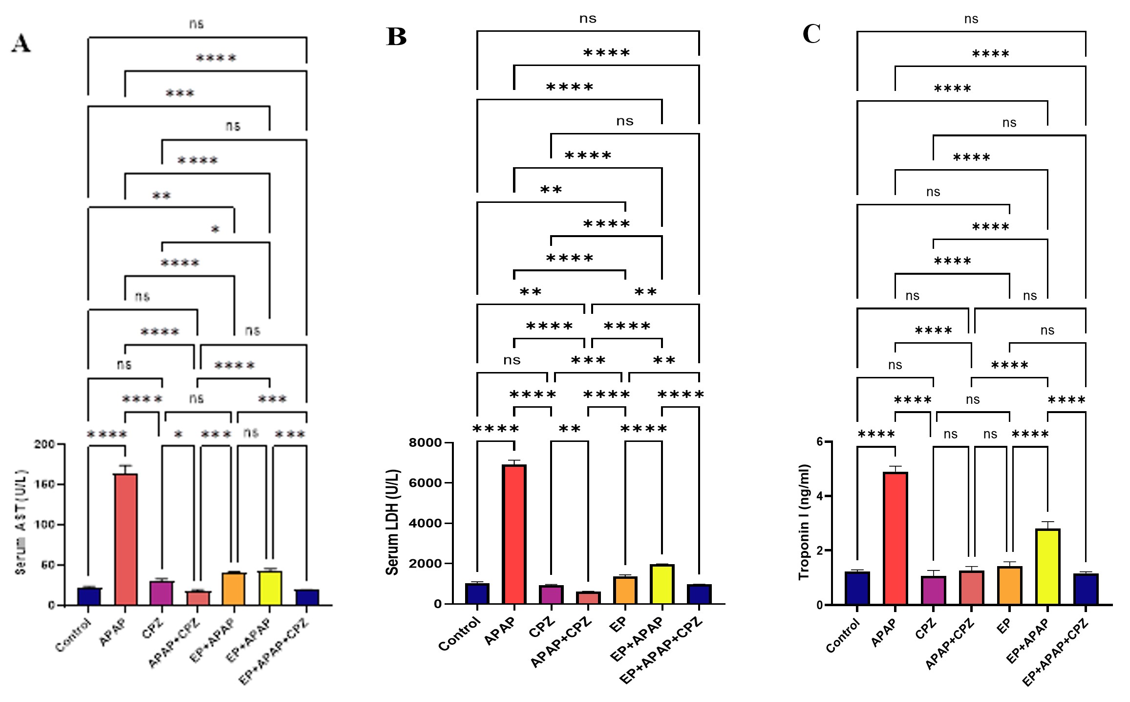
Serum biomarkers of cardiac injury in APAP-intoxicated mice. Intraperitoneal administration of a toxic dose of APAP significantly elevated serum markers of acute myocardial injury: (A) aspartate aminotransferase (AST; p < 0.0001), (B) lactate dehydrogenase (LDH; p < 0.0001), and (C) cardiac troponin I (cTnI; p < 0.0001). Daily pre-treatment with EP and/or subtoxic CPZ significantly attenuated these elevations. Bars represent mean ± SD (n = 3 independent experiments). Data were analyzed by two-way ANOVA followed by Tukey's post-hoc test. *p < 0.05, **p < 0.01, ***p < 0.001, ****p < 0.0001; ns, non-significant.
Furthermore, APAP intoxication severely compromised endogenous antioxidant defenses in heart tissue. Levels of GSH, SOD activity, and Nrf2 protein expression were significantly depleted compared to control values (p < 0.0001 for all; Figure 3A–C). In contrast, cardiac TNF-α levels were significantly increased (p < 0.0001; Figure 3D), indicating pronounced cardiac inflammation. Histological examination of heart tissue from control mice (Group I) revealed normal myocardial architecture (Figure 6A, B). In contrast, heart sections from APAP-treated mice (Group II) exhibited localized intramyocardial hematomas devoid of surrounding endothelium, along with hypereosinophilic, homogeneous sarcoplasm and small pyknotic nuclei in cardiomyocytes (Figure 6C, D).
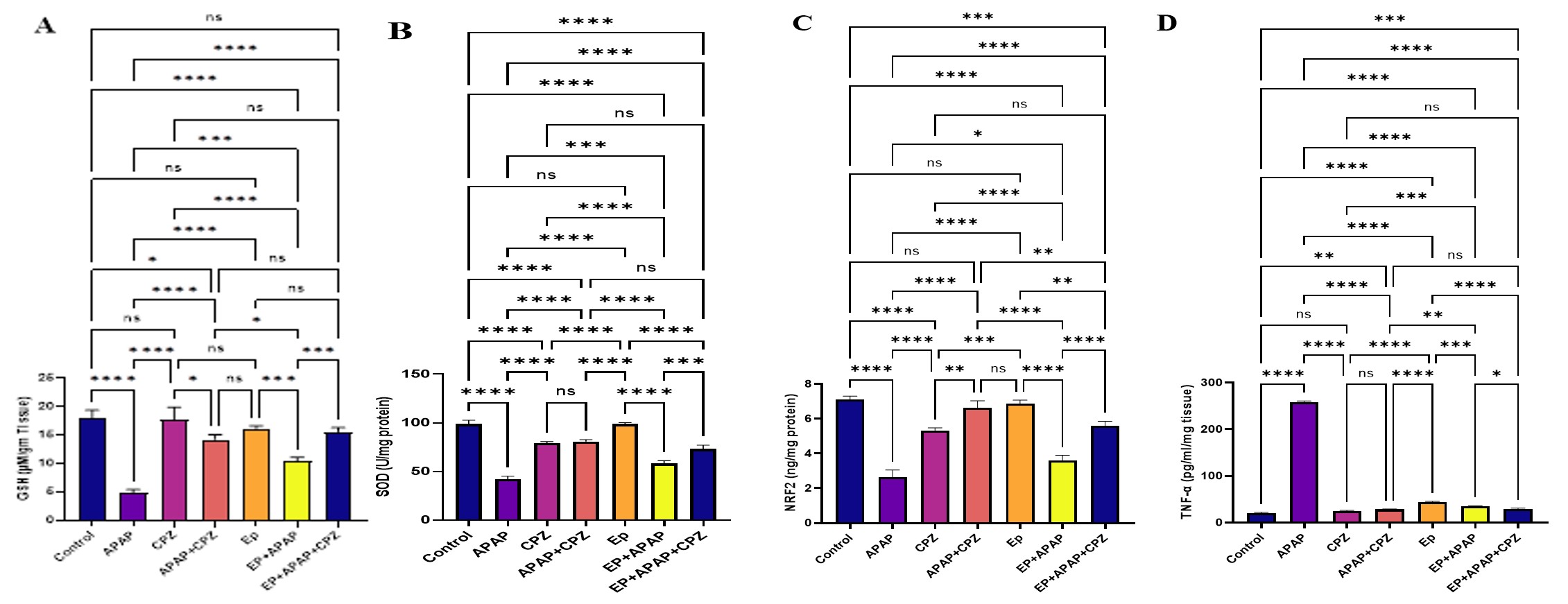
Cardiac antioxidant defenses and inflammatory markers in APAP-intoxicated mice. APAP intoxication caused significant depletion of myocardial antioxidant markers: (A) reduced glutathione (GSH; p < 0.0001), (B) superoxide dismutase activity (SOD; p < 0.0001), and (C) nuclear factor erythroid 2-related factor 2 expression (Nrf2; p < 0.0001), along with marked elevation of the pro-inflammatory cytokine (D) tumor necrosis factor-alpha (TNF-α; p < 0.0001) compared with control mice (Group I). Pre-treatment with EP, alone or combined with CPZ, significantly restored antioxidant levels and suppressed TNF-α. Bars represent mean ± SD (n = 3 independent experiments). Data were analyzed by two-way ANOVA followed by Tukey's post-hoc test. *p < 0.05, **p < 0.01, ***p < 0.001, ****p < 0.0001; ns, non-significant.
Epinephrine administration enhanced cardiac thioredoxin system expression
To upregulate the thioredoxin system, mice were treated daily with EP (0.2 mg/kg, s.c.) for 14 days 27. EP treatment caused no observable behavioral changes or mortality. Serum liver biomarkers remained within normal ranges (data not shown), whereas serum AST and LDH showed slight, non-pathological increases (p < 0.01; Figure 2A, B). Importantly, chronic low-dose EP administration significantly preserved cardiac antioxidant markers (GSH, SOD, Nrf2) and reduced TNF-α levels compared with APAP-treated mice (p < 0.0001; Figure 3A–D). Furthermore, EP alone produced no histological abnormalities in myocardial tissue (Figure 7A, B).
Epinephrine boosted thioredoxin-related protein expression
To confirm that EP upregulates the thioredoxin system in myocardial tissue, mRNA and protein expression of Trx-1, TrxR1, Trx-L1, and TXNIP were measured. qRT-PCR analysis demonstrated that EP treatment induced significant fold-increases in mRNA levels of Trx-1 (p < 0.001), TrxR1 (p < 0.0001), and Trx-L1 (p < 0.0001) compared with controls, whereas TXNIP mRNA expression remained unchanged (Figure 4A, B). ELISA assays (Figure 4C, D) and Western blot analysis (Figure 5A) confirmed significant upregulation of Trx-1 and TrxR1 protein levels relative to control values.
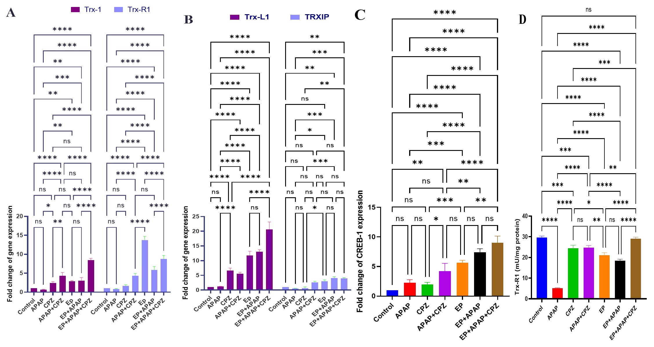
Expression of thioredoxin system components and downstream transcriptional regulators. Fold-changes in mRNA levels of (A)
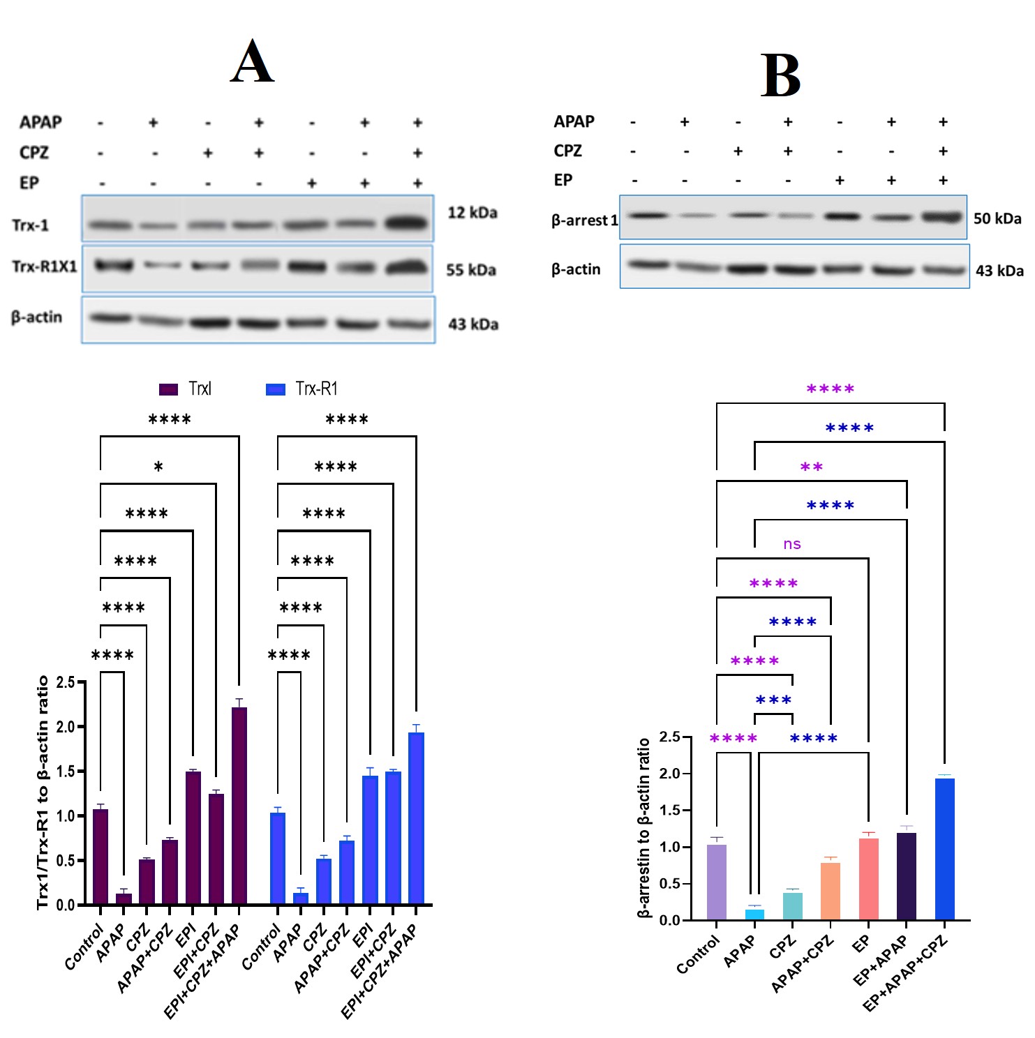
Immunoblotting analysis of thioredoxin system proteins, apoptotic markers, and β-arrestin-1 in cardiac tissue homogenates. Representative Western blot images and densitometric quantifications for (A) Trx-1 and TrxR1, and (B) β-arrestin-1. Densitometric band intensities were normalized to β-actin loading controls. Bars represent mean ± SD (n = 3). Data were analyzed by two-way ANOVA followed by Tukey's post-hoc test. *p < 0.05, **p < 0.01, ***p < 0.001, ****p < 0.0001; ns, non-significant.
Cardioprotective efficacy of EP-enhanced thioredoxin system
We next evaluated whether EP-mediated upregulation of the Trx system protected against APAP cardiotoxicity. Comparison between the APAP group (Group II) and the EP + APAP group (Group VI) revealed that EP pre-treatment significantly attenuated APAP-induced elevations in serum AST (p < 0.0001), LDH (p < 0.0001), and cTnI (p < 0.0001) (Figure 2). Additionally, EP pre-treatment restored myocardial redox status, as evidenced by significant increases in GSH (p < 0.001; Figure 3A), SOD activity (p < 0.001; Figure 3B), and Nrf2 levels (p < 0.05; Figure 3C) compared to APAP alone. EP pre-treatment also suppressed cardiac TNF-α elevation (p < 0.0001; Figure 3D) and markedly reduced myocardial histological lesions (Figure 7C, D).
Additive cardioprotection of thioredoxin enhancement and chlorpromazine
To assess potential synergistic or additive effects between Trx enhancement and CME inhibition, mice were treated with CPZ alone (Group III), APAP + CPZ (Group IV), or EP + APAP + CPZ (Group VII). Single administration of CPZ alone altered neither baseline cardiac biomarkers (AST, LDH, cTnI; p > 0.05; Figure 2) nor tissue histology (Figure 6E, F). In APAP-treated mice, CPZ administration partially restored cardiac antioxidant levels and suppressed inflammation (Figure 3). Remarkably, combined triple treatment (EP + APAP + CPZ; Group VII) exerted an additive cardioprotective effect, completely normalizing serum cardiac injury markers, restoring antioxidant levels, reducing TNF-α, and fully resolving myocardial histological damage (Figure 7E, F). The combined EP + APAP + CPZ regimen provided superior protection compared to either EP or CPZ monotherapy.
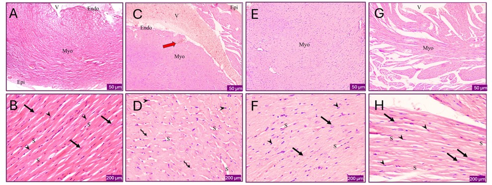
Histological analysis of myocardial tissue in control, APAP-, CPZ-, and APAP + CPZ-treated mice (H&E staining). (A, B) Control group showing normal myocardial architecture: endocardium (Endo), myocardium (Myo), and epicardium (EP). High magnification (B) shows branching, anastomosing cardiac muscle fibers with slit-like spaces (S), eosinophilic sarcoplasm, oval central vesicular nuclei (arrows), and interstitial fibroblasts (arrowheads). V, ventricular lumen. (C, D) APAP-treated group showing intramyocardial hematoma lacking endothelium (red arrow) and cardiomyocytes with hypereosinophilic sarcoplasm and pyknotic nuclei (curved arrows). (E, F) CPZ-treated group showing normal myocardial morphology. (G, H) APAP + CPZ-treated group showing preserved myocardial structure with minimal histological damage. Scale bars: A, C, E, G = 50 µm; B, D, F, H = 200 µm.
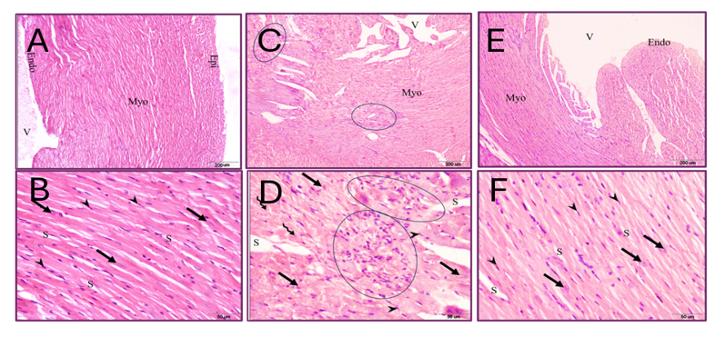
Histological analysis of myocardial tissue in EP-, EP + APAP-, and EP + APAP + CPZ-treated mice (H&E staining). (A, B) EP-treated group (Group V) showing normal myocardial histology. (C, D) EP + APAP-treated group (Group VI) showing focal areas of inflammatory cell infiltration (circles) and scattered cardiomyocytes with pyknotic nuclei (curved arrows) alongside normal cardiomyocytes (straight arrows). (E, F) EP + APAP + CPZ-treated group (Group VII) showing complete restoration of normal myocardial architecture. Endo, endocardium; Myo, myocardium; V, ventricular lumen; S, slit-like spaces; arrows, normal cardiomyocytes; arrowheads, fibroblasts.
Discussion
This study demonstrates that pharmacological enhancement of the cytosolic thioredoxin system protects against APAP-induced acute cardiotoxicity and additively enhances the cardioprotective efficacy of chlorpromazine. Chronic administration of low-dose epinephrine increased the expression of key thioredoxin system components—including Trx-1, Trx-L1, and TrxR1—in cardiac tissue. Upregulation of these redox regulators attenuated APAP-induced myocardial injury and overcame the partial efficacy limitations of CPZ monotherapy. Concomitant administration of low-dose EP and CPZ normalized cardiac biochemical markers, restored myocardial redox balance, suppressed cardiac inflammation, and preserved normal myocardial architecture.
The protective role of the augmented thioredoxin system has been demonstrated across several cardiovascular and metabolic disease models. For instance, transgenic mice overexpressing Trx-1 (Trx-1) exhibit reduced focal ischemic brain damage 10, improved functional recovery following chronic myocardial infarction 11, and enhanced neovascularization during hindlimb ischemia 12. Similarly, activation of TrxR1 decreases ROS production and apoptosis in murine models of neurodegeneration 29. Rather than relying on genetic overexpression or viral gene therapy vectors, our study achieved functional reinforcement of the Trx system through pharmacological administration of low-dose epinephrine. Other pharmacological and natural approaches—such as Korean Red Ginseng extract—have similarly been reported to upregulate TrxR1, protecting endothelial cells 30 and attenuating oxidative stress in pulmonary artery smooth muscle cells 31. Additional physiological regulators of the Trx system include estrogens, prostaglandins, and intracellular cAMP. In our model, daily EP administration induced 2.9-, 13.7-, and 5.5-fold increases in cardiac Trx-1, TrxR1, and Trx-L1 mRNA levels, respectively. Although the precise molecular mechanisms governing EP-mediated Trx gene induction require further investigation, EP likely acts through α- and β-adrenergic receptor signaling 32. Furthermore, EP has been shown to modulate stress-response and antioxidant gene networks via activation of the AP-1 signaling pathway 33. To minimize toxicities associated with high-dose catecholamines 34, we utilized a low-dose EP regimen (0.2 mg/kg/day) that preserved myocardial redox balance without triggering cardiomyocyte apoptosis or structural damage.
Interestingly, EP-mediated upregulation of the Trx system was accompanied by increased expression of β-arrestin-1 and CREB1 (Figures 4 and 5). β-Arrestin-1 is a key multifunctional adaptor protein that regulates G-protein-coupled receptor (GPCR) desensitization, clathrin-mediated endocytosis, and downstream intracellular kinase cascades 35. CREB1 functions downstream of β-adrenergic receptor activation and cAMP elevation to directly transactivate Txn1 and Txnrd1 promoter regions 36. These findings align with previous reports demonstrating that EP suppresses cellular invasion via cAMP/CREB pathway modulation 37. Additionally, EP may influence Trx system activity through inducible nitric oxide synthase (iNOS) modulation, as NO signaling can suppress TXNIP expression, thereby relieving TXNIP-mediated inhibition of Trx-1 38.
In parallel, EP-induced Trx system activation was associated with marked upregulation of endogenous antioxidant pathways (Nrf2, GSH, and SOD) in heart tissue. APAP toxicity induces severe myocardial oxidative stress through NAPQI formation, mitochondrial damage, and rapid GSH depletion 39,40. In our model, APAP intoxication caused a 3.6-fold decrease in GSH, a 2.3-fold decrease in SOD activity, and a 2.7-fold decrease in Nrf2 expression in myocardial tissue. EP-mediated enhancement of the Trx system successfully counteracted APAP-induced oxidative stress, producing 4-fold, 3-fold, and 2-fold increases in GSH, SOD, and Nrf2 levels, respectively, relative to untreated APAP mice. Furthermore, co-administration of subtoxic CPZ further normalized myocardial redox balance and resolved tissue lesions. Although CPZ is not a standard agent in cardiovascular therapy, its broad pharmacological properties—including antiviral 42, anti-inflammatory 43, and antinecrotic 41,44 activities—have been widely documented. In this study, subtoxic CPZ acted as a complementary antioxidant, anti-inflammatory, and anti-endocytic agent, mitigating APAP-induced myocardial injury alongside EP pre-treatment. The involvement of β-arrestin-1 and CREB1 further accords with CPZ's recognized role as a CME inhibitor 22 and modulator of receptor trafficking, autophagy, and JNK signaling 26.
Conclusion
Chronic administration of low-dose epinephrine upregulates the cytosolic thioredoxin system (Trx-1/TrxR1) in cardiac tissue without inducing cardiotoxicity or structural damage. Enhancing the thioredoxin system significantly attenuates acetaminophen-induced acute cardiotoxicity by reducing oxidative stress, suppressing inflammation, and preserving myocardial histological integrity. Furthermore, combining epinephrine-mediated thioredoxin enhancement with chlorpromazine provides an additive cardioprotective effect.
Study Limitations
This study focused primarily on the cytosolic thioredoxin system (Trx-1/TrxR1) in an acute drug-induced cardiotoxicity model; whether these protective mechanisms extend to chronic cardiac conditions (e.g., ischemic cardiomyopathy, heart failure) remains to be investigated. Furthermore, the potential contributions of mitochondrial thioredoxin isoforms (Trx-2/TrxR2), mitochondrial dynamics, and off-target protein interactions warrant future study. Finally, cardiac evaluation relied on biochemical markers and histopathological analyses; future studies incorporating in vivo hemodynamic and echocardiographic assessments will further validate functional cardiac recovery.
Declarations
Abbreviations
APAP: Acetaminophen; ALP: Alkaline phosphatase; ALT: Alanine aminotransferase; AST: Aspartate aminotransferase; BSA: Bovine serum albumin; cAMP: Cyclic adenosine monophosphate; CME: Clathrin-mediated endocytosis; CPZ: Chlorpromazine; CREB1: cAMP response element-binding protein 1; cTnI: Cardiac troponin I; CYP1A2: Cytochrome P450 1A2; CYP2E1: Cytochrome P450 2E1; DILI: Drug-induced liver injury; ECL: Enhanced chemiluminescence; ELISA: Enzyme-linked immunosorbent assay; EP: Epinephrine; GAPDH: Glyceraldehyde 3-phosphate dehydrogenase; GPCR: G-protein-coupled receptor; GSH: Reduced glutathione; H&E: Hematoxylin and eosin; HRP: Horseradish peroxidase; iNOS: Inducible nitric oxide synthase; JNK: c-Jun N-terminal kinase; LDH: Lactate dehydrogenase; MI: Myocardial infarction; mRNA: Messenger RNA; NAPQI: N-acetyl-p-benzoquinone imine; NIH: National Institutes of Health; NK: Natural killer; Nrf2: Nuclear factor erythroid 2-related factor 2; PBS: Phosphate-buffered saline; PVDF: Polyvinylidene difluoride; qRT-PCR: Quantitative reverse transcription polymerase chain reaction; ROS: Reactive oxygen species; SD: Standard deviation; SDS-PAGE: Sodium dodecyl sulfate-polyacrylamide gel electrophoresis; SOD: Superoxide dismutase; TBST: Tris-buffered saline with Tween-20; TNF-α: Tumor necrosis factor-alpha; Trx-1: Thioredoxin-1; Trx-L1: Thioredoxin-like protein 1; TrxR1: Thioredoxin reductase 1; TXNIP: Thioredoxin-interacting protein.
Acknowledgments
This work was kindly supported by the Science, Technology and Innovation Funding Authority (STDF), Egypt (Grant ID: 21000152; Grantee: Mohamed Hessien). The authors gratefully acknowledge Prof. Nilanjana Maulik for her critical review and valuable suggestions.
Author’s contributions
SK and TD contributed to visualization, methodology, and experimental investigations. ES, MHammady, and MMS performed animal experimentation, biochemical assays, and histological analysis. MHessien was responsible for conceptualization, formal analysis, project administration, funding acquisition, resources, and manuscript writing, review, and editing. All authors read and approved the final manuscript.
Funding
This study was supported by a US-Egypt joint collaboration grant (Grant ID: 21000152) funded by the Science, Technology and Innovation Funding Authority (STDF), Egypt.
Availability of data and materials
The datasets generated and analyzed during the current study are available from the corresponding author upon reasonable request.
Ethics approval and consent to participate
All animal procedures and experimental protocols were approved by the Institutional Animal Care and Use Committee of the Faculty of Science, Tanta University, Egypt (Approval No. IACUC-SCI-TU-0243) and complied with the NIH Guide for the Care and Use of Laboratory Animals. Consent to participate is not applicable as this study did not involve human participants.
Consent for publication
Not applicable.
Declaration of generative AI and AI-assisted technologies in the writing process
None.
Competing interests
The authors declare that they have no competing interests.