


Clinical Features, Mechanisms, and Renal Outcomes of Hantavirus-Associated Kidney Injury: A Narrative Review

- Department of Nephrology & Transplant Immunology, People’s Hospital 115, Ho Chi Minh City, Vietnam
- Department of Infectious Diseases, Faculty of Medicine, Pham Ngoc Thach University of Medicine, Ho Chi Minh City, Vietnam
- Department of Infection B, Hospital for Tropical Diseases, Ho Chi Minh City, Vietnam
- International PhD Program in Medicine, College of Medicine, Taipei Medical University, Taipei City, Taiwan
- Intensive Care Unit, Military Hospital 175, Ho Chi Minh City, Vietnam
- College of Nursing, Taipei Medical University, Taipei City, Taiwan
- Cardiac Surgery Intensive Care Unit, Cho Ray Hospital, Ho Chi Minh City, Vietnam
- Tumor Immunology Analysis Core, College of Medical Science and Technology, Taipei Medical University, Taipei City, Taiwan
Abstract
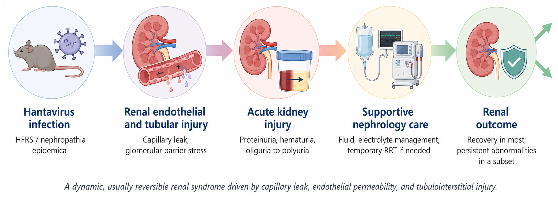
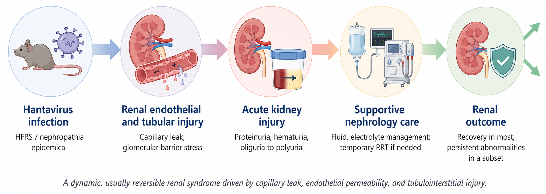
Hantavirus infection is an underrecognized infectious etiology of acute kidney injury (AKI), with hemorrhagic fever with renal syndrome (HFRS) defining its dominant renal phenotype. Kidney involvement spans from transient urinary abnormalities to severe AKI with oliguria, electrolyte and acid-base disturbances, and a requirement for temporary renal replacement therapy in selected cases. Available clinicopathologic and translational evidence supports an integrated model in which tubulointerstitial injury, microvascular inflammation, tubular epithelial stress, glomerular barrier disruption, and hemodynamic vulnerability interact dynamically to produce an acute, albeit typically reversible, form of kidney injury. Clinical management remains supportive but requires structured vigilance: meticulous assessment of volume status, urine output, electrolytes, acid-base balance, blood pressure, bleeding risk, and the evolving phase of illness is essential for optimal nephrology care. Complete functional renal recovery is common—particularly in nephropathia epidemica and other survivable forms of HFRS—yet normalization of serum creatinine does not consistently equate to full physiologic recovery. Persistent proteinuria, hematuria, hypertension, or subtle tubular dysfunction may occur in subsets of patients, although large long-term cohorts indicate that progressive chronic kidney disease is not the typical outcome. A robust clinicopathologic framework integrating urinary sediment findings, AKI severity staging, renal histopathology, phase-oriented supportive care, and risk-stratified post-discharge follow-up can significantly enhance the clinical recognition and management of hantavirus-associated kidney injury in daily practice.
Graphical Abstract
INTRODUCTION
Hantavirus infection occupies a distinctive position in clinical nephrology. Whereas acute kidney injury (AKI) in most viral diseases occurs as a downstream consequence of sepsis, systemic hemodynamic compromise, or nephrotoxin exposure, in hemorrhagic fever with renal syndrome (HFRS)—and in nephropathia epidemica (NE), its milder European expression—kidney injury constitutes a defining, central feature of the pathogenesis 1,2. Patients typically present with abrupt onset of fever, flank or abdominal pain, thrombocytopenia, capillary leak, urinary abnormalities, and rapidly deteriorating kidney function. Globally, hantavirus causes an estimated 150,000–200,000 cases of HFRS annually. China and Korea account for the largest documented burden, driven predominantly by Hantaan and Seoul viruses, whereas Puumala virus is responsible for NE across Northern and Central Europe, and Dobrava-Belgrade virus drives more severe disease in the Balkans 3,4,5.
Viral species, geographical distribution, and host factors collectively dictate clinical severity. Hantaan and Dobrava-Belgrade viruses are associated with severe HFRS; Puumala virus with NE; and Seoul virus with a globally distributed, generally milder renal syndrome 2,4,6. For the consulting nephrologist, the diagnosis of HFRS must be translated into phase-specific risk stratification: oliguria, dialysis necessity, bleeding diathesis, electrolyte derangement, and recovery trajectory (Table 1). The differential diagnosis of febrile AKI accompanied by thrombocytopenia and hematuria is broad, encompassing sepsis-associated AKI, leptospirosis, severe fever with thrombocytopenia syndrome, thrombotic microangiopathy, acute glomerulonephritis, systemic vasculitis, and drug-induced acute interstitial nephritis 1,2. Hantavirus infection should be prioritized when a compatible exposure history is present, proteinuria is disproportionate to the initial serum creatinine elevation, thrombocytopenia occurs in the absence of overt disseminated intravascular coagulation, and the clinical course transitions characteristically from oliguria toward polyuria 7. Hantavirus-associated nephropathy is increasingly recognized in core nephrology curricula as a unique infectious cause of AKI requiring tailored diagnostic and management considerations 8.
Major Hantavirus species associated with renal disease and typical severity
| Virus species | Main geographic distribution | Main clinical syndrome | Typical renal involvement | Mortality pattern |
|---|---|---|---|---|
| Puumala virus (PUUV) | Scandinavia, Russia, Central Europe | Nephropathia epidemica (mild HFRS) | Mild-to-moderate AKI; proteinuria appears early and is common; KDIGO stage 3 reported in approximately 34% of hospitalized patients in the Tampere PUUV cohort | Low case fatality (<1% in most published series) |
| Hantaan virus (HTNV) | China, Korea, Far East Russia | Severe HFRS | Severe AKI with prominent oliguria; high rates of KDIGO stage 3 disease | Higher than PUUV; historical hospitalized estimates approximately 1-5% |
| Dobrava-Belgrade virus (DOBV) | Balkans, Central and Eastern Europe | HFRS (severity varies by viral lineage) | Moderate-to-severe AKI; lineage-dependent (Dobrava lineage > Kurkino lineage) | Intermediate-to-high (Dobrava lineage); Kurkino lineage typically milder |
| Seoul virus (SEOV) | Worldwide via Rattus norvegicus | Mild-to-moderate HFRS | Variable; generally milder than HTNV; AKI may occur | Low case fatality |
| Sin Nombre / Andes virus | Americas (North and South) | Hantavirus cardiopulmonary syndrome (HCPS/HPS) | Renal involvement mild and secondary to cardiopulmonary failure | High case fatality (HCPS; historical estimates approximately 35–40%) |
A foundational nephrology-focused framework was established by Koehler et al. 1. The present narrative review expands upon that work by synthesizing literature published through May 2026, with focused emphasis on integrin receptor tropism, endothelial glycocalyx degradation, interleukin-6 (IL-6) trans-signaling, immune checkpoint dynamics, coagulation monitoring, structured biomarker risk stratification, and long-term renal sequelae. Our primary objective is to provide a comprehensive clinicopathologic model to guide bedside nephrology assessment, renal replacement therapy (RRT) decision-making, and long-term post-discharge follow-up.
Relevant literature was identified through systematic searches of PubMed, Scopus, and Google Scholar utilizing search terms including hantavirus, hemorrhagic fever with renal syndrome, nephropathia epidemica, acute kidney injury, proteinuria, and renal outcomes. Priority was given to clinical cohort studies, renal pathology reports, mechanistic translational studies, and long-term renal outcome evaluations; review articles were cited selectively for context and synthesis. All primary human studies cited were conducted in accordance with the Declaration of Helsinki and received appropriate institutional ethical approvals.
CLINICAL SPECTRUM OF KIDNEY INJURY
The renal presentation in hantavirus infection is often abrupt. In European Puumala virus cohorts, elevated serum creatinine and Kidney Disease: Improving Global Outcomes (KDIGO) stage 2–3 AKI are frequent among hospitalized patients, though mortality and irreversible kidney failure are rare when supportive care is accessible 9,10,11,12. Long-term cohort evaluations reveal a nuanced clinical picture: although most patients achieve complete normalization of serum creatinine, a distinct subset exhibits persistent microscopic hematuria, proteinuria, hypertension, or tubular proteinuria years after the acute illness 13,14,15. Dobrava-Belgrade virus infection induces a significantly more severe AKI phenotype than Puumala virus, characterized by higher rates of anuria and RRT in comparative studies 16,17. In East Asian HFRS caused by Hantaan virus genetic variants, severe AKI develops in approximately one in seven hospitalized cases, accompanied by universal thrombocytopenia and prominent hepatic transaminase elevations that can complicate initial diagnostic triage 18. Seoul virus continues to cause sporadic renal disease via contact with pet or laboratory rats in urban settings, with recent cases from Korea and Germany illustrating the expanding geographic reach of Seoul virus-associated AKI 7,19.
The clinical distinction between Puumala-driven NE and Hantaan-driven HFRS carries direct bedside relevance (Figure 1). European NE cohorts document dialysis requirements ranging from 1% to 15% among hospitalized patients, minimal case fatality (<1%), and near-universal creatinine recovery 9,10,11,12. In contrast, hospitalized patients with Hantaan virus HFRS exhibit a markedly higher risk profile: shock, anuria, severe coagulopathy, and multiorgan failure are substantially more common; RRT is required in approximately 20–40% of severe East Asian cases 4,18; and historical case fatality reached 5–15% in resource-limited settings 4,6. Dobrava-Belgrade virus occupies an intermediate-to-high severity tier in European comparative cohorts, causing more frequent anuria and dialysis dependency than Puumala virus within the same geographic regions 16. These species-specific gradients should directly inform clinical triage: a patient returning from a Hantaan-endemic region in East Asia or the Balkans with febrile AKI and thrombocytopenia has a substantially higher a priori risk of severe oliguria and dialysis than a patient presenting similarly in a Puumala-endemic European setting. Nevertheless, viral species should serve as a prior probability guide rather than a substitute for individualized bedside assessment, given the broad clinical variability within each viral phenotype.
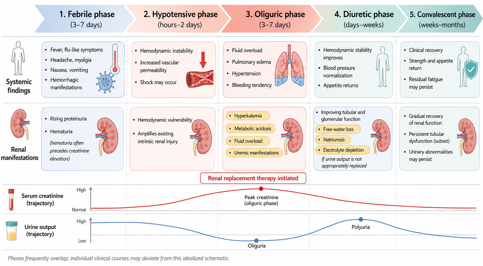
Schematic overview illustrating the five classic sequential clinical phases of hemorrhagic fever with renal syndrome (HFRS)—febrile, hypotensive, oliguric, diuretic, and convalescent phases—along with their approximate clinical durations and hallmark manifestations. Upper panels: Systemic clinical features across phases, progressing from early constitutional symptoms and headache (febrile phase) to hemodynamic instability and shock (hypotensive phase), fluid overload and pulmonary edema (oliguric phase), hemodynamic stabilization (diuretic phase), and eventual functional convalescence. Middle panels: Corresponding renal manifestations across phases, demonstrating that heavy proteinuria and microscopic hematuria often precede marked serum creatinine elevation. The oliguric phase carries the highest clinical risk, characterized by hyperkalemia, metabolic acidosis, fluid overload, and uremic symptoms; renal replacement therapy (RRT) is most frequently initiated during this window (red highlight). The diuretic phase brings functional tubular recovery but introduces significant risks of brisk natriuresis, solute wasting, and dehydration if urinary losses are not adequately replaced. Lower panels: Idealized serial clinical trajectories for serum creatinine (peaking during the oliguric-to-diuretic transition) and urine output (reaching a trough during oliguria and peaking during polyuria). Clinical phases frequently overlap, and individual patient trajectories may deviate from this idealized timeline. Abbreviations: AKI, acute kidney injury; HFRS, hemorrhagic fever with renal syndrome; RRT, renal replacement therapy.
Serum creatinine elevation alone inadequately captures the full spectrum of the syndrome. Staging of AKI must be integrated with serial monitoring of urine output, fluid balance, body weight, blood pressure, oxygenation requirements, and urine chemistry. A patient with a stable serum creatinine plateau but escalating pulmonary edema presents greater immediate clinical risk than one with a rising creatinine whose urine output is recovering 20,21.
Proteinuria warrants meticulous clinical appraisal due to its characteristically dramatic yet transient nature. Studies of acute Puumala virus infection describe a "flash-like" albuminuric surge that frequently precedes peak serum creatinine and rapidly resolves as the polyuric phase commences 22,23. Microscopic hematuria and leukocyturia routinely accompany acute disease (Table 2). Identified host risk factors—including active smoking, lymphopenia, dyspnea at presentation, and an elevated neutrophil-to-lymphocyte ratio—identify patients at elevated risk for a complicated clinical course 24,25,26,27.
Renal manifestations of Hantavirus infection and their nephrologic interpretation
| Manifestation | Typical clinical features | Nephrologic interpretation |
|---|---|---|
| Acute kidney injury | Rising creatinine, oliguria, fluid retention; severity ranges from mild to dialysis-requiring; KDIGO stage 3 in roughly one-third of hospitalized PUUV patients | Intrinsic AKI; tubulointerstitial injury with microvascular component, not pure prerenal disease |
| Proteinuria | Often appears early; dipstick ≥2+ may precede peak creatinine; mixed glomerular and tubular components; nephrin and IgG correlate with severity | Marker of renal parenchymal involvement; glomerular proteinuria correlates with AKI severity in PUUV |
| Hematuria | Microscopic hematuria common during acute phase; persists in ~25% at 7–35 months and ~6% at long-term follow-up | Reinforces parenchymal injury; persistence after recovery warrants reassessment |
| Tubular dysfunction | Impaired urinary concentration, polyuria, electrolyte disturbance, raised urinary α1-microglobulin and KIM-1 | Reflects tubular injury; may lag behind creatinine recovery; explains diuretic-phase risks |
| Oliguria-polyuria transition | Oliguria during acute phase, often followed by brisk polyuria during recovery | Dynamic injury–repair pattern; both phases carry distinct risks (overload vs depletion) |
| Glomerular barrier stress | Urinary nephrin elevation, foot-process effacement, increased serum suPAR; in vitro disruption of ZO-1 and VE-cadherin | Self-limiting glomerulopathy; supports barrier dysfunction without classic primary glomerulonephritis |
| Hypertension | May appear or worsen during the acute episode; reported in subsets at follow-up; high prevalence in long-term cohorts | Causality with prior infection uncertain; reflects vascular and volume changes plus baseline risk |
A frequent diagnostic pitfall is misinterpreting heavy acute proteinuria as primary nephrotic syndrome; during active hantavirus infection, proteinuria reflects reversible disruption of the glomerular and tubular filtration barriers 1. Persistent proteinuria following the acute phase, active urine sediment that fails to clear, or a progressive decline in estimated glomerular filtration rate (eGFR) should prompt investigation for superimposed or infection-triggered glomerular pathology 9. The clinical course is best interpreted across defined clinical phases (Figure 1), recognizing that fluid balance requirements change rapidly: an edematous, oliguric patient with severe capillary leak and thrombocytopenia requires fundamentally different management than a volume-depleted patient with uncomplicated prerenal azotemia. Hyponatremia, hyperkalemia, and metabolic acidosis during the oliguric phase frequently give way to solute wasting, hypokalemia, and intravascular depletion during the polyuric recovery phase 1,9,11.
RENAL PATHOLOGY AND MECHANISTIC INTERPRETATION
The predominant histopathologic pattern in hantavirus nephropathy is tubulointerstitial injury accompanied by microvascular inflammation, rather than immune-complex glomerulonephritis. Contemporary renal pathology series describe acute tubular injury/necrosis, marked interstitial edema, medullary hemorrhage, and prominent cortical peritubular capillaritis 28. These structural lesions account for the clinical presentation of sudden AKI, impaired tubular concentration ability, and subsequent polyuria during recovery 15,29. In severe cases, microvascular congestion and endothelial activation exacerbate hypoxic tubular stress beyond what would occur from systemic hemodynamic instability alone 28,30.
Glomerular involvement is increasingly supported by translational research (Figure 2). Pathogenic Old World hantaviruses directly infect human glomerular endothelial cells, tubular epithelial cells, and podocytes, leading to the disassembly of intercellular tight junction and adherens junction proteins, including zonula occludens-1 (ZO-1) and vascular endothelial cadherin (VE-cadherin) 31. Elevated urinary excretion of nephrin, immunoglobulin G (IgG), and other markers of glomerular filtration barrier breakdown are detectable during severe acute disease 32. Furthermore, biopsy-confirmed reports demonstrating podocyte foot-process effacement in patients with massive proteinuria provide ultrastructural confirmation of a reversible viral glomerulopathy 33. Hantaviruses replicate within human mesangial cells without inducing immediate cytolysis, reinforcing the concept that functional filtration barrier failure—rather than direct parenchymal cell death—drives the proteinuric phenotype 34. Glycocalyx shedding, tight-junction disassembly, cytokine exposure, and complement activation collectively impair vascular and podocyte permeability in a self-limiting manner that aligns with the rapid onset and clearance of albuminuria seen clinically 35,36,37,38.
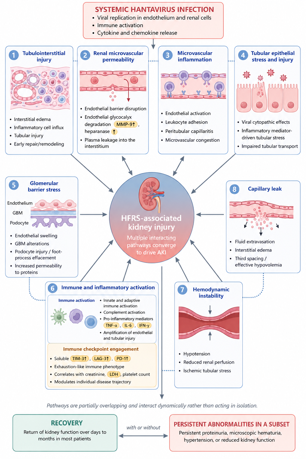
Comprehensive mechanistic diagram detailing the eight major interacting cellular and molecular pathways triggered by systemic hantavirus infection that converge to drive HFRS-associated acute kidney injury (AKI): (1) Tubulointerstitial injury: Interstitial edema, inflammatory cell infiltration, tubular necrosis, and early tissue remodeling. (2) Renal microvascular hyperpermeability: Endothelial barrier disruption, MMP-9 and heparanase degradation of the endothelial glycocalyx, and plasma extravasation. (3) Microvascular inflammation: Microvascular endothelial activation, leukocyte adhesion, cortical peritubular capillaritis, and vascular congestion. (4) Tubular epithelial stress and injury: Viral cytopathy, inflammatory cytokine-driven tubular stress, and impaired solute transport. (5) Glomerular barrier stress: Glomerular endothelial swelling, basement membrane alterations, podocyte foot-process effacement, ZO-1/VE-cadherin disassembly, and heavy proteinuria. (6) Immune and inflammatory activation: Innate/adaptive immune activation, cytokine storm (TNF-α, IL-6, IFN-γ), complement cascade activation (SC5b-9), and immune checkpoint upregulation (soluble TIM-3, LAG-3, PD-1) reflecting T-cell exhaustion dynamics. (7) Hemodynamic instability: Systemic hypotension, reduced renal blood flow, and ischemic tubular injury. (8) Capillary leak syndrome: Widespread vascular leakage, fluid extravasation, third-spacing, and effective intravascular hypovolemia. These pathways interact dynamically rather than in isolation. Following the acute phase, most patients achieve complete functional renal recovery, whereas a subset exhibits persistent proteinuria, microscopic hematuria, hypertension, or subtle tubular dysfunction. Abbreviations: AKI, acute kidney injury; GBM, glomerular basement membrane; HFRS, hemorrhagic fever with renal syndrome; IFN-γ, interferon-gamma; IL-6, interleukin-6; LAG-3, lymphocyte-activation gene 3; LDH, lactate dehydrogenase; MMP-9, matrix metalloproteinase-9; PD-1, programmed cell death protein 1; TIM-3, T-cell immunoglobulin and mucin-domain containing-3; TNF-α, tumor necrosis factor-alpha.
A critical molecular determinant of pathogenic hantaviruses is their selective utilization of β3 integrins for cell entry. Pathogenic Old World and New World strains enter endothelial cells and platelets via αvβ3 and αIIbβ3 integrins, whereas non-pathogenic strains (such as Prospect Hill and Tula viruses) utilize β1 integrins (α5β1) 39. This receptor preference is mechanistically crucial: endothelial β3 integrins modulate vascular permeability downstream of vascular endothelial growth factor (VEGF) signaling. Co-option of β3 integrins by pathogenic hantaviruses hyper-sensitizes endothelial cells to VEGF-mediated capillary leak—an effect absent in cells infected by β1 integrin-binding non-pathogenic strains 40. Pathogenic hantaviruses selectively dysregulate β3 integrin-dependent endothelial barrier function without altering β1 integrin pathways, directly linking viral entry receptor choice to the systemic vascular permeability phenotypes of both HFRS and hantavirus cardiopulmonary syndrome 39,40. Furthermore, because αIIbβ3 is the primary fibrinogen receptor on platelets, this integrin tropism links vascular hyperpermeability directly to the thrombocytopenia and platelet dysfunction characteristic of severe HFRS.
Endothelial pathology extends beyond integrin signaling. Elevated levels of urinary heparanase and matrix metalloproteinase-9 (MMP-9)—key enzymes responsible for endothelial glycocalyx degradation—are measurable in HFRS patients and correlate directly with disease severity 35,36. IL-6 trans-signaling mediates proinflammatory cytokine secretion and endothelial barrier breakdown in infected tissues, independently predicting AKI severity and proteinuria magnitude in clinical cohorts 37,41,42. Complement system activation, evidenced by elevated terminal complement complex (SC5b-9) and reduced C3 levels, correlates strongly with disease severity metrics during acute Puumala virus infection 38. Neutrophil activation triggered by infected microvascular endothelial cells—with elevated plasma myeloperoxidase, elastase, and IL-8—amplifies inflammatory tissue damage without requiring widespread viral cytolysis 30. Furthermore, elevated serum concentrations of soluble immune checkpoint molecules (TIM-3, LAG-3, and PD-1) indicate that an exhaustion-like immune state coexists with systemic inflammation, potentially modulating individual clinical trajectories 43.
Biomarkers of hantavirus nephropathy can be stratified into two clinical tiers based on diagnostic readiness. At the established tier, serum/plasma cystatin C and urinary α1-microglobulin reliably detect tubular and filtration impairment prior to peak serum creatinine, while plasma or urinary IL-6 correlates strongly with AKI severity 41,42,44. At the investigational tier, urinary nephrin 27,32, soluble urokinase plasminogen activator receptor (suPAR) 45, plasma thrombomodulin 46,47, coagulation indices (such as fibrinogen and prothrombin time) 48, and soluble immune checkpoint proteins 43 offer mechanistic insight and cohort-level prognostic value, though external validation is required before routine clinical implementation.
MANAGEMENT FROM A NEPHROLOGY PERSPECTIVE
Management of hantavirus-associated AKI remains primarily supportive, but supportive care must be proactive and highly structured (Table 3). Initial clinical evaluation should document exposure history, day of illness, blood pressure trends, body weight changes, urine output, urinalysis with microscopy, serum creatinine trajectory, serum potassium, bicarbonate, sodium, albumin, platelet count, coagulation profile, and evidence of cardiopulmonary involvement. Standard AKI principles—avoidance of nephrotoxic medications, maintenance of renal perfusion, and timely application of RRT—are paramount. However, HFRS introduces two unique management challenges: systemic capillary leak renders intravascular volume status difficult to assess using single static parameters, and profound thrombocytopenia increases the risk of vascular access procedures 20,21.
Supportive nephrology care in hantavirus-associated acute kidney injury
| Phase / problem | Key monitoring and intervention | Pitfalls to avoid |
|---|---|---|
| Capillary leak / early hypovolemia | Cautious volume resuscitation; serial blood pressure, urine output, lactate; track hemoconcentration and platelets | Persistent aggressive fluids after physiology shifts toward retention |
| Oliguric AKI | Daily creatinine, potassium, bicarbonate, fluid balance; early correction of hyperkalemia and acidosis; consider RRT per KDIGO indications | Forced diuresis as a substitute for assessment; delaying RRT when conventional indications are present |
| Renal replacement therapy | Modality choice driven by hemodynamic tolerance, bleeding risk, and local expertise; standard AKI thresholds apply; favorable outcome despite KDIGO stage 3 | Treating the etiologic diagnosis as an indication or contraindication in itself |
| Diuretic phase | Replace ongoing losses; monitor sodium, potassium, magnesium; avoid abrupt volume contraction | Assuming polyuria equals full recovery |
| Bleeding risk | Platelet count and bleeding tendency considered before vascular access and during anticoagulation | Routine systemic anticoagulation without weighing thrombocytopenia and active bleeding |
| Antiviral / immunomodulatory therapy | Supportive care remains the foundation; ribavirin reduced mortality and oliguria in a Chinese HFRS RCT but not generalizable to all syndromes | Treating case-report agents (e.g., icatibant) as standard therapy |
| Early biomarker risk stratification | Plasma cystatin C and urinary α1-microglobulin rise earlier than creatinine and predict severe AKI; high IL-6 identifies severe NE | Relying solely on creatinine in the first 24-48 h |
| Post-discharge follow-up | Reassess creatinine/eGFR, urinalysis, quantified proteinuria, and blood pressure, especially after dialysis-requiring or persistent abnormalities; apply ADQI acute kidney disease framework | Equating a normalized creatinine with full physiologic recovery |
Early nephrology consultation is most effective prior to crossing the dialysis threshold to determine whether AKI represents an isolated renal insult or part of a systemic capillary leak syndrome, verify that urinary findings align with hantavirus nephropathy, and establish an appropriate monitoring plan 1,2. In healthcare facilities lacking on-site hemodialysis capabilities, patient transfer should be arranged before severe hyperkalemia, refractory pulmonary edema, or progressive coagulopathy render transport hazardous.
A structured monitoring protocol updated at each phase transition should incorporate: urine output recorded hourly in critically ill patients or per shift in stable ward patients; daily serum creatinine, electrolytes (potassium, sodium, bicarbonate, phosphate), and fluid balance; serial blood pressure and body weight measurements; complete blood count and coagulation profile; and serial pulse oximetry accompanied by careful pulmonary examination 20,21. Indications for clinical escalation include KDIGO stage 2 or higher AKI, oliguria (<0.5 mL/kg/h for >6 hours), serum potassium >5.5 mmol/L, serum bicarbonate <18 mmol/L with clinical deterioration, or new pulmonary infiltrates in a fluid-overloaded oliguric patient 20,21.
Fluid administration represents the most delicate bedside decision. Judicious crystalloid resuscitation may be required during the early vascular leak phase to maintain systemic organ perfusion; however, aggressive fluid boluses accelerate pulmonary edema once oliguria and capillary leak predominate 1,2. Dynamic monitoring—including serial oxygenation requirements, chest imaging or point-of-care lung ultrasound, hematocrit trends, and small fluid challenge responsiveness—is substantially more reliable than static volume assessments.
Initiation of RRT should follow standard clinical indications: refractory hyperkalemia, severe metabolic acidosis, medically refractory volume overload/pulmonary edema, uremic complications, or progressive oligoanuria with severe biochemical derangement 20,21. RRT requirement is almost universally temporary among survivors 9,10,12. Coagulopathy and thrombocytopenia necessitate individualized anticoagulation strategies: intermittent hemodialysis without systemic anticoagulation is suitable for hemodynamically stable patients, whereas continuous renal replacement therapy (CRRT) is preferred when shock, severe fluid overload, or high bleeding risk complicates management 1,20,21. Coagulation abnormalities require vigilant parallel monitoring: in a retrospective cohort of 395 HFRS patients, disseminated intravascular coagulation (DIC) was documented in 27.3% of cases, and prolonged prothrombin time, reduced fibrinogen, and elevated serum bilirubin were independent predictors of in-hospital mortality 48. Consequently, coagulation parameters should be routinely integrated into monitoring bundles alongside renal and hematologic tests, particularly in Hantaan or Dobrava-Belgrade virus cases with stage 3 AKI.
Evidence regarding specific antiviral therapy remains limited and species-dependent. A double-blind, placebo-controlled randomized trial of intravenous ribavirin in 242 Chinese patients with Hantaan virus-associated HFRS demonstrated significant reductions in mortality and risk of oliguria when treatment was initiated within seven days of fever onset 49. Conversely, a subsequent randomized trial involving 73 patients with Puumala virus HFRS in Russia showed no significant effect on viral load kinetics, alongside higher rates of adverse events including hemolysis, hyperbilirubinemia, and bradycardia in the ribavirin arm 50. This discrepancy highlights key biological differences: Hantaan virus replicates to significantly higher viremic titers during the early febrile phase than Puumala virus, creating a broader therapeutic window for a nucleoside analogue; differences in patient acuity, sample size, and timing of intervention further preclude direct comparison. Therefore, ribavirin efficacy in Hantaan virus HFRS cannot be extrapolated to Puumala-dominated NE. Currently, no specific antiviral agent is approved for hantavirus treatment by major regulatory authorities 51.
From a pathophysiological standpoint, complement activation in HFRS provides a rationale for complement-targeted therapies (such as eculizumab), familiar to nephrologists from atypical hemolytic uremic syndrome 38; however, clinical trial data in HFRS are lacking. Endothelial barrier-stabilizing agents, including the fibrin-derived peptide FX06 (which preserves VE-cadherin integrity via Fyn kinase-mediated RhoA inhibition), have shown promise in reducing capillary leak in animal models of dengue hemorrhagic fever and endotoxemia 52, but remain unstudied in human HFRS. Bradykinin receptor blockade with icatibant remains speculative based on isolated case reports 53. This review focuses on supportive nephrology management; detailed appraisals of experimental antiviral and immunomodulatory agents are available in specialized infectious disease literature 2,51.
RENAL OUTCOMES AND FOLLOW-UP
The short-term renal prognosis of hantavirus-associated AKI is highly favorable among patients who survive the acute phase. Serum creatinine typically recovers over days to weeks, proteinuria resolves during the polyuric phase, and RRT can almost always be successfully discontinued. The Acute Disease Quality Initiative (ADQI) framework distinguishes acute kidney disease (AKD) from complete recovery, emphasizing the necessity of structured post-episode monitoring 21. In the 556-patient Tampere Puumala virus cohort, KDIGO stage 3 AKI was associated with higher serum creatinine at one month, but no significant difference in median creatinine was observed at one year; after five years, all but one patient had returned to baseline renal function 10.
Long-term outcome studies present a more nuanced perspective. Progression to end-stage kidney disease following a single episode of Puumala virus infection is rare 11,12. However, elevated blood pressure 14, persistent urinary sediment abnormalities 15, subtle tubular protein wasting 17, and circulating endothelial dysfunction markers detectable up to 14 years post-infection have been reported in survivor subsets 54. In a 20-year follow-up cohort of 156 NE survivors, chronic kidney disease (CKD) was identified in 8%, persistent hematuria in 6%, and hypertension in 78%; however, the authors concluded that these rates did not support an increased risk of incident CKD attributable to prior hantavirus infection 12. These observations suggest that prior hantavirus AKI represents a renal risk state warranting structured long-term health surveillance, rather than a cause of progressive nephropathy 21.
Post-AKI follow-up should be risk-stratified. Patients with a history of severe AKI, RRT requirement, prolonged oliguria, persistent proteinuria or hematuria, uncontrolled hypertension, advanced age, diabetes mellitus, pre-existing CKD, or infection with Hantaan or Dobrava-Belgrade viruses should undergo structured nephrology follow-up 11. A practical follow-up schedule includes evaluation at 4–12 weeks post-discharge, followed by repeat assessment at 6–12 months if abnormalities persist. Evaluation should include blood pressure measurement, serum creatinine/eGFR, urine albumin-to-creatinine ratio (UACR), and urine sediment microscopy, with formal nephrology referral indicated for unresolving proteinuria, hematuria accompanied by declining eGFR, or persistent tubular dysfunction 11,15,21. Survivors of New World hantavirus infections who experience documented AKI or proteinuria during their acute illness should also be included in renal follow-up programs, regardless of their primary cardiopulmonary presentation 55.
KNOWLEDGE GAPS AND FUTURE DIRECTIONS
Significant knowledge gaps remain in hantavirus nephropathy. The field lacks prospective, biopsy-correlated longitudinal cohorts that simultaneously evaluate viral species, urine sediment kinetics, novel glomerular and tubular biomarkers, endothelial shed markers, and long-term renal outcomes 28,29. Most mechanistic studies to date are limited by small sample sizes and single-center designs. The most immediate priority is the establishment of integrated nephrology registries to determine whether early measurement of proteinuria 22,23, cystatin C and α1-microglobulin 44, urinary nephrin 27,32, suPAR 45, thrombomodulin 46,47, or IL-6 trans-signaling markers 37,41 improves prediction of RRT requirement and non-recovery beyond standard clinical variables.
Comparative research spanning the full geographic and viral spectrum—including East Asian Hantaan and Seoul viruses, Balkan Dobrava-Belgrade strains, and American New World hantaviruses—is essential. Standardized renal phenotyping across endemic regions will help differentiate universal pathophysiological mechanisms from species-specific features and health-system variables, establishing a necessary foundation for validating novel prognostic biomarkers and targeted therapeutic interventions 1,2.
CONCLUSIONS
Hantavirus-associated kidney injury is a complex, dynamic renal syndrome resulting from the interplay of systemic capillary leak, integrin-mediated endothelial permeability signaling, tubulointerstitial inflammation, tubular epithelial stress, glomerular barrier breakdown, and cytokine-mediated inflammatory amplification. Its clinical signature comprises febrile AKI, severe thrombocytopenia, marked transient proteinuria, hematuria, an oliguric-to-polyuric transition, and generally favorable—though occasionally incomplete—long-term functional recovery. A dedicated nephrology-focused management approach optimizes diagnostic accuracy, ensures safe fluid and electrolyte management, guides RRT timing, and structures long-term post-discharge surveillance. Future clinical research must advance from descriptive biomarker studies toward validated renal risk-prediction models and mechanism-targeted therapeutic trials.
DECLARATIONS
Abbreviations
ADQI — Acute Disease Quality Initiative; AKI — acute kidney injury; CKD — chronic kidney disease; CRRT — continuous renal replacement therapy; DIC — disseminated intravascular coagulation; DOBV — Dobrava-Belgrade virus; eGFR — estimated glomerular filtration rate; HCPS — hantavirus cardiopulmonary syndrome; HFRS — hemorrhagic fever with renal syndrome; HTNV — Hantaan virus; IgG — immunoglobulin G; IL-6 — interleukin-6; KDIGO — Kidney Disease: Improving Global Outcomes; KIM-1 — kidney injury molecule-1; MMP-9 — matrix metalloproteinase-9; NE — nephropathia epidemica; PD-1 — programmed cell death protein 1; PUUV — Puumala virus; RRT — renal replacement therapy; SC5b-9 — terminal complement complex; SEOV — Seoul virus; suPAR — soluble urokinase plasminogen activator receptor; TIM-3 — T-cell immunoglobulin and mucin-domain containing-3; VE-cadherin — vascular endothelial cadherin; VEGF — vascular endothelial growth factor; ZO-1 — zonula occludens-1
Acknowledgments
None.
Author’s contributions
Conceptualization: Hoai Vy Nguyen-Thi, Thang Nguyen-Ngoc, Thuan Thien Vo-Nguyen, Anh Dinh-The, Hong Vu Le-Thi, Phuong Thao Phan-Ngoc, Cong Nguyen-Thanh. Methodology: Hoai Vy Nguyen-Thi, Thang Nguyen-Ngoc, Phuong Thao Phan-Ngoc, Cong Nguyen-Thanh, Thuan Thien Vo-Nguyen, Anh Dinh-The. Supervision: Hong Vu Le-Thi, Cong Nguyen-Thanh. Visualization: Phuong Thao Phan-Ngoc, Thang Nguyen-Ngoc, Thuan Thien Vo-Nguyen, Anh Dinh-The. Writing – original draft: Hoai Vy Nguyen-Thi, Thang Nguyen-Ngoc, Thuan Thien Vo-Nguyen, Anh Dinh-The, Phuong Thao Phan-Ngoc. Writing – review & editing: Hoai Vy Nguyen-Thi, Thang Nguyen-Ngoc, Cong Nguyen-Thanh. All authors read and approved the final manuscript.
Funding
The author(s) received no financial support for the research, authorship, and/or publication of this article.
Availability of data and materials
No new primary data were generated for this narrative review. All cited references are available through their respective DOI links or PubMed records.
Ethics approval and consent to participate
This review did not involve collection of primary human or animal data and did not require ethics committee approval. No patient-identifiable information is presented. Consent to participate: Not applicable.
Consent for publication
Not applicable.
Declaration of generative AI and AI-assisted technologies in the writing process
An artificial intelligence language model (Claude, Anthropic, version Sonnet 4.6, accessed via Claude Code) was used solely for proofreading purposes, including grammar, vocabulary, and language polishing of this manuscript. No other artificial intelligence tool was used at any stage of this work. In particular, no AI tool was used to generate, create, or manipulate any research data, images, or figures; all figures in this manuscript were designed and created by the authors using standard illustration software (BioRender). AI was not used to generate primary data, conduct autonomous literature searches, or produce clinical recommendations independent of the authors' judgment. The AI tool is not listed as an author and holds no authorship rights, in accordance with the journal's publishing policy. The authors bear full responsibility for the accuracy and integrity of all content.
Competing interests
The authors declare that they have no competing interests.