
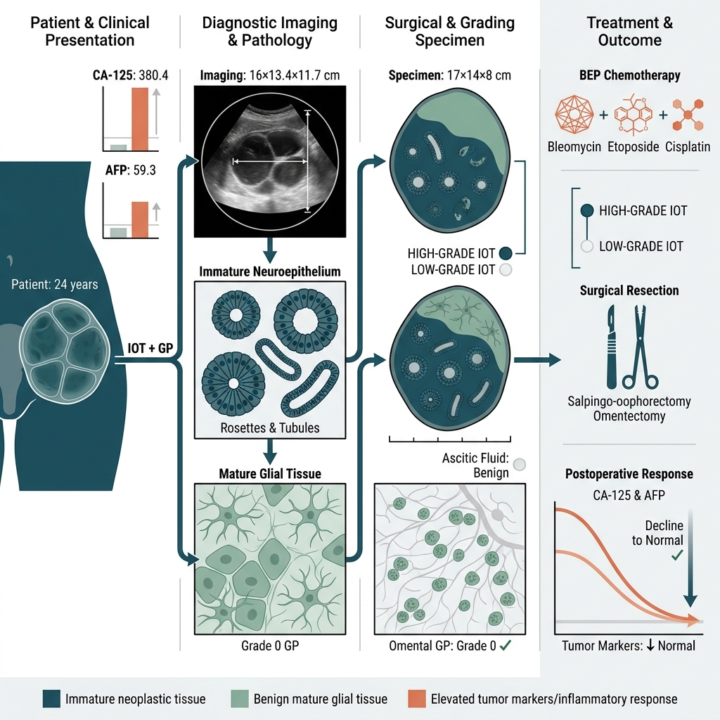


Immature Teratoma with Gliomatosis Peritonei: A Unique Case Report

- Adesh Institute of Medical Sciences & Research, Bathinda, Punjab 151101, India
- Department of Pathology, Maharishi Markandeshwar Institute of Medical Sciences & Research (MMIMSR), Mullana -Ambala 133207, India
- National Institute of Medical Sciences & Research, Jaipur, Rajasthan 303121, India
Abstract
Immature ovarian teratomas (IOTs) are rare malignant germ cell tumours that account for fewer than 1% of all ovarian teratomas, predominantly affecting women in their reproductive years, particularly during the first two decades of life. IOTs are rarely associated with a condition known as gliomatosis peritonei (GP), which is characterized by implants of mature glial tissue within the peritoneum. GP was first described in 1906 by Neuhäuser et al. and remains a rare entity, with only a limited number of reported cases. The presence of GP, regardless of its extent, is usually not associated with adverse outcomes; however, it has been reported to undergo transformation into malignant glial neoplasms. The pathogenesis of GP is also poorly understood. This condition can further complicate clinical management and necessitates careful monitoring and long-term follow-up, particularly given the risks of growing teratoma syndrome (GTS) and potential malignant transformation.
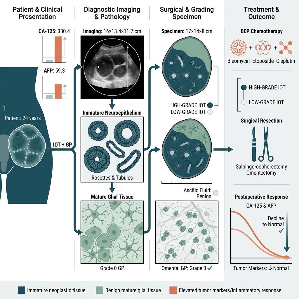
Graphical abstract
INTRODUCTION
Immature ovarian teratomas (IOTs) are rare malignant germ cell tumours that account for fewer than 1% of all ovarian teratomas, predominantly affecting females in their reproductive years, particularly during the first two decades of life. These tumours are characterised by the presence of immature neuroectodermal elements and can present with a variety of clinical manifestations, including abdominal pain, abdominal distension, and hormonal imbalances.1,2,3
IOTs are rarely associated with a condition known as gliomatosis peritonei (GP), which is characterised by mature glial tissue implants in the peritoneum. Pathologically, GP is considered to be a grade 0 teratoma according to the World Health Organization (WHO) grading system used for immature teratomas.1,2 The presence of GP, regardless of its extent, is usually not associated with adverse outcomes; however, it has been reported to undergo malignant transformation into glial neoplasms.4 The pathogenesis of GP is also poorly understood.5 This condition can further complicate clinical management and necessitates careful monitoring and intervention. Growing teratoma syndrome (GTS) is another rare outcome that may occur following treatment, characterised by the growth of residual mature teratoma elements despite normalised tumour markers, highlighting the need for vigilant follow-up in affected patients.6,7
CASE REPORT
A 24-year-old unmarried female presented to the gynaecology outpatient department of our institute with a chief complaint of lower abdominal pain lasting for two days. Per-abdominal examination revealed a non-tender, palpable mass measuring approximately 14 cm in size, which was mobile from side to side. Her menstrual history indicated regular cycles with dysmenorrhea. There was no family history of malignancy, and she had no history of prior surgeries or chronic illnesses.
Ultrasonography demonstrated a complex pelvic-abdominal mass with both solid and cystic components. Magnetic resonance imaging (MRI) revealed a well-defined, multiloculated left-sided pelvic-abdominal cystic mass measuring 16 × 13.4 × 11.7 cm. T1-hyperintense signal intensity was observed, suggestive of proteinaceous, mucinous, or haemorrhagic contents, along with T2-hypointense areas indicative of calcification within the lesion. Associated findings included omental thickening and free fluid in the peritoneal recesses. The radiological impression was suggestive of an ovarian teratoma with metastatic omental thickening and ascites.
Preoperative tumour markers were as follows: cancer antigen 125 (CA-125), 380.4 U/mL (reference range: <35 U/mL); alpha-fetoprotein (AFP), 59.3 IU/mL (reference range: <8.1 IU/mL); lactate dehydrogenase (LDH), 293.3 U/L (reference range: 140–280 U/L); carcinoembryonic antigen (CEA), 0.93 ng/mL (reference range: <5 ng/mL); and beta-human chorionic gonadotropin (β-hCG), <2 mIU/mL (reference range: <5 mIU/mL). Ascitic fluid aspiration was performed and submitted to the cytology section of our department for evaluation.
Microscopic examination of the ascitic fluid revealed numerous singly scattered and clustered benign mesothelial cells exhibiting reactive changes against a haemorrhagic background. No atypical cells were identified on the smears. The patient subsequently underwent a left salpingo-oophorectomy and omentectomy, and the resected specimen was received in our department for histopathological evaluation.
Gross examination revealed a left ovarian mass measuring 17 × 14 × 8 cm. The outer surface was smooth and bosselated, and the capsule was intact. Cut sections showed the mass to be predominantly solid with cystic areas filled with seromucinous fluid. Focal areas containing hair, sebaceous material, and calcification were also noted. A separately received fallopian tube measured 4 cm in length, with an identifiable lumen on cut section. A grey-yellow soft tissue piece labelled as omentum, measuring 9.5 × 6 × 0.5 cm, was received in a separate container.
Histopathological examination of the ovarian mass, which had an intact capsule, demonstrated elements from all three germ layers. Immature neuroepithelial tissue was identified in five low-power fields, forming rosettes and tubules (Figure 1, 2, and 3). Applying the updated two-tier grading system, in which low-grade tumours show ≤1 low-power field of immature neuroepithelium and high-grade tumours show >1 low-power field, the presence of immature neuroepithelium in five low-power fields classified this tumour as a high-grade immature ovarian teratoma.8 Mature components included skin with adnexal structures, intestinal, respiratory, and mucinous epithelia, along with bone, cartilage, muscle, adipose tissue, and mature neuroepithelium. Sections from the omentum showed fibroadipose tissue with mature glial proliferation and congestion (Figure 4), while the fallopian tube was unremarkable. A final histopathological diagnosis of left-sided high-grade immature ovarian teratoma (IOT) with gliomatosis peritonei (GP) was rendered.1,2,9
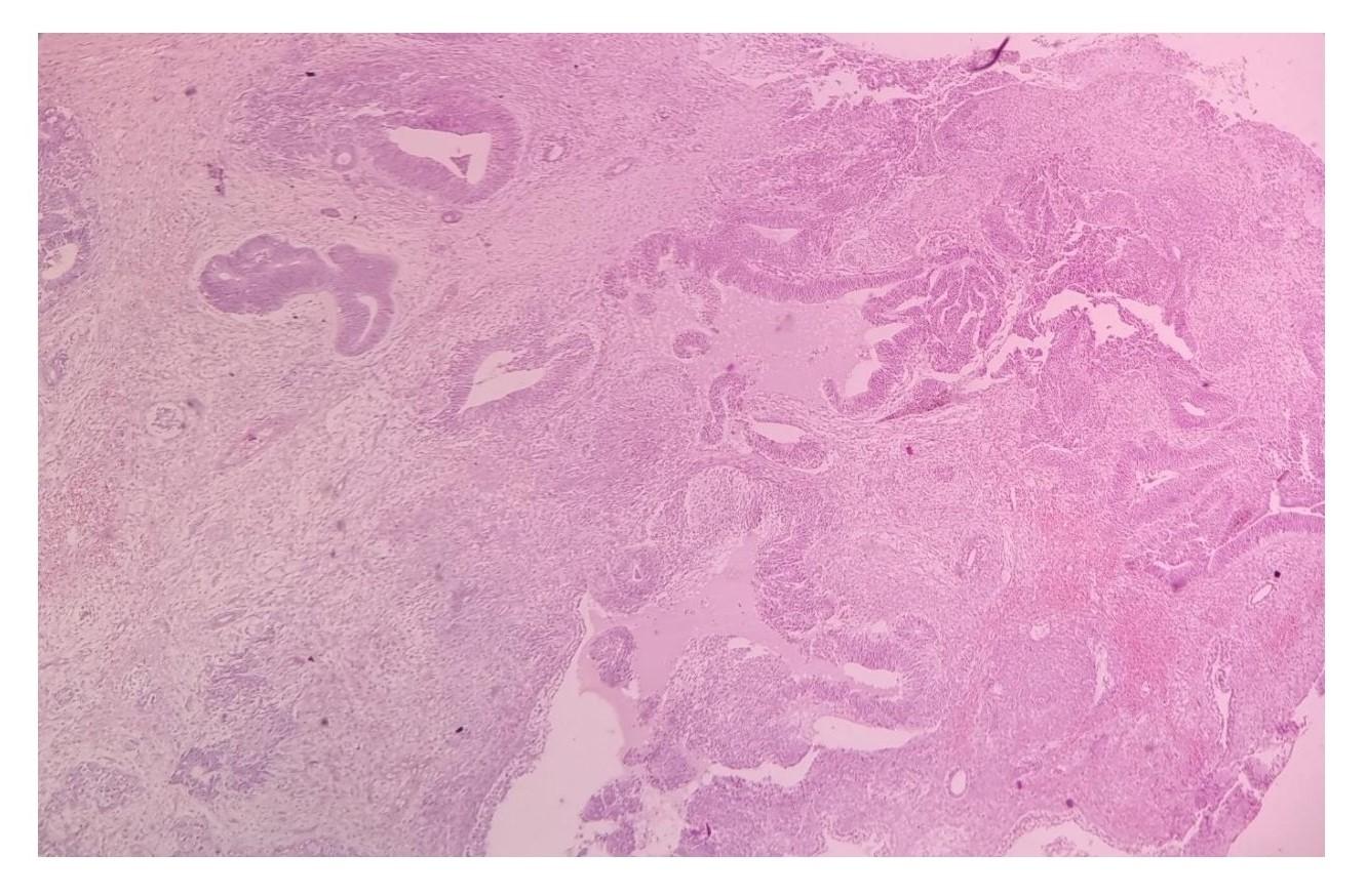
Immature neuroectodermal tissue predominantly arranged as primitive neural rosettes (Hematoxylin and Eosin [H&E]; original magnification ×100). Scale bar represents 100 μm.
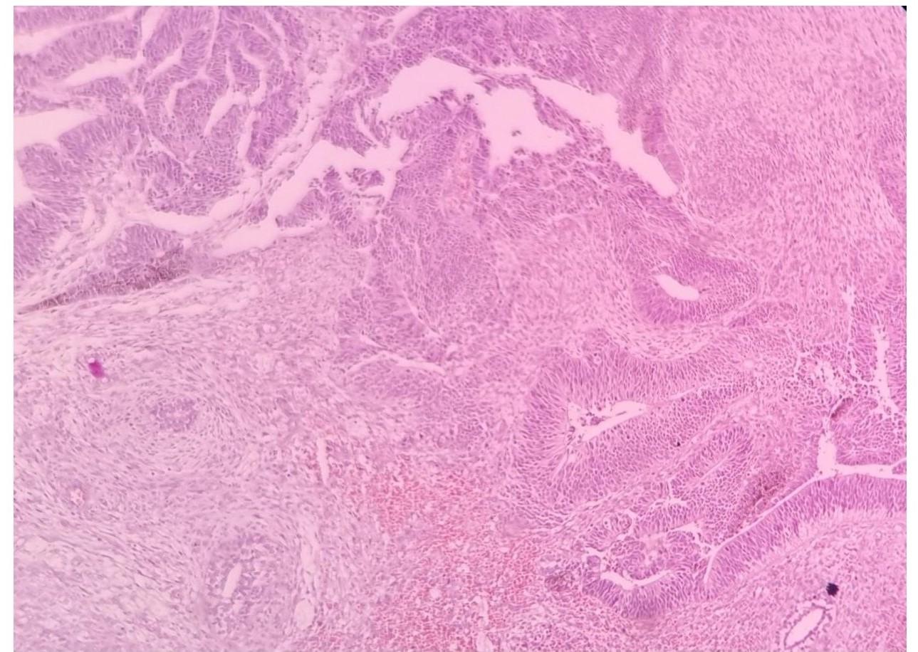
Immature neuroectodermal tissue predominantly arranged as primitive neuroepithelial tubules (Hematoxylin and Eosin [H&E]; original magnification ×100). Scale bar represents 100 μm.
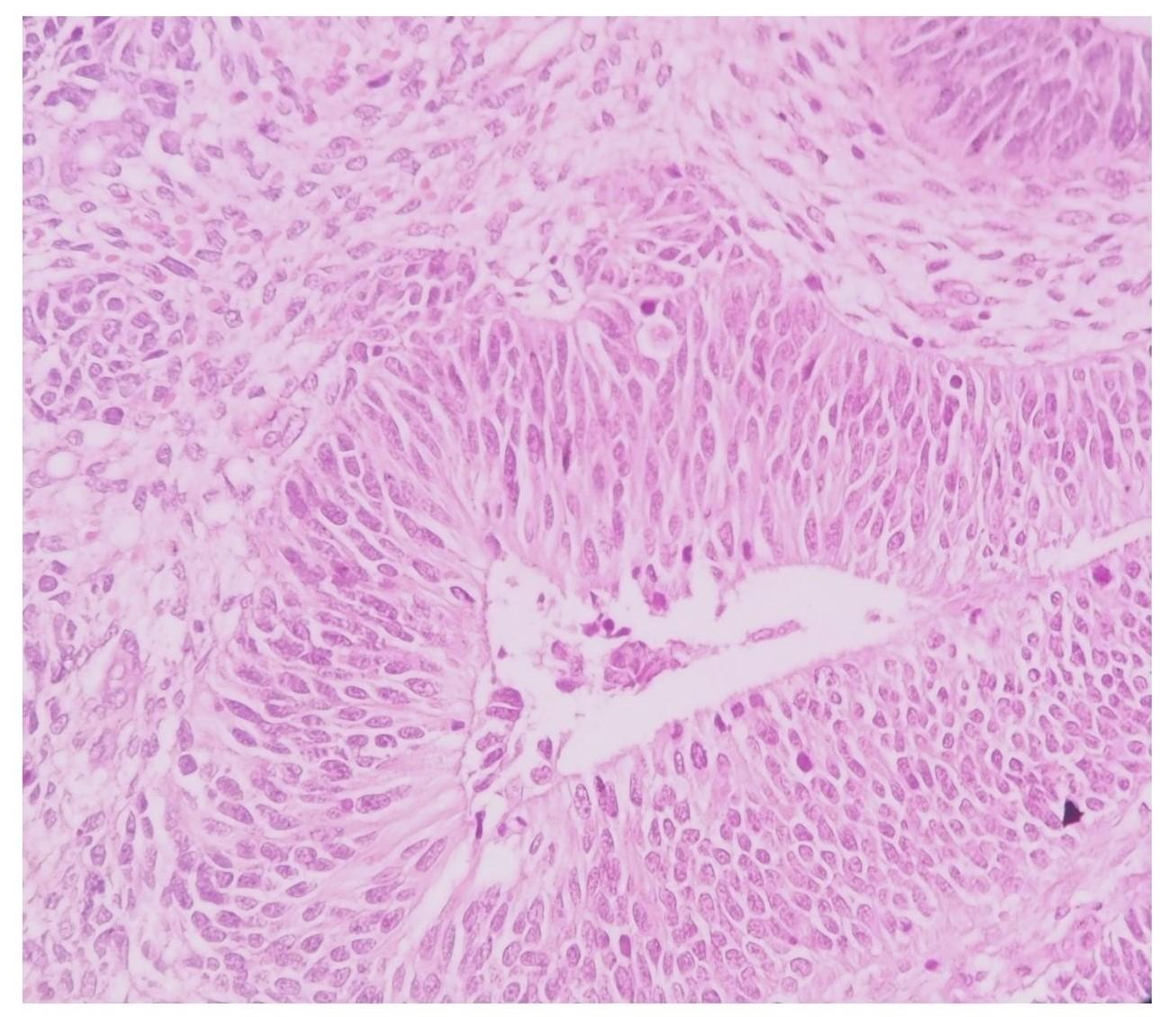
Neuroectodermal tissue arranged as tubules (Hematoxylin and Eosin [H&E]; original magnification ×400). Scale bar represents 25 μm.
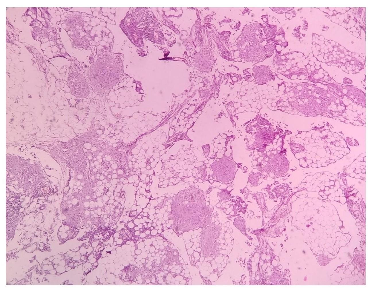
Adipose tissue (omentum) demonstrating mature glial proliferation (Hematoxylin and Eosin [H&E]; original magnification ×100). Scale bar represents 100 μm.
Following histopathological diagnosis, the case was discussed at a multidisciplinary team (MDT) meeting. In view of the high-grade histology and elevated preoperative tumour markers, adjuvant chemotherapy with the BEP regimen (bleomycin, etoposide, and cisplatin) was recommended. Postoperatively, the patient's tumour markers were reassessed and demonstrated a significant decline toward normal levels, consistent with an adequate surgical response. The patient was counselled regarding the need for long-term surveillance, given the known risks of growing teratoma syndrome and the rare potential for malignant transformation of gliomatosis peritonei.4,6,7
DISCUSSION
Immature ovarian teratomas (IOTs), while uncommon, are significant germ cell tumours typically presenting in the ovaries of young women, particularly in their second or third decade of life.1 These malignancies are characterised histologically by the presence of immature tissues, such as primitive neuroepithelial elements. When IOTs are accompanied by gliomatosis peritonei (GP)—a rare phenomenon marked by mature glial tissue implants in the peritoneum—the clinical scenario becomes notably more complex.
GP, associated predominantly with IOTs, is considered a benign secondary implantation of mature glial cells. It was first described in 1906 by Neuhäuser et al. and remains rare, with only a limited number of reported cases. The aetiology of GP is debated, with prevailing theories suggesting a derivation either from the maturing cells of the teratoma itself or from peritoneal or subperitoneal mesenchymal cells that transdifferentiate under the influence of secreted factors from the primary tumour.10 Although considered benign, GP's capacity for rare malignant transformation and recurrence underscores the necessity for vigilant long-term monitoring, often via imaging modalities such as computed tomography (CT) scans.4,7,11
Clinically, patients with IOTs and GP typically present with abdominal pain and a palpable pelvic-abdominal mass, as was observed in the present case. Ultrasonography generally demonstrates a complex adnexal mass, though this finding is non-specific.10 Calcifications may also be present.
On MRI, the presence of a distinct solid component with areas of calcification and small pockets of fat is highly suggestive of an immature teratoma. Cystic regions may contain serous, mucinous, or fatty sebaceous material, and occasional haemorrhagic changes may also be present.12,13 In the present case, MRI demonstrated a left-sided, well-defined, multiloculated pelvic-abdominal cystic mass (16 × 13.4 × 11.7 cm) with T1-hyperintense areas suggestive of proteinaceous, mucinous, or haemorrhagic content. Hypointense T1 and T2 areas indicated calcification within the lesion. Associated findings included omental thickening and free peritoneal fluid. Surgical excision remains the cornerstone of treatment for IOTs with GP, supported by histopathological evaluation to ensure complete resection and the absence of malignant implants.
Histopathologically, IOTs contain a mix of mature and immature elements, primarily neuroectodermal tissue. Immature neuroepithelium, which may form rosettes, pseudorosettes, or primitive tubules, is characterised by scant cytoplasm, hyperchromatic nuclei, and frequent mitoses. Grading is based on low-power field (4× objective) counts of immature neuroepithelium per slide.2,9 The updated two-tier system—introduced to improve interobserver reproducibility—classifies IOTs as low-grade (≤1 low-power field) or high-grade (>1 low-power field), combining the former grades 2 and 3 into a single high-grade category.8 This approach is now recommended over the older three-tier system. Gliomatosis peritonei, represented as mature glial nodules in the peritoneum, is classified as grade 0 but requires thorough sampling to exclude immature components, which would worsen the prognosis.2,9 The presence of associated yolk sac tumour or embryonal carcinoma would necessitate reclassification as a mixed germ cell tumour.2
Histological evaluation of GP reveals glial nodules that may closely mimic metastases, necessitating careful differentiation to avoid overtreatment. This differentiation hinges on strict adherence to diagnostic criteria, emphasising the exclusive presence of mature glial tissues without malignant features. In the present case, the ovarian mass with an intact capsule revealed a tumour comprising elements from all three germ layers. Immature neuroepithelial tissue was identified in five low-power fields, arranged as immature rosettes and tubules, consistent with high-grade classification under the updated two-tier system. Mature elements included skin epidermal lining with adnexal structures, intestinal, mucinous columnar, and respiratory lining epithelia, mature neuroepithelium, bone, adipose tissue, cartilage, and muscle. Omental sections demonstrated fibroadipose tissue with areas of mature glial proliferation and congestion. Sections from the fallopian tube were unremarkable.
In view of the high-grade histology, the case was referred for oncological consultation. Adjuvant chemotherapy with the BEP protocol was recommended and discussed with the patient, in keeping with published guidelines for high-grade IOTs.6,9 Given the patient’s young age and reproductive potential, fertility-sparing surgery—unilateral salpingo-oophorectomy with preservation of the uterus and the unaffected contralateral ovary—is the standard treatment of choice for young women with immature ovarian teratomas, including high-grade cases, provided the contralateral ovary is uninvolved.9 The postoperative decline in tumour markers toward normal levels provided evidence of an adequate initial surgical response and established a meaningful baseline for future monitoring. The prognosis is generally favourable when GP is strictly composed of mature glial tissue, correlating with lower tumour grades and adherence to treatment protocols (e.g., FIGO staging).14 Ongoing long-term surveillance—including serial imaging and tumour marker assessment—is essential given the documented risks of GTS and the rare malignant transformation of GP.4,6,7 Some studies have highlighted the potential utility of biomarkers such as OCT4 to identify more aggressive teratoma variants, which may further inform prognosis and tailored therapeutic strategies.14
Ongoing research into the molecular and immunohistochemical profiles of IOTs and GP may provide deeper insights into their pathophysiology and contribute to refined management approaches.
CONCLUSION
Gliomatosis peritonei (GP) is a rare condition often associated with immature ovarian teratomas (IOTs). Radiological features such as fatty speckles may suggest immaturity or malignancy, prompting evaluation of peritoneal and omental lesions. These should be excised along with the ovarian mass for histopathological examination. Thorough sampling of the ovarian mass is essential to detect immature neuroepithelium or mesodermal components and to assign the histological grade using the updated two-tier system, which directly guides subsequent clinical management. A multidisciplinary approach combining clinical, radiological, and histological findings is essential to confirm GP, differentiate it from malignant mimickers, and plan adjuvant therapy where indicated. Long-term postoperative surveillance, including serial tumour marker assessment and imaging, remains indispensable to monitor for GTS and the rare but recognised risk of malignant transformation, thereby optimising outcomes in this predominantly young patient population.
DECLARATIONS
Abbreviations
AFP: Alpha-fetoprotein; BEP: Bleomycin, etoposide, and cisplatin; β-hCG: Beta-human chorionic gonadotropin; CA-125: Cancer antigen 125; CEA: Carcinoembryonic antigen; CT: Computed tomography; FIGO: International Federation of Gynecology and Obstetrics; GP: Gliomatosis peritonei; GTS: Growing teratoma syndrome; H&E: Hematoxylin and eosin; IOT / IOTs: Immature ovarian teratoma / Immature ovarian teratomas; LDH: Lactate dehydrogenase; MDT: Multidisciplinary team; MRI: Magnetic resonance imaging; OCT4: Octamer-binding transcription factor 4; WHO: World Health Organization
Acknowledgments
None.
Author’s contributions
SP, AKR, and PC were involved in clinical and pathological data acquisition, case management, histopathological evaluation, literature review, and drafting and editing of the manuscript. All authors read and approved the final manuscript.
Funding
None.
Availability of data and materials
Not applicable. All relevant clinical, radiological, and histopathological data are included in this published article.
Ethics approval and consent to participate
Not applicable. Institutional Review Board approval is waived for single case reports at our institution.
Consent for publication
Written informed consent was obtained from the patient for publication of this case report and any accompanying images. A copy of the written consent is available for review by the Editor-in-Chief of this journal.
Declaration of generative AI and AI-assisted technologies in the writing process
None.
Competing interests
The authors declare that they have no competing interests.