

Exploring Potential of Azolla pinnata and Calcium Oxide Nanoparticles against Osteosarcoma Therapy: A Comprehensive Review
- Department of Pharmacology, Saveetha College of Pharmacy, SIMATS, India
- Department of Pharmacy Practice, Saveetha College of Pharmacy, Saveetha Institute of Medical and Technical Sciences, Thandalam, Chennai, India
- Department of Pharmacy Practice, Saveetha College of Pharmacy, SIMATS, India
Abstract
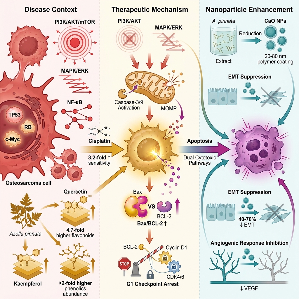
Osteosarcoma is a highly aggressive primary bone malignancy characterized by a poor prognosis due to treatment-related toxicity and chemoresistance. This clinical challenge has spurred growing interest in natural adjuncts, particularly plant-derived therapeutic agents. Azolla pinnata, an aquatic fern, is recognized for its potential antioxidant, anti-inflammatory, and anticancer properties. It is rich in bioactive constituents, including phenolics, flavonoids, tannins, and terpenoids. Key phytochemicals, such as gallic acid, kaempferol, and quercetin, have been shown to modulate redox balance and tumor suppression pathways. These compounds can induce cell-cycle arrest and trigger apoptosis through both extrinsic and intrinsic (mitochondrial) pathways—characterized by caspase-3/9 activation and BCL-2 downregulation—while concurrently inhibiting oncogenic signaling cascades, such as PI3K/AKT/mTOR, MAPK/ERK, and NF-κB. Furthermore, the modulation of VEGF, MMP-2, and MMP-9 suggests potential anti-angiogenic and anti-metastatic effects. The utilization of A. pinnata extracts in the green synthesis of CaO nanoparticles is proposed to offer therapeutic advantages, including pH-responsive dissolution, ROS generation, and bone-targeted delivery; however, these hypothesized effects require rigorous experimental validation. Despite promising preliminary data, significant hurdles remain, including inconsistent reproducibility in batch-to-batch green synthesis, a paucity of comprehensive in vivo toxicity data, and an incomplete understanding of the underlying molecular mechanisms. In summary, while current evidence suggests that A. pinnata and A. pinnata-derived CaO nanoparticles constitute promising theoretical platforms for osteosarcoma therapy, further in vivo validation, mechanistic elucidation, and translational development are critically warranted. Notably, the integration of A. pinnata bioactive compounds with CaO nanoparticles remains a conceptual framework, currently lacking direct experimental evidence to confirm synergistic efficacy.
Introduction
Osteosarcoma is a highly prevalent malignant bone tumor occurring predominantly in children and adolescents 1. The impact of age on disease prognosis remains incompletely understood. Approximately 25% of patients with osteosarcoma possess a germline pathogenic variant in a recognized cancer-susceptibility gene, with the highest frequency observed in children younger than 10 years 2. The most frequent primary site of tumor presentation is the metaphysis of long bones, specifically the femur, tibia, and humerus. The histological hallmark of osteosarcoma is the presence of immature osteoid or bone formed by neoplastic cells 3. The TP53 gene is the most frequently altered gene in osteosarcoma. Furthermore, among genes altered in more than 10% of cases, c-Myc plays a critical role in the onset of the disease and enhances cell invasion by initiating the MEK–ERK pathways. Several genomic studies have revealed that the RB gene is frequently mutated in pediatric osteosarcoma patients. Additionally, mutations in NOTCH1, FOS, NF2, WIF1, BRCA2, APC, PTCH1, and PRKAR1A have been identified. The pathogenic activity of the MAPK and PI3K/Akt signaling pathways in osteosarcoma is regulated by various miRNAs, such as miR-21, -34a, -143, -148a, -195a, -199a-3p, and -382. CD133+ cells represent the subpopulation of osteosarcoma stem-like cells capable of initiating tumor growth; these cells express stemness-related genes, play a vital role in metastasis, and exhibit therapeutic resistance 4.
Natural products regulate multiple oncogenic signaling pathways through their ability to alter the expression and activity of specific molecular targets. The low toxicity, chemical diversity, and widespread availability of these compounds render them attractive alternatives to standard chemotherapy 5,6. Bioactive phytochemicals, such as terpenoids and alkaloids, exhibit broad therapeutic potential by interfering with cancer development processes involving the PI3K/AKT, MAPK, and NF-κB pathways, which subsequently activate apoptotic pathways 7,8.
The floating aquatic fern A. pinnata is indigenous to the subtropical and tropical regions of Africa, Asia, and the Americas. This plant possesses a nutrient-rich composition resulting from its symbiotic relationship with Anabaena azollae cyanobacteria. It contains bioactive secondary metabolites that demonstrate antibacterial, anti-inflammatory, antioxidant, and anticancer effects. These phytochemicals include flavonoids (e.g., quercetin and kaempferol), phenolic acids (e.g., caffeic acid and gallic acid), terpenoids, and tannins 9,10,11. A. pinnata represents a practical solution for cost-effective osteosarcoma treatment development due to its ease of cultivation, sustainable growth, and rich bioactive profile. The synergistic activity of these compounds functions to protect against free radicals while regulating the expression of apoptotic proteins and modulating cancer-related signaling mechanisms. A. pinnata was selected for this review because its high flavonoid and phenolic content has been proven to target essential pathways governing tumor growth and cell death. Specifically, the presence of quercetin and kaempferol reduces cancer cell proliferation and promotes programmed cell death by interfering with multiple signaling cascades, including the PI3K/AKT, MAPK/ERK, and NF-κB pathways. Quercetin preserves normal cell functions while inhibiting cancer cell growth through the blockade of PI3K/AKT pathways, activation of MAPK pathways, increased ROS production, and induction of mitochondrial damage 12,13. Flavonoids regulate biological processes by upregulating pro-apoptotic proteins, such as Bax, and downregulating anti-apoptotic protein levels 14. They can also induce cell cycle arrest by altering cyclins and CDKs. Although A. pinnata phytochemicals and CaO nanoparticles have been studied individually for their anticancer effects, there are no studies demonstrating their combined or synergistic potential in osteosarcoma. Therefore, this review proposes a conceptual and hypothesis-driven approach for integrating these two components based on their complementary mechanisms, although direct experimental evidence for such synergy is currently lacking.
This review aims to explore the development of biodegradable nanoparticles incorporating CaO nanoparticles and A. pinnata plant extracts as a solution to create safer and more effective therapeutic interventions for osteosarcoma. The research investigates the benefits of green synthesis methods, targeted delivery systems, and reduced systemic toxicity in sustainable biomedical applications. This comprehensive review highlights the mechanisms of action of A. pinnata against osteosarcoma, summarizes the available evidence regarding its anticancer prospects, and discusses the anti-proliferative and cytotoxic effects of its constituents in vitro and in vivo. Furthermore, it details how these bioactive compounds modulate major molecular pathways integral to tumor suppression. Finally, to advance the clinical application of A. pinnata in osteosarcoma therapy, this review identifies existing research gaps and outlines future directions for experimental and clinical investigation.
Phytochemical constituents of A. pinnata
Primary and Secondary Metabolites
A. pinnata is being actively investigated as a pharmacological and nutraceutical agent due to its relatively high levels of primary metabolites. For example, a proximate analysis conducted by El Naggar and El-Mesery (2022) indicated that dried A. pinnata contains high levels of crude protein, essential amino acids, lipids, crude fiber, and ash, rendering it a valuable feed supplement and potential source of bioactive metabolites. The study highlighted that A. pinnata dry biomass possesses a significant proportion of organic matter and carbohydrates, underscoring its metabolic versatility and suitability for both nutritional and pharmacological applications 15. In a greenhouse study, A. pinnata exhibited greater concentrations of crude protein, lipids, and essential amino acids compared to A. caroliniana, with palmitic acid, oleic acid, and lignoceric acid being the predominant fatty acids 16. These findings indicate that A. pinnata has a metabolite profile rich in proteins and lipids, with levels varying based on growth conditions, which supports the derivation of secondary bioactive compounds. Table 1 lists the primary and secondary metabolites of A. pinnata, including essential components such as phenolics, flavonoids (kaempferol, quercetin), and fatty acids, as well as changes in their composition under stress. It highlights their pharmacological relevance to the treatment of osteosarcoma through their antioxidant and apoptotic effects and their modulation of signaling pathways.
In addition to other classes of compounds (including tannins, terpenoids, saponins, and alkaloids), the secondary metabolite profile of A. pinnata is characterized by exceptionally high levels of phenolic compounds and flavonoids. For instance, A. pinnata exhibited more than twice the concentration of total phenols, up to 4.7-fold higher flavonoids, and up to 2.7-fold higher condensed tannins when assessed under combined environmental and nutritional stresses, while also showing increased carbohydrate levels and a reduction in both protein and lipid levels 17. Based on a comparative analysis, A. pinnata contained higher concentrations of phenolics and flavonoid compounds than A. caroliniana, which was associated with improved growth performance and antioxidant capacity 16. A recent in vivo study in broiler chickens found that A. pinnata extract demonstrated hepatic protection and immune-stimulatory gene expression, as well as the presence of significant concentrations of phenolics and flavonoid compounds 18. This evidence supports the conclusion that many of the biological activities (antioxidant, cytotoxic, and immunomodulatory) of A. pinnata are attributable to its specific secondary metabolites.
Phytochemical Profile and Pharmacological Relevance of
| Metabolite Class | Key Compounds Identified | Content/Characteristics | Pharmacological Relevance |
|---|---|---|---|
| Primary Metabolites | Crude protein, essential amino acids, lipids, crude fiber, ash, carbohydrates | High crude protein (up to superior levels vs. other | Nutritional base; supports metabolite derivation for bioactivity |
| Phenolics | Caffeic acid, gallic acid, total phenols | >2x higher under stress; positively correlates with antioxidant assays (DPPH, FRAP) | Free radical scavenging; modulates apoptotic pathways in osteosarcoma |
| Flavonoids | Quercetin, kaempferol, rutin | Up to 4.7x higher; highest in ethanol/methanol extracts | Induces apoptosis, cell cycle arrest (G1/S phase); inhibits PI3K/AKT, MAPK |
| Other Secondary Metabolites | Condensed tannins, terpenoids, saponins, alkaloids | Elevated under environmental stress; confirmed via HPLC, GC-MS | Anti-inflammatory, antimicrobial; anti-metastatic via MMP/VEGF inhibition |
Analytical indication methods
HPLC is employed for the quantitative analysis of phenolic compounds, flavonoids, and amino acids in A. pinnata, providing acceptable sensitivity and reproducibility for bioactive metabolite detection 19. GC-MS analysis facilitates the identification of volatile compounds (e.g., fatty acids, esters, and terpenoids) within A. pinnata, allowing for the assessment and structural determination of phytochemicals with both antibacterial and antioxidant activities 20. Functional groups within A. pinnata, such as hydroxyl, carboxyl, and amide moieties, are typically characterized by FTIR spectroscopy. Characteristic peaks corresponding to phenolic OH groups are observed in the region between 3200 and 3500 cm⁻¹, whereas proteins and flavonoids are differentiated based on C=O stretching in the 1600–1700 cm⁻¹ region 21. NMR spectroscopy, especially ¹H and ¹³C NMR, corroborates the molecular structure of secondary metabolites extracted from A. pinnata. NMR is particularly advantageous for the structural elucidation of sterols and isolated flavonoids 22. UV–Vis spectroscopy serves as a preliminary technique to confirm the presence of conjugated systems, such as flavonoids and chlorophyll derivatives. Flavonoids typically exhibit maximum absorbance at 270–290 nm, while chlorophyll displays maxima at 650–680 nm 23.
Pharmacological activities of A. pinnata
Anti-oxidant properties
Extracts from A. pinnata exhibit significant in vitro and in vivo antioxidant activity, primarily attributed to their high flavonoid and phenolic content. An abundance of antioxidant enzymes is also a documented outcome of secondary metabolite accumulation (due to UV, herbicide, salinity, and nutrient stress) in Azolla species 24. In summary, A. pinnata demonstrates substantial antioxidant activity, providing a robust molecular basis for its potential therapeutic application. Total phenolic and flavonoid contents exhibit a positive correlation with DPPH, ferric-reducing antioxidant power (FRAP), and ABTS activity in ethanol and methanol extracts 25,26. In a rat model of lead-induced hepatotoxicity, the ethanolic extract of A. pinnata significantly improved oxidative stress markers (by decreasing lipid peroxidation and increasing GSH and antioxidant enzyme activities), which corroborates the protective biological activity mediated by its antioxidant constituents 27. These protective effects have been consistently observed across various animal and cell models.
Anti-inflammatory effect
The anti-inflammatory properties of A. pinnata have been demonstrated in many in vitro and in vivo experimental models. The administration of A. pinnata ethanolic extract in rats with liver damage attenuates serum liver injury and reduces the expression of pro-inflammatory cytokines and mediators, such as TNF-α, IL-6, and COX-2, demonstrating an anti-inflammatory mechanism that likely enhances its antioxidant activity 27. Comparative phytochemical studies have reported similar actions attributed to its phenolic and flavonoid components, such as quercetin, kaempferol derivatives, and phenolic acids, which are believed to influence NF-κB and MAPK signaling in inflammatory cells 28,29. Furthermore, Azolla biomass has been reported to enhance immunological markers and mitigate inflammatory responses in poultry and aquaculture models, thus indicating the practical importance of these anti-inflammatory characteristics.
Antimicrobial and cytoprotective activities
Several investigations have highlighted the antimicrobial properties of A. pinnata extracts against both Gram-positive and Gram-negative bacteria, as well as certain fungal strains; typically, ethanol and methanol extracts demonstrate the highest potency due to the superior extraction of phenolics and flavonoids 30,31. Azolla extracts are rich sources of fatty acids, terpenoids, and phenolic derivatives, the antimicrobial efficacy of which has been established through GC-MS and LC-MS profiling and verified by reported zones of inhibition and minimum inhibitory concentration (MIC) values in standard microbiological assays 32. Beyond their antimicrobial effects, A. pinnata exhibits the capacity to safeguard animal models from the adverse effects of chemical stressors, such as lead exposure 27. Treated subjects exhibited elevated levels of antioxidant enzymes compared to untreated counterparts, along with reduced markers of apoptosis and preserved hepatic histology 27. Given that anti-inflammatory and antioxidant agents are the primary mediators of these cytoprotective effects, A. pinnata contributes not only to detoxification but also to the enhancement of tissue resilience, representing significant therapeutic benefits of the plant 27.
Anticancer mechanism of A. pinnata against osteosarcoma
Introduction of apoptosis
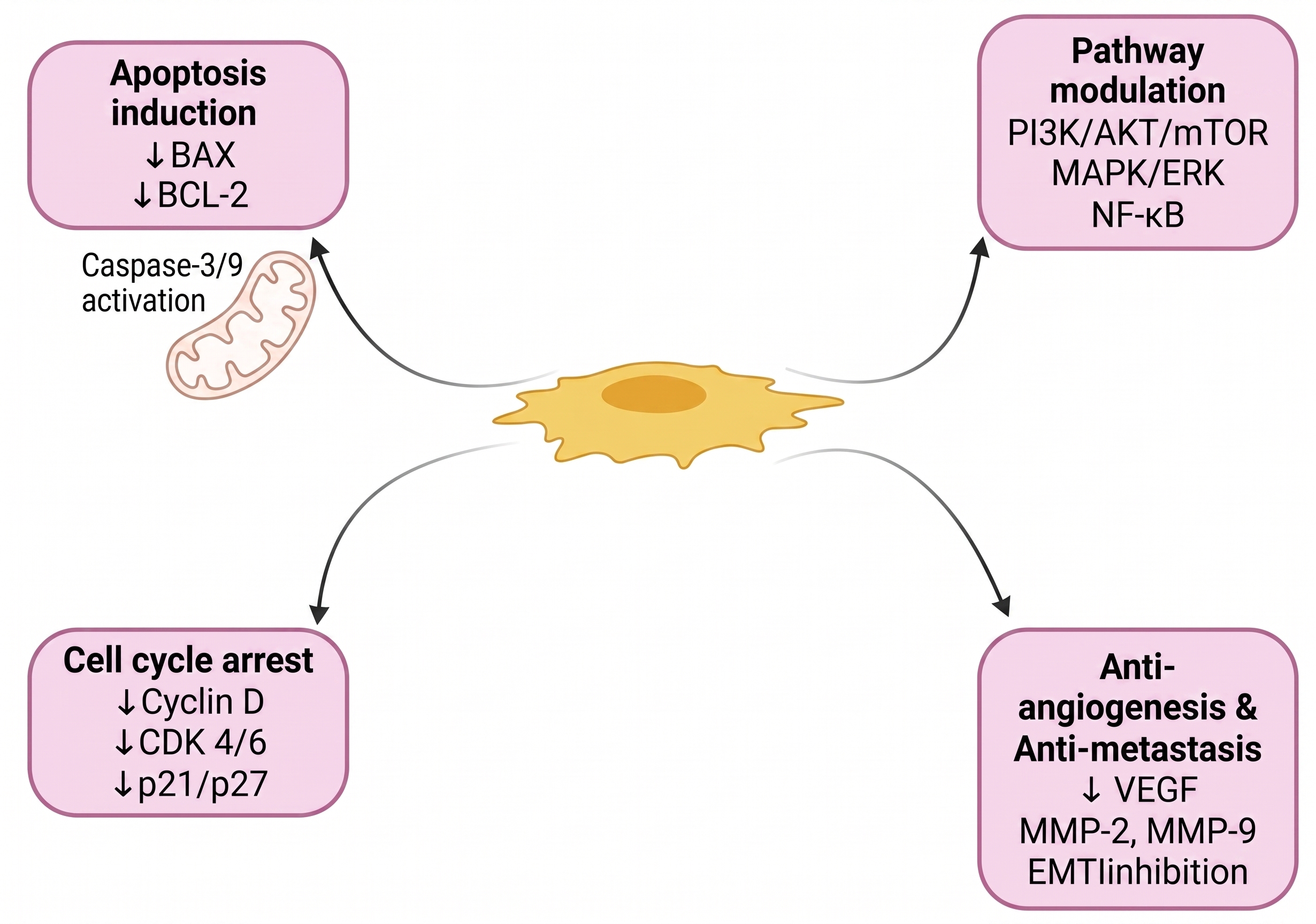
Apoptosis, or programmed cell death, is a tightly regulated process facilitated by the removal of damaged or malignant cells, representing one of the primary therapeutic objectives in oncology. The therapeutic application of plant-based compounds aims to trigger apoptosis in tumor cells via both death-receptor (extrinsic) and mitochondrial (intrinsic) pathways; these processes involve DNA fragmentation, caspase activation, and subsequent cell death 33,34. Two flavonoids present in A. pinnata, quercetin and kaempferol, have been shown to increase the Bax/BCL-2 ratio, leading to depolarization of the mitochondrial membrane and, in osteosarcoma models, the activation of caspase-3/9 and the induction of apoptosis 35,36. The phytochemicals of A. pinnata likely induce apoptosis in osteosarcoma cells through a dual-pathway mechanism involving both death receptor activation and mitochondrial signaling. The combination of gallic acid, phenolic acids, quercetin, and kaempferol may promote an altered Bax/BCL-2 ratio, leading to mitochondrial outer membrane permeabilization (MOMP). This process results in the translocation of cytochrome C into the cytosol, where it binds with Apaf-1 and procaspase-9 to form the apoptosome complex, subsequently activating initiator caspase-9. The subsequent activation of executioner caspase-3 triggers chromatin condensation, DNA fragmentation, and the formation of apoptotic bodies 37,38. Although the capacity of the total A. pinnata extract to activate these processes in osteosarcoma models requires further validation, these mechanisms are well-documented in studies involving isolated flavonoids. The antioxidant compounds within A. pinnata may modulate reactive oxygen species (ROS) levels, which serve as secondary messengers to amplify apoptotic signals while protecting healthy cells from oxidative damage. Additionally, the extrinsic pathway may be initiated through death receptors, including Fas, CD95, and TRAIL receptors, which recruit FADD and procaspase-8 to further promote caspase-3 activation 39. Depending on the dosage and cellular microenvironment, a balance between the pro-oxidant and antioxidant activities of these phytochemicals may modulate ROS-mediated apoptotic signaling. A. pinnata phytochemicals may overcome resistance mechanisms that protect osteosarcoma cells from single-target treatments by engaging multiple pro-apoptotic signaling pathways. Moreover, the antioxidant compounds derived from A. pinnata could be administered at higher, selectively cytotoxic doses without significant harm to normal cells; these nutrients regulate redox-sensitive apoptotic signaling, thereby permitting the activation of redox-sensitive apoptotic triggers by acute ROS levels while reducing chronic oxidative damage that promotes tumor survival 40,41. Even though the induction of apoptosis via caspase-dependent and mitochondrial processes has been demonstrated in osteosarcoma models, the specific molecular contributions of the entire A. pinnata phytochemical complex warrant further investigation 34,42.
Regulation of cell cycle progression
Dysregulation of cell-cycle checkpoints plays a pivotal role in cancer progression. Several natural flavonoids are known to block cyclins and cyclin-dependent kinases (CDKs), consequently inducing G0/G1 or G₂/M arrest in cancer cells (Figure 1) 43. Notably, quercetin and kaempferol have been shown to inhibit the expression of cyclin D1 and CDK4/6 while increasing the levels of CDK inhibitors (p21, p27), thereby blocking passage through the G1 checkpoint in osteosarcoma models and reducing clonogenic survival 44,45. The p21 protein directly inhibits Cyclin D1-CDK4/6 and Cyclin E-CDK2 complexes during periods of cellular stress or DNA damage when p53 proteins are stabilized. Subsequently, Rb hypophosphorylation leads to cell cycle arrest. Furthermore, even in p53-mutant conditions, the cell cycle can be blocked as p27 levels increase through p53-independent mechanisms, including reduced AKT-mediated degradation and FoxO transcription factor activation. Multi-targeted therapeutic agents that impact both p53-dependent and independent checkpoints offer significant therapeutic potential, as these intersecting pathways ensure effective proliferation control while driving cancer cell death through apoptosis 46,47,48. Such regulatory systems require rigorous validation in whole-plant extract systems, as current evidence is primarily based on isolated phytochemicals. A. pinnata extracts are expected to exert similar cell-cycle modulatory effects due to their high levels of flavonoids and related phenolics, which contribute to decreased proliferation rates and enhanced osteosarcoma cell sensitivity to apoptotic signals and chemotherapeutics 49,50. Although direct experimental validation of A. pinnata extracts in osteosarcoma models is currently limited, their potential to modulate cell-cycle regulators is supported by their established flavonoid content.
Modulation of key signaling pathways
Various oncogenic signaling pathways, such as the p53-associated networks, MAPK/ERK pathway, NF-kB system, and PI3K/AKT/mTOR pathway, control the development, progression, and therapeutic resistance of osteosarcoma (Figure 1). Flavonoids in A. pinnata have been reported to affect the following pathways: they thwart PI3K/AKT signaling, reduce the phosphorylation of AKT and its downstream mTOR signaling, blunt the activation of MAPK/ERK signaling, and suppress the nuclear translocation of NF-κB, which decreases survival signaling and induces apoptosis 51. For example, kaempferol is able to recover PTEN activity in order to re-inhibit PI3K signaling, while quercetin specifically suppresses/dephosphorylates AKT or promotes p53-dependent responses in osteosarcoma cells 52,53. Although flavonoids such as quercetin and kaempferol may be able to modify the PI3K/AKT, MAPK/ERK, and NF-kB pathways in cancer models, the establishment of their bioactive concentration and abundance in A. pinnata extracts is required before a definitive mechanistic attribution can be made 54,55.
More than 65–75% of osteosarcoma cases exhibit activation of the PI3K/AKT/mTOR pathway, which contributes to the poor response of patients toward doxorubicin and cisplatin treatment. The viability of osteosarcoma cells decreases by approximately 40–50% when researchers use pharmacological methods to inhibit AKT, but ERK activation prevents cells from undergoing apoptosis. Research has found that dual inhibition studies demonstrated a >60% reduction in clonogenic survival together with a 2–3-fold increase in caspase-3 activation when compared to single treatment methods 56. The MAPK/ERK pathway promotes DNA repair activities while driving cell proliferation processes. The hyperactivation of ERK1/2 results in enhanced expression of multidrug resistance proteins (e.g., MDR1) and cyclin D1. In osteosarcoma cell lines, dual PI3K–ERK inhibition has been demonstrated to increase cisplatin sensitivity by around 3.2 times, and in xenograft models, it effectively inhibits tumor development 57,58. The NF-κB signaling pathway controls pro-inflammatory survival genes, including BCL-XL and XIAP. In osteosarcoma cells that exhibit chemoresistance, NF-κB maintains its active state. Inhibition of this pathway reduces IL-6 and TNF-α secretion by 50 to 70 percent, thereby decreasing metastatic potential and increasing apoptotic sensitivity. Contemporary oncology supports network-based treatment methods, as the simultaneous suppression of the PI3K/AKT/mTOR, MAPK/ERK, and NF-κB pathways disrupts survival mechanisms while preventing adaptive resistance and extending apoptosis duration 59. The phytochemical profile of A. pinnata indicates potential multi-target interactions, although, at present, any synergistic effects remain speculative. Quercetin demonstrates the ability to block osteosarcoma cell growth by arresting the cell cycle and triggering cell death through the mitochondrial pathway, which begins with Bax upregulation and ends with the combined activation of caspase-3 and caspase-9 60. This process results in diminished mitochondrial membrane potential, leading to increased apoptosis in both MG-63 and U2OS cells. Quercetin increases the effectiveness of cisplatin chemotherapy by altering the miR-217-KRAS pathway, and it decreases the ability of HOS and MG-63 cell lines to invade and migrate by reducing the expression of VEGF, HIF-1α, MMP-2, and MMP-9. Although there is limited evidence regarding the effects of kaempferol and gallic acid in osteosarcoma studies, research on flavonoid activity demonstrates that kaempferol hinders cell migration by blocking MMP-2 activity while inducing cell death in carcinoma cells through a dose-dependent mechanism that activates caspase-3 (e.g., in squamous carcinoma cells, the IC₅₀ value is approximately 40 µM) 61. The use of whole-plant extracts, such as A. pinnata, for multi-targeting osteosarcoma treatment is justified because their biological activity allows for the regulation of multiple cancer pathways through PI3K/AKT, MAPK/ERK, NF-κB, and apoptotic cascades, which may provide greater anticancer effects than the use of single chemical substances.
Anti-metastatic and anti-angiogenic effects
The advancement of osteosarcoma tumor growth and its ability to disseminate depend on two primary processes: angiogenesis and extracellular matrix (ECM) remodeling. Vascular endothelial growth factor (VEGF) promotes tumor-associated angiogenesis through its effects on endothelial cell proliferation, vascular permeability, and hypoxic stress adaptation 62. The degradation of type IV collagen in the basement membrane by matrix metalloproteinases (MMPs), specifically MMP-2 and MMP-9, facilitates the invasion of tumor cells into surrounding tissues and their subsequent spread to the lungs. Clinical studies indicate that osteosarcoma patients with elevated VEGF and MMP-9 levels have a poor prognosis, which correlates with an increased risk of pulmonary metastases exceeding 60% 63. The simultaneous interruption of neovascularization and the reduction of nutrient supply increase tumor cell susceptibility to chemotherapy, potentially enhancing treatment efficacy. The downregulation of both MMP-2 and MMP-9 delays the epithelial-mesenchymal transition (EMT) because this inhibition reduces the capacity of osteosarcoma cells to degrade the ECM, migrate, and infiltrate tissues by 40% to 70% 64,65. This significant reduction has been observed in cancer models. The tumor microenvironment undergoes a vital transformation from a pro-metastatic condition to a restrictive state as the combined inhibition of VEGF and MMP signaling obstructs tumor-stromal interactions, resulting in reduced metastatic spread. The most effective approach to mitigating metastatic spread through microenvironment interaction in osteosarcoma requires the blockade of VEGF and MMP signaling pathways 64,65. Angiogenesis and metastasis are the primary causes of mortality in osteosarcoma patients. Natural products can reduce the risk of metastasis by decreasing VEGF-driven angiogenesis, inhibiting EMT markers, and reducing the activity of matrix metalloproteinases (MMP-2, MMP-9) 66,67. Flavonoids (e.g., quercetin and kaempferol) have been shown to inhibit VEGF-induced endothelial responses, reduce MMP expression and activity, and suppress cell migration and invasion in vitro. A. pinnata may influence the VEGF and MMP pathways, which are important metastasis regulators, due to the presence of flavonoids and phenolic acids 68,69.
Anti-oxidant mediated cytoprotection
Two representative antioxidant components of A. pinnata, polyphenols and flavonoids, exert a dual effect: at physiological levels, they mitigate mutagenic damage and chronic oxidative stress in healthy tissues, whereas in cancer models, they have been shown to induce significant cytotoxicity. Polyphenols modulate the vulnerability of cancer cells to apoptosis by acting as pro-oxidants or antioxidants, depending on the specific cellular conditions and dosage 70. The A. pinnata ethanolic extract has been shown to enhance enzyme antioxidant defense mechanisms, reduce lipid peroxidation, and replenish glutathione levels in animal models subjected to toxic insults 27. Although these effects are generally observed in systemic toxicity models, their relevance to osteosarcoma-specific cellular environments warrants further exploration. Researchers 54,71 posit that this capacity to modulate oxidative stress may preserve non-malignant tissues while simultaneously sensitizing tumor cells to apoptotic treatments, thereby establishing a potential therapeutic window for additive phytochemical interventions in osteosarcoma.
Experimental and computational evidence
In vitro studies
In vitro studies substantiate the biological activity of A. pinnata, particularly its antioxidant, cytoprotective, and anticancer properties. Methanolic and ethanolic extracts of A. pinnata have exhibited significant DPPH, ABTS, FRAP, hydroxyl radical scavenging, and lipid peroxidation inhibitory activities, confirming the presence of robust antioxidant phytochemicals such as flavonoids, phenolics, and tannins 72. Furthermore, these extracts possess anti-inflammatory effects, including the reduction of TNF-α and IL-6 in LPS-stimulated macrophages and the inhibition of NO production 73. Recent studies using human cancer cell lines have shown that plant-derived polyphenolic extracts—including those chemically similar to those found in A. pinnata—induce apoptosis, ROS-mediated cytotoxicity, and cell-cycle arrest in osteosarcoma cells (MG-63, U2OS) 74. Similar potential pathways are indicated by related ferns and flavonoid-rich extracts which induce caspase activation, mitochondrial membrane depolarization, and reduced cell invasion, although direct osteosarcoma studies on A. pinnata remain limited.
In silico studies
In silico approaches provide valuable mechanistic insights into the medicinal potential of bioactive compounds derived from A. pinnata, encompassing molecular docking, ADME prediction, and network pharmacology. Some flavonoids present in A. pinnata, including quercetin, kaempferol, rutin, and gallic acid, have shown binding affinity to osteosarcoma targets, such as BCL-2, caspase-3, MMP-9, VEGF, AKT1, and NF-kB 75. These substances regulate key pathways that are crucial to the pathophysiology of osteosarcoma, including PI3K/AKT, MAPK, apoptotic signaling, and oxidative stress regulation 76. Moreover, ADME/tox predictions suggest the potential of these main metabolites as therapeutic agents, as they exhibit favorable drug-likeness and satisfactory bioavailability scores 76.
Green nanotechnology approaches
Green nanotechnology offers a promising strategy for enhancing the medicinal properties of plant-derived substances. Through the use of phytochemicals as reducing and capping agents, several studies have demonstrated the effectiveness of ZnO, CaO, and bimetallic oxide nanoparticles produced via green methods using plant extracts. Extracts of A. pinnata have been successfully used to synthesize ZnO and CaO nanoparticles with strong antioxidant, antibacterial, and cytotoxic properties 77,78. Similarly, the use of Boerhavia diffusa, which you are considering for the development of a Zn-CaO composite, has been comprehensively reported as an effective reducing agent for the synthesis of metal and metal-oxide nanoparticles with anti-inflammatory, anticancer, and anti-diabetic effects 78. ZnO-CaO composites produced via green synthesis are promising for the treatment of osteosarcoma and the promotion of diabetic wound healing because of their higher biocompatibility, ROS-controlling properties, and capacity to enhance osteoblast functions 78,79.
There are two ways to create CaO nanoparticles: conventional chemical procedures and green synthesis methods. The chemical method generates particles in the size range of 50 to 200 nanometers through the combination of calcium salts, such as CaCl₂ and Ca(NO₃)₂, with strong bases and high-temperature calcination (Table 2). In contrast, green synthesis uses biological extracts from algae and plant leaves as both reducing and stabilizing agents. This process produces particles with uniform sizes while requiring lower temperatures and achieving better biocompatibility. The A. pinnata extracts contain flavonoids, phenolics, and terpenoids, which act as natural capping and reducing agents to control the growth of CaO nanoparticles while producing other bioactive compounds 80.
Comparison of Green vs Chemical synthesis of CaO nanoparticles
| Synthesis Method | Precursors | Reducing / Stabilizing Agent | Typical Particle Size | Notes / Advantages |
|---|---|---|---|---|
| Chemical | CaCl₂, Ca(NO₃)₂ | NaOH, high-temperature calcination | 50–200 nm | Fast and reproducible; may require toxic chemicals; broad size distribution |
| Green (Plant-based) | CaCl₂, Ca(NO₃)₂ | Plant extracts (e.g., | 20–80 nm | Eco-friendly, biocompatible, bioactive capping; narrower size distribution; mild reaction conditions |
Potential synergistic and therapeutic applications
Combination therapy with chemotherapeutic/ Nanocomposites
Combination therapy involves the simultaneous administration of phytochemicals or nanoparticles (NPs) with conventional anticancer regimens, such as chemotherapy, to exploit synergistic effects. This approach aims to reduce systemic toxicity and improve therapeutic efficacy by allowing for lower drug dosages and enhanced site-specific targeting 81,82. Co-delivery techniques can modulate the signaling pathways that facilitate tumor survival (e.g., PI3K/AKT and NF-κB) by sensitizing tumor cells to chemotherapy, overcoming multidrug resistance (MDR) by blocking efflux pumps and disrupting redox homeostasis, or enabling the spatiotemporal delivery of therapeutic agents (e.g., administering a chemotherapy-sensitizing agent followed by a cytotoxic drug) 81,82. Nanoparticles (NPs) such as liposomes, polymeric NPs, and metallic NPs—including zinc oxide-based formulations—have been extensively investigated for co-packaging phytochemicals and/or drugs, either through encapsulation or surface conjugation 81,82.
Chemical modifications to nanoparticles intended for osteosarcoma treatment can enhance their ability to deliver chemotherapeutics directly to bone tissue. These methods include the attachment of bone-targeting ligands, such as alendronate, lanthanides, or tetracycline derivatives, which facilitate the ligation and/or conjugation of nanoparticles to the osteosarcoma tissue, thereby enhancing both phytochemical and therapeutic activity 83. In addition, the use of bimetallic or composite oxide nanoparticles, such as Zn–CaO, which promote cell death through the production of reactive oxygen species (ROS) and the integration of plant-derived active agents (e.g., volatile compounds), enhances therapeutic efficacy 83.
Numerous studies suggest that combination therapies incorporating traditional chemotherapeutics (e.g., doxorubicin and/or cisplatin) have been successfully utilized to treat surgically resected and/or chemoresistant osteosarcoma 83. Standard osteosarcoma chemotherapy regimens, which utilize high-dose methotrexate, doxorubicin, and cisplatin (MAP), have increased survival rates over the years; however, they continue to present significant treatment limitations 84. Osteosarcoma frequently develops chemoresistance to these drugs through mechanisms involving increased DNA repair, drug efflux, and anti-apoptotic signaling, which leads to treatment failure and disease recurrence 84. A large percentage of patients receiving cisplatin experience nephrotoxicity and electrolyte imbalance—systemic toxicities that limit dose administration—while the cardiotoxicity of doxorubicin becomes a clinical concern at cumulative doses exceeding 300–450 mg/m² 85. Furthermore, all three drugs are associated with myelosuppression, nausea, and organ dysfunction, while methotrexate specifically leads to acute renal injury and neurotoxicity 85. These adverse effects necessitate the development of new treatment methods that improve both safety profiles and patient outcomes.
When developing bimetallic or composite material-based combination therapies, it is critical to identify an optimal drug-to-nanocomposite ratio, define the release kinetics (e.g., burst vs. sustained release), and conduct complete combination index studies (Chou–Talalay) to evaluate whether the interactions produce a synergistic or merely additive effect 86. There is currently no scientific evidence confirming that A. pinnata bioactive compounds work synergistically with CaO nanoparticles; however, their combination in a nanocomposite formulation may engage distinct anticancer pathways 86. The phytochemicals in A. pinnata, including flavonoids, phenolics, and terpenoids, have been proven to reduce oxidative stress, induce cell death, and disrupt multiple cancer-related signaling pathways 86. The acidic tumor microenvironment facilitates CaO nanoparticle-mediated generation of ROS and Ca²⁺ overload, which triggers mitochondria-mediated apoptosis 86. This combination system activates dual processes that promote cell death and modulate cellular signaling; consequently, future experimental studies are warranted to determine whether the synergy between these two components is both safe and effective 86.
Formulation strategies for enhanced delivery
The synthesis of CaO nanoparticles remains highly dependent on the preparation methods employed, as this dependency influences crystallinity, particle shape, and size, which typically range from 10 to 150 nm 87. Unpredictable physicochemical characteristics often lead to variable anticancer treatment outcomes by affecting reactive oxygen species (ROS) production, material dissolution, and cellular uptake 87. To improve property consistency and reliability in biological outcomes, researchers have developed controlled synthesis methods, including sol-gel techniques, hydrothermal procedures, and green synthesis utilizing plant extracts 87. When engineering Calcium Oxide Nanoparticles (CaO NPs) for biological applications, it is essential to consider their high reactivity and rapid conversion to Ca(OH)₂ upon exposure to humidity and moisture. To control the delivery of these nanoparticles, they are generally encapsulated within a polymeric or lipid-based carrier system (e.g., Poly(lactic-co-glycolic acid) [PLGA], Polyethylene Glycol (PEG), Chitosan, Alginate, Liposomes, Niosomes), providing a stabilizing shell that extends circulation times and modulates Ca²⁺ release, thereby reducing the risk of premature seeding into tissues prior to dissolution (Figure 2) 88.
Functionalizing the surface of a CaO NP with targeted bone-delivery ligands (e.g., alendronate, tetracycline derivatives, or hydroxyapatite-binding peptides) enhances therapeutic selectivity for osteosarcoma or the local bone microenvironment 89. Tumor-targeting ligands (e.g., folate or Arg-Gly-Asp [RGD] peptides) can also be employed to more efficiently and effectively direct these nanoparticles to cancer cells. Through their intrinsic pH sensitivity, CaO can enable the development of drug-release systems where acidic tumor microenvironments accelerate both hydration and the release of ions to achieve higher local cytotoxicity 89. While CaO nanoparticles have been reported to induce apoptosis in cancer cells, the mechanism underlying their selective toxicity remains unclear. Solid tumors typically maintain an extracellular pH of approximately 6.5 to 7.0 due to abnormal proton extrusion systems and glycolytic metabolism (the Warburg effect) 91. The acidic environment of tumor tissues accelerates calcium ion (Ca²⁺) and ROS release due to the pH-dependent dissolution of calcium-based nanoparticles 89,90. Research indicates that pH-responsive CaO₂-based systems produce ROS at their highest levels when tested under acidic TME pH conditions, which leads to selective apoptosis of tumor cells while sparing normal tissue at neutral pH 89,90. This occurs because acidic environments accelerate hydrolysis (CaO₂ + 2H⁺ → Ca²⁺ + H₂O₂), which increases ROS formation and causes calcium overload 89,90.
CaO NPs can also be used as vehicles for either chemotherapeutics or bioactive compounds derived from plants via adsorption or core/shell assembly, allowing for both sequential and/or synergistic drug release 90. To ensure the stability of CaO NPs during manufacturing and storage, their synthesis should avoid moisture and utilize reproducible, controlled green synthesis methods 90. Sterilization methods, such as gamma radiation or dry heat, should be used to maintain the physical and chemical properties of the NPs. The osteosarcoma tumor microenvironment exhibits moderately acidic conditions (pH 6.5–7.0) due to reduced blood flow and heightened glycolytic activity. Calcium oxide nanoparticles hydrate more rapidly in acidic conditions than at physiological pH, resulting in the release of calcium ions and the production of ROS. This pH-dependent disintegration method enables targeted tumor destruction while protecting normal bone tissue. Ultimately, calcium oxide nanoparticles serve as microenvironment-responsive therapeutic systems for osteosarcoma because they exhibit high affinity for hydroxyapatite in mineralized bone, facilitating localized drug delivery and enhanced treatment efficacy 91,92.
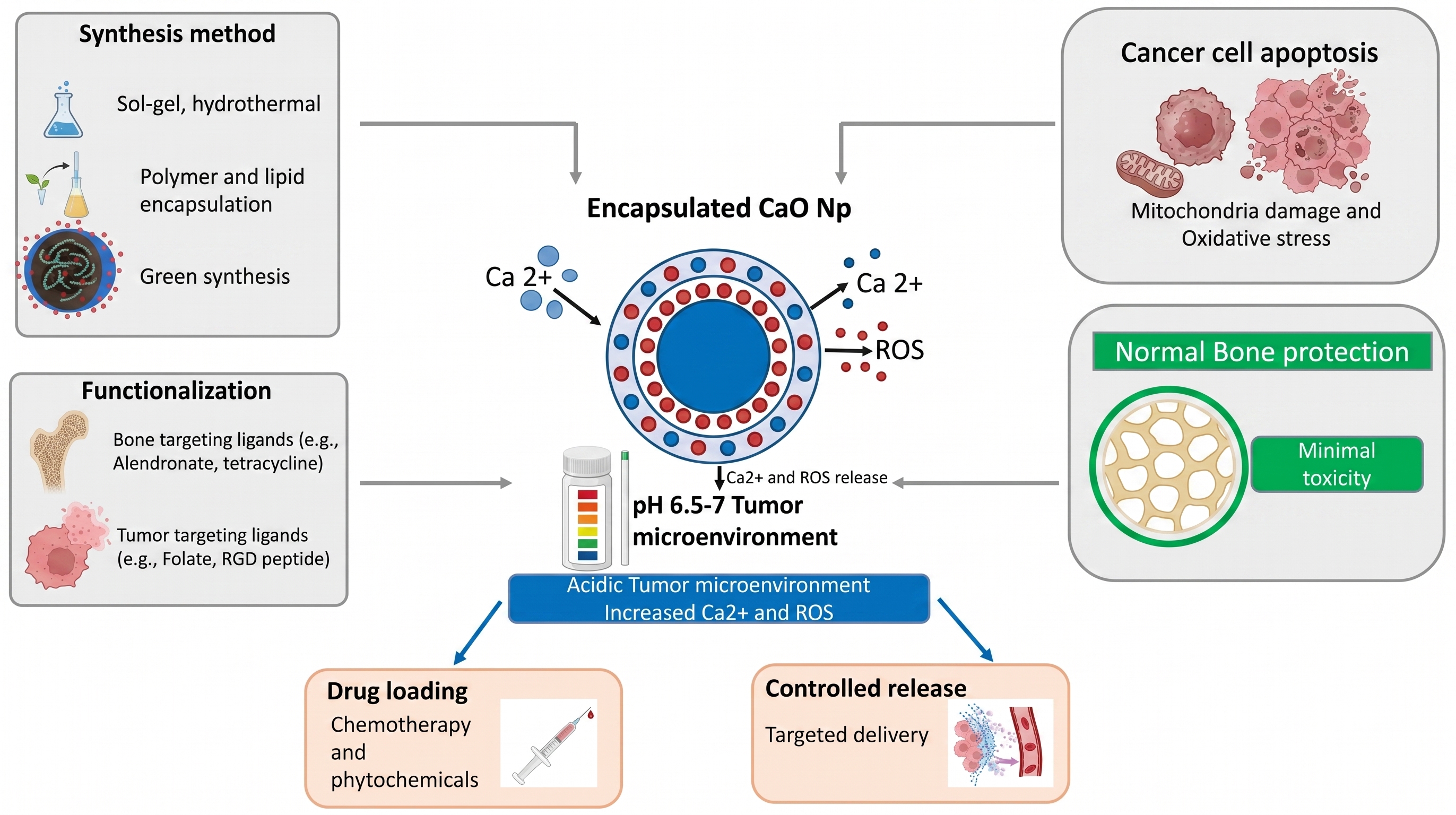
Schematic representation of osteosarcoma with CaO nanoparticles based targeted therapy- The design and suggested therapeutic mechanism of encapsulated CaO nanoparticles are depicted in the image. The CaO NPs may be chemically synthesized (sol-gel, hydrothermal) or synthesized through a green synthesis method. They can also be further stabilized by coating them with polymers or lipids. Selective accumulation at osteosarcoma sites may be improved by surface functionalization with ligands that target bones (like alendronate) or tumors (like folate, RGD peptides). It is postulated that CaO nanoparticles are hydrated in the acidic tumor microenvironment (pH 6.5-7.0) which leads to controlled release of Ca2+ ions and generation of ROS. An increase in intracellular Ca2+ and ROS levels can lead to apoptosis and malfunction of mitochondria in cancer cells. Whereas dashed arrows indicate proposed or design-based processes such as targeted delivery and controlled release, solid arrows are used to indicate nanoparticlesproperties (such ROS production and cytotoxicity) that have been validated by experiments. Even though these advantages require additional in vivo confirmation, encapsulation and phytochemical capping generated through green synthesis could lead to improved biocompatibility, reduced excessive production of ROS, and altered biodistribution of nanoparticles. The therapeutic potential and the need to conduct additional translational studies are both discussed in the figure.
Safety and toxicology evaluation
Due to the specific reactivity and solubility characteristics of CaO nanoparticles, as well as their ability to induce localized alkalinity, it is imperative to evaluate their safety profile. Uncoated CaO NPs have been reported to cause oxidative stress, membrane destruction, and mitochondrial failure. Therefore, cytotoxicity testing, the assessment of reactive oxygen species (ROS) production, the measurement of inflammatory cytokine release, and the evaluation of compatibility with osteoblasts, fibroblasts, and hepatocytes should be prioritized 93,94. In animal models, CaO NPs primarily accumulate in the kidneys, spleen, liver, and bones. An excess of Ca²⁺ in these areas may disrupt calcium homeostasis and induce oxidative stress within the kidneys or liver. Toxicity has been reduced with either polymer-coated or plant-derived CaO NPs through improved compatibility with living tissue and controlled dissolution rates 95,96. Bioactive capping agents, such as flavonoids (quercetin, kaempferol), phenolic acids (gallic acid), proteins, and polysaccharides, are introduced through plant-mediated synthesis. These agents adsorb to the surface of the nanoparticle and reduce its physicochemical reactivity. This phytochemical surface coating contributes to surface passivation by minimizing direct membrane contact and reducing excessive ROS formation. Moreover, such capping agents prevent a sudden influx of intracellular Ca²⁺—which may lead to mitochondrial malfunction—by modulating the kinetics of calcium ion release. Pharmacokinetically, these surface-functionalized nanoparticles may exhibit increased colloidal stability, reduced aggregation, and altered patterns of biodistribution, such as slower clearance and diminished accumulation in reticuloendothelial organs such as the liver and spleen. However, these effects have yet to be rigorously validated in vivo.
A full toxicological profile requires testing for genotoxicity (Ames test, comet assay, micronucleus test), an assessment of systemic toxicity (for example, complement activation and macrophage response), and studies of how calcium hydroxide nanoparticles are distributed in the body—utilizing ICP-MS or radioactive labeling—to determine their clearance routes, potential long-term retention, and accumulation in target versus non-target tissues 97. These assessments are essential for determining systemic toxicity and therapeutic safety. Possible long-term toxicities that must be considered include irritation from alkaline by-products, chronic inflammation, and potential immunological activation arising from the nanoparticle synthesis process. For these reasons, strategies to minimize the risk of CaO NPs will focus on polymeric encapsulation, ensuring that particles are produced free of endotoxins, optimizing size (30 to 100 nm), and administering the NPs locally to minimize systemic absorption. Upon completion, these evaluations will help to ascertain whether CaO NPs are sufficiently safe for potential therapeutic applications 98. Existing research has evaluated the effects of rhombohedral/fusiform calcium carbonate nanoparticles on bone marrow-derived mesenchymal stem cells and their potential to enhance bone formation (by 10–37%) and inhibit adipogenesis 99. The study necessitates that researchers conduct direct tests of CaO NPs on human fetal osteoblasts (hFOBs) and bone marrow-derived mesenchymal stem cells (BMSCs), because the findings indicate that calcium-based systems maintain normal bone cell functions. Clinicians must assess nanotherapeutics by testing their toxicity, pharmacokinetic properties, and biodistribution patterns. Preclinical studies show that nanoparticles, when administered via intraperitoneal dosing over two months, result in no significant decrease in body weight or development of organ disease despite their accumulation in the liver, spleen, and bone marrow. The pharmacokinetic (PK) analyses demonstrate that nanoparticles maintain a dose-dependent relationship between their half-lives, which range from 13.8 hours at 10 mg/kg to 92.9 hours at 90 mg/kg, and their systemic exposure levels. Animal studies demonstrate that bone-targeting delivery methods are necessary because the body rapidly clears medications through reticuloendothelial organs. In accordance with ICH GCP criteria, all regulatory translation processes must complete safety assessments in conjunction with immunotoxicity studies and pharmacodynamic evaluations.
Current Challenges and Future Perspectives
Research gaps and limitations
The presence of flavonoids and phenolic compounds—including quercetin, kaempferol, and vitexin—in A. pinnata extracts demonstrates potential cytotoxic and antioxidant properties; however, no single component has been definitively proven to eradicate osteosarcoma 74. The existing evidence, which largely demonstrates general cytotoxicity in other cancer models, represents a significant research gap. Future research should concentrate on isolating and assessing specific phytochemicals against osteosarcoma cells using bioactivity-guided fractionation. The primary anti-osteosarcoma properties of A. pinnata are believed to originate from its isolated flavonoids. According to Liang et al. (2013), quercetin (25–100 µM) induces MG-63 cell apoptosis by caspase-3/9 activation, mitochondrial depolarization, and Bax/Bcl-2 modulation 37. Furthermore, kaempferol (40–80 µM) induces mitochondrial apoptosis, thereby reducing tumor cell survival 37. A critical research gap exists due to the paucity of studies utilizing whole-plant extracts, which necessitates further dose-response data and mechanistic insights. The adoption of CaO nanotherapies based on plant-mediated synthesis to treat osteosarcoma will be limited until more rigorous research is conducted. Currently, no studies have provided a comprehensive examination of the integration of CaO nanoparticles with phytochemicals derived from A. pinnata. The majority of existing research regarding CaO nanoparticles has concentrated on ZnO, CaP, or mixed calcium-phosphate systems, with limited focus on the utility of CaO for osteosarcoma 100,101. Furthermore, there is a lack of consensus regarding the effective treatment window for osteosarcoma, as the vast majority of studies evaluating the nanotoxicity and anticancer activity of CaO nanoparticle formulations have been conducted using non-bone malignancies or non-malignant cell lines.
Plant extraction methods, pH levels, precursor concentrations, calcination conditions, and particle size and shape all significantly influence the dissolution kinetics of calcium ions from nanoparticles 102,103. This variability restricts access to uniform protocols for producing nanoparticle formulations through green synthesis methods 102,103. The paucity of in vivo studies pertaining to the immunogenicity of nanoparticles, cumulative dosing, biodistribution, and interactions between bone and the tumor microenvironment contributes to a limited understanding of the mechanism(s) of action of nanoparticles in primary and secondary tumor sites—especially considering that factors such as moisture sensitivity and the rapid conversion of calcium oxide to calcium hydroxide can introduce challenges for systemic administration 104. Finally, there is a deficiency in the development of calcium oxide-based nanoparticles that target multiple conditions, including osteosarcoma and metabolic disorders. The development of successful osteosarcoma treatments utilizing herbal extracts and nanoparticles faces substantial challenges because of their low bioavailability at target sites, which compels clinicians to administer high systemic doses to achieve therapeutic bone tissue levels. The effectiveness of many phytochemicals from herbal sources is limited for systemic administration because their oral bioavailability often falls below 10% due to low solubility and rapid first-pass metabolism 105. Unmodified nanoparticles exhibit rapid elimination through the reticuloendothelial system, with only 5% of the delivered dose reaching tumor sites when standard administration methods are used 106. Research has yielded bone-targeted delivery technologies through nanoencapsulation—including injectable hydrogels, scaffold-based platforms, bisphosphonate conjugates, and hydroxyapatite-binding ligands—to enhance local retention while minimizing systemic exposure 107,108. These methods address the bioavailability challenges faced by both herbal medicines and inorganic nanoparticles by enabling bone-specific drug uptake and controlled drug release, which enhances therapeutic efficacy 107,108.
The effectiveness and reproducibility of experimental results depend on the phytochemical composition of A. pinnata, which changes according to geographical origin and the methods used for cultivation, harvesting, and extraction. Untargeted metabolomics (utilizing LC-MS/MS and NMR) and analytical fingerprinting methods (utilizing HPLC/UPLC/SPLC) require standardization to track global phytochemical profiles and measure key bioactive flavonoids, such as quercetin, kaempferol, and gallic acid. Bioactivity-guided validation ensures consistent therapeutic effects, facilitating clinical translation and reproducible formulation development. Direct proof in osteosarcoma models is still lacking, despite the potential anti-osteosarcoma action of the phytochemicals (quercetin, kaempferol, gallic acid) and CaO nanoparticles derived from A. pinnata. The available research includes only a few in vivo studies regarding biodistribution, immunological responses, toxicity, and pharmacokinetics, while sufficient in vitro studies evaluating cytotoxic effects on osteosarcoma cell lines are lacking. This disparity necessitates extensive research to establish safety, efficacy, and tumor-targeting capabilities before clinical application. The research demonstrates that hydroxide forms exert biological effects through the cytotoxicity of Ca(OH)₂ nanoparticles, which possess an IC₅₀ value between 271.9 and 291.8 µg/mL and induce DNA damage through specific ROS mechanisms that affect cancer cells differently than normal cells 109,110. While findings demonstrate that Ca(OH)₂ nanoparticles induce cancer cell death by generating specific types of ROS, the physiological impact of this transformation product on various body systems or cancerous environments requires further investigation 109,110.
Future research perspectives
The future of research in this field will focus on the systematic production of nanoparticles from A. pinnata using environmentally friendly techniques to achieve homogeneity in particle size, crystallinity, morphology, and dissolution characteristics. Another promising application of combining A. pinnata metabolomics and nanoparticle production is the identification of stabilizing agents that enhance the antioxidant and anticancer properties of the resulting nanostructures. Therefore, it is important to develop CaO nanoparticles as "multifunctional platforms" that target the major tumor signaling pathways in osteosarcoma—such as PI3K/AKT, Wnt/β-catenin, and MAPK—via a combination of phytochemicals, chemotherapeutics, or gene-regulatory therapies. Novel 3D in vitro models mimicking osteosarcoma spheroids, along with bone-like scaffolds, will be utilized to more accurately replicate the tumor microenvironment and the cytotoxic activity of the ions 101. The use of orthotopic or patient-derived xenograft (PDX) models in future in vivo studies will provide an opportunity to further evaluate biodistribution, long-term toxicity, immune responses, and tumor-bone interactions 111,112. The use of precision targeting methods, such as hydroxyapatite-binding peptides or bisphosphonate conjugates, is expected to improve bone specificity, while local delivery techniques—including hydrogels, injectable pastes, or bone cement composites—should reduce systemic toxicity 113,114. Successful translation from the laboratory to the clinic must include considerations of standardized, large-scale production, regulatory compliance concerning toxicology studies, and validation via pharmacokinetic, pharmacodynamic, and safety profiles. Future studies will evaluate the use of CaO nanoplatforms for the treatment of osteosarcoma and other comorbidities, such as diabetes or cachexia. Recent progress in bone tissue engineering highlights the critical importance of scaffold design, material selection, and fabrication methods 115. Guo et al. (2025) created PLA/NbC scaffolds which exhibited shape memory and photothermal properties to achieve 90% inhibition of HOS osteosarcoma cells 115. Through FDM technology, Guo et al. (2023) developed TPMS PLA/0.1% GO scaffolds which enhanced BMSC adhesion and osteogenic differentiation and produced a strength increase of approximately 35% in both tensile and compressive measures 116. Guo et al. (2025) and Guo et al. (2024) demonstrated that TPMS hydroxyapatite scaffolds containing bioglass or bredigite as dopants achieved improved compressive strength (reaching 33.5 MPa) along with enhanced biomineralization and ion release of Si, Ca, and P 117,118. The dual-scale PLA-pearl/chitosan scaffolds developed by Guo et al. (2025) showed superior mechanical and biological results because the researchers demonstrated that they could achieve optimal scaffold performance for bone repair through the process of co-designing materials with structural elements and fabrication methods 119.
Discussion
A. pinnata research primarily focuses on isolated flavonoids rather than the use of complete plant extracts. Despite the extensive demonstration of flavonoid-mediated apoptosis in osteosarcoma model systems, there is currently no standardized way to extrapolate such findings on isolated chemicals to whole-plant systems. The available research regarding whole plants is limited due to the lack of quantitative data explaining their mechanistic function.
Calcium oxide (CaO) nanoparticles—which can be produced chemically or through green synthesis—are becoming a common modality in oncological nanomedicine, especially for treating challenging malignancies such as osteosarcoma. Recent studies describe various therapeutic effects of CaO nanoparticles, including their potential to impact oncogenic pathways, induce selective cell death, stimulate ROS-mediated apoptosis, and trigger mitochondrial dysfunction 120. In light of these potential effects, it appears that these strategies could take advantage of the inherent metabolic weaknesses displayed by osteosarcoma cells—specifically, their elevated level of oxidative stress and altered mitochondrial respiration—which would allow these cells to be highly sensitive to additional increases in ROS levels.
In addition, the affinity of calcium oxide (CaO) nanoparticles for bone tissue provides a unique therapeutic opportunity. Upon exposure to physiological conditions, the nanoparticles undergo hydration, resulting in an increase in the concentration of hydroxyl ions (OH⁻) and a simultaneous increase in local pH. Additionally, the dissolution of CaO generates ionic calcium (Ca²⁺), which may play a key role in promoting the accumulation of tumor-specific materials via interaction with components of the bone matrix. As calcium oxide nanoparticles can be delivered directly to the bone, they offer potential benefits, such as reduced systemic toxicity, increased tumor penetration, and localized delivery, representing a promising but still experimental treatment for osteosarcoma. Nonetheless, these benefits must be further confirmed in in vivo and clinical experiments 121.
Combination chemotherapy with drugs like methotrexate, doxorubicin, and cisplatin constitutes a significant portion of the modern treatment of osteosarcoma. Despite their established ability to improve survival rates, the use of these regimens is restricted by severe side effects such as nephrotoxicity, cardiotoxicity, and the development of chemoresistance. In contrast, a multi-targeted strategy that may mitigate systemic toxicity and enhance tumor selectivity is provided by the suggested combination of phytochemicals and CaO nanoparticles, although this hypothesis remains to be verified by experimental validation.
Moreover, due to their intrinsic affinity for bone tissue, calcium oxide nanoparticles (which transform into nano-sized hydrated calcium hydroxide upon contact with physiological solutions) 122,123 produce an immediate change in the acid-base balance of the local environment. This releases calcium ions that could potentially interact with the extracellular matrix of local bone tissue, thereby causing an increase in the locally deposited calcium content and facilitating neo-bone formation at the site of the osteogenic tumor (i.e., osteosarcoma). CaO nanoparticles are therefore presented as a potential therapy for osteosarcoma primarily because they could be delivered directly to bone tissue, where local delivery, increased penetration of tumor cells, and minimized systemic toxicity are clinically required. A new consideration with respect to nano-bio interaction will be how different characteristics of the nanoparticles affect cellular uptake, escape from lysosomes, and the generation of oxidative stress within a cell 124. Also, while there is considerable in vitro evidence that CaO nanoparticles are effective anticancer agents, little information is available regarding their effectiveness in vivo. Thus, there is currently a lower level of confidence regarding the translational potential of CaO nanoparticles as anticancer agents.
The interaction between CaO nanoparticles and A. pinnata phytochemicals has not been scientifically examined. The plant-mediated nanoparticles in this study exhibited lower IC₅₀ values than their respective ZnO nanoparticles, which displayed IC₅₀ values above 500 µg/mL. Therefore, the study remains uncertain about the potential combined treatment effectiveness of these substances against osteosarcoma.
Another major gap in the research is the understanding of the safety and toxicity of materials. High levels of CaO in soft tissues may result in unregulated production of reactive oxygen species (ROS), provoking an inflammatory response and subsequently damaging the tissue 125. Excessive levels of CaO or Ca(OH)₂ nanoparticles create uncontrollable production of ROS, which results in cell death, genomic destruction, and oxidative damage to cells. The need to balance the dosage of ROS-mediated toxicity with increased doses of nanoparticles is also emphasized by these safety concerns. The high levels of ROS in the study caused organ damage, including to the liver and bone marrow, raising safety concerns for high-dose treatment 109. The long-term fate within the body (biodistribution, clearance, potential for immunogenicity) and the risk of Ca²⁺ accumulation in tissues not directly associated with the treatment have not been investigated in adequate detail 126,127. Therefore, before application to the treatment of osteosarcoma, it will be necessary to conduct extensive optimization, standardization, and preclinical evaluation of the nanocarrier systems developed with CaO, even though the results from the initial studies are promising.
Ko et al. (2025) tested CD47 as a potential immunotherapy target for osteosarcoma using samples from 24 patients. The researchers detected CD47 expression in 20.8% of samples, which showed a strong connection with both the age of patients and their metastasis status at diagnosis. The anti-CD47 antibody (B6H12) showed that CD47 plays a critical role in immune system evasion and the process of metastasis because the antibody boosted macrophage phagocytosis, while CD47 blockade failed to stop tumor cell death and their normal cell functions 128. Cancer remains the world's second most frequent cause of death because standard treatments, such as chemotherapy, radiation, and surgical procedures, demonstrate limited success for patients with advanced disease. The CD47 protein functions as an immunological checkpoint because its excessive presence on cancer cells prevents macrophages from performing their phagocytic functions. CD47 therapeutic inhibition serves as an effective cancer immunotherapy method because it activates T cells and myeloid cells and macrophages to boost anticancer immune activity 129. Additional in vivo, pharmacokinetic, and safety tests are required even when preclinical studies show promise for anticancer applications.
Conclusion
Calcium Oxide (CaO) nanoparticles functionalized with A. pinnata extracts have demonstrated significant potential for the treatment of osteosarcoma due to their natural affinity for bone tissue, the ROS-mediated cytotoxic effects of CaO, and their ability to undergo pH-responsive dissolution. Green synthesis of CaO nanoparticles improves their compatibility with human cells and incorporates phytochemicals that provide additional anticancer effects, thereby addressing challenges associated with standardization, stability, and toxicity. However, further research utilizing more robust delivery systems and experimental models is required to investigate these products for clinical therapeutic use before they can be considered a viable treatment option for osteosarcoma. Overall, CaO nanoparticles represent an attractive, emerging, and as-yet-unvalidated technology for cancer therapy.
Abbreviations
ADME: Absorption, Distribution, Metabolism, and Excretion; AKT: Protein Kinase B; Bcl-2: B-cell lymphoma 2; BMSCs: Bone Marrow-derived Mesenchymal Stem Cells; Ca²⁺: Calcium Ion; CaCl₂: Calcium Chloride; Ca(NO₃)₂: Calcium Nitrate; Ca(OH)₂: Calcium Hydroxide; CaO: Calcium Oxide; CaO NPs: Calcium Oxide Nanoparticles; CDK: Cyclin-Dependent Kinase; COX-2: Cyclooxygenase-2; ECM: Extracellular Matrix; EMT: Epithelial-Mesenchymal Transition; ERK: Extracellular Signal-Regulated Kinase; FADD: Fas-Associated protein with Death Domain; FTIR: Fourier-Transform Infrared Spectroscopy; GCP: Good Clinical Practice; GSH: Glutathione; HIF-1α: Hypoxia-Inducible Factor 1-alpha; HPLC: High-Performance Liquid Chromatography; ICP-MS: Inductively Coupled Plasma Mass Spectrometry; ICH: International Council for Harmonisation; IC₅₀: Half-maximal inhibitory concentration; IL-6: Interleukin-6; LC-MS/MS: Liquid Chromatography with tandem Mass Spectrometry; MAP: Methotrexate, Doxorubicin, and Cisplatin; MAPK: Mitogen-Activated Protein Kinase; MDR: Multidrug Resistance; MMP: Matrix Metalloproteinase; MOMP: Mitochondrial Outer Membrane Permeabilization; NF-κB: Nuclear Factor kappa-light-chain-enhancer of activated B cells; NMR: Nuclear Magnetic Resonance; NO: Nitric Oxide; NP(s): Nanoparticle(s); OS: Osteosarcoma; PDX: Patient-Derived Xenograft; PEG: Polyethylene Glycol; PI3K: Phosphatidylinositol 3-Kinase; PK: Pharmacokinetic; PLA: Polylactic Acid; PLGA: Poly(lactic-co-glycolic acid); PTEN: Phosphatase and Tensin Homolog; RGD: Arg-Gly-Asp; ROS: Reactive Oxygen Species; TME: Tumor Microenvironment; TNF-α: Tumor Necrosis Factor-alpha; TPMS: Triply Periodic Minimal Surface; UPLC: Ultra-Performance Liquid Chromatography; UV–Vis: Ultraviolet–Visible; VEGF: Vascular Endothelial Growth Factor; ZnO: Zinc Oxide.
Acknowledgments
None.
Author’s contributions
All authors read and approved the final manuscript.
Funding
None.
Availability of data and materials
Not applicable.
Ethics approval and consent to participate
Not applicable.
Consent for publication
Not applicable.
Declaration of generative AI and AI-assisted technologies in the writing process
The authors declare that they have not used generative AI (a type of artificial intelligence technology that can produce various types of content including text, imagery, audio and synthetic data).
Competing interests
The authors declare that they have no competing interests.